| [1] |
Bray F, Laversanne M, Sung H, et al. Global cancer statistics 2022: GLOBOCAN estimates of incidence and mortality worldwide for 36 cancers in 185 countries[J]. CA Cancer J Clin, 2024, 74(3): 229-263.
|
| [2] |
Tan Y, Wang Z, Xu M, et al. Oral squamous cell carcinomas: state of the field and emerging directions[J]. Int J Oral Sci, 2023, 15(1): 44.
|
| [3] |
Radaic A, Kamarajan P, Cho A, et al. Biological biomarkers of oral cancer[J]. Periodontol 2000, 2024, 96(1): 250-280.
|
| [4] |
Kladney RD, Bulla GA, Guo L, et al. GP73, a novel Golgi-localized protein upregulated by viral infection[J]. Ge-ne, 2000, 249(1/2): 53-65.
|
| [5] |
Xia Y, Zhang Y, Shen M, et al. Golgi protein 73 and its diagnostic value in liver diseases[J]. Cell Prolif, 2019, 52(2): e12538.
|
| [6] |
Liu Y, Hu X, Liu S, et al. Golgi phosphoprotein 73: the driver of epithelial-mesenchymal transition in cancer[J]. Front Oncol, 2021, 11: 783860.
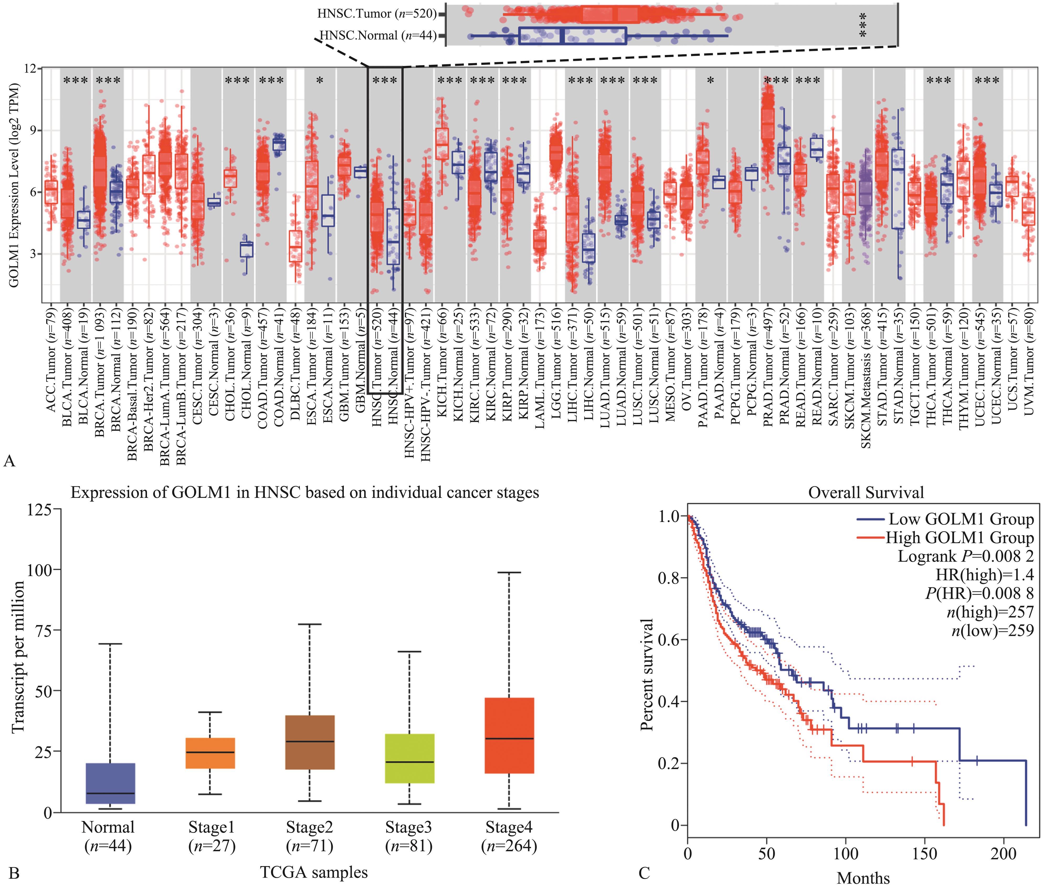
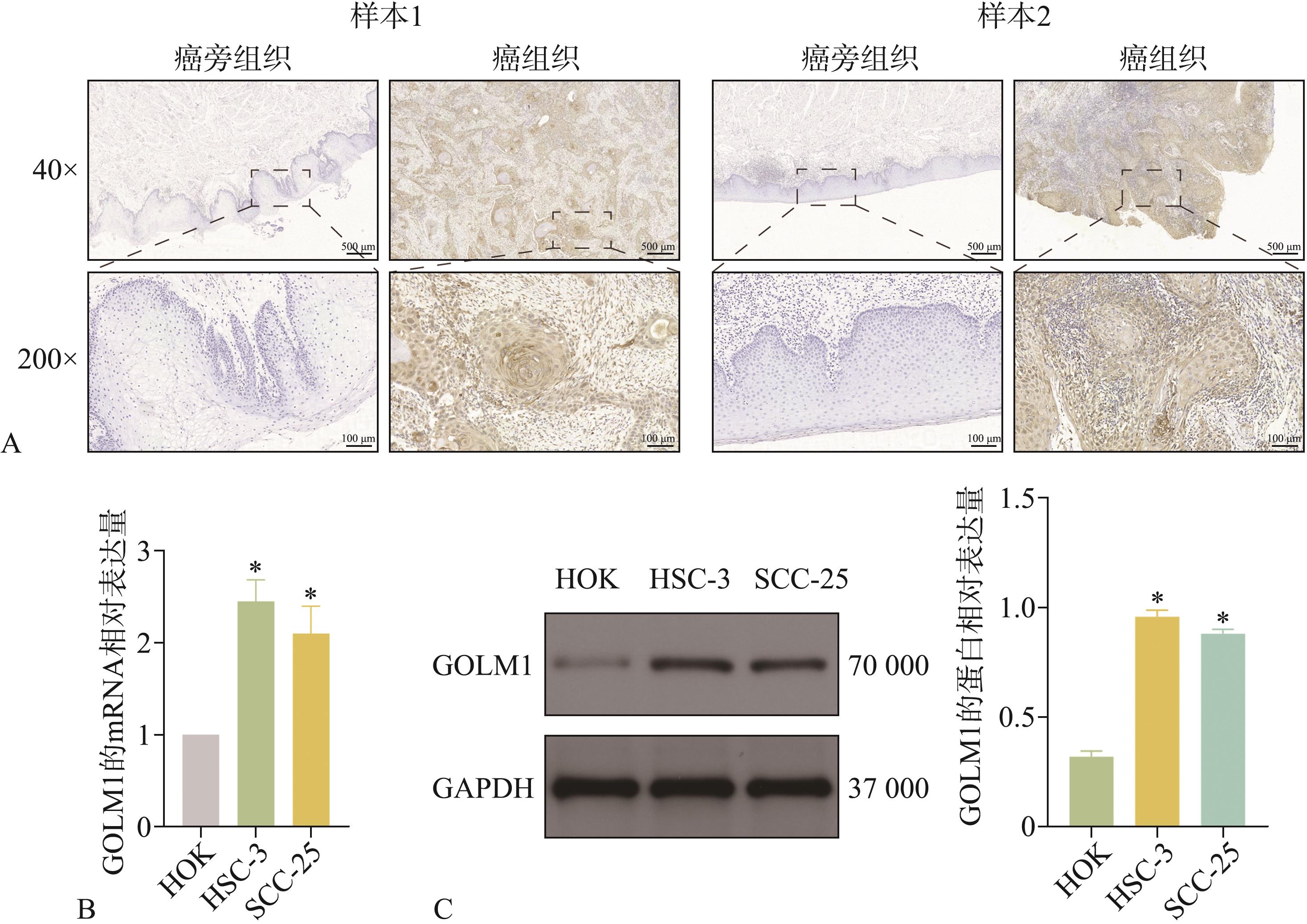
|
| [7] |
Feng P, Hu X, Zhou S, et al. Golgi protein 73: the driver of inflammation in the immune and tumor microenvironment[J]. Front Immunol, 2025, 15: 1508034.
|
| [8] |
Xie P, Wu M, Wang H, et al. GOLM1 dictates acquired Lenvatinib resistance by a GOLM1-CSN5 positive feedback loop upon EGFR signaling activation in hepatocellular carcinoma[J]. Oncogene, 2024, 43(42): 3108-3120.
|
| [9] |
Wang S, Zhang T, Zhou Y, et al. GP73-mediated secretion of PKM2 and GP73 promotes angiogenesis and M2-like macrophage polarization in hepatocellular carcinoma[J]. Cell Death Dis, 2025, 16(1): 69.
|
| [10] |
Yan G, Zhu T, Zhou J, et al. GOLM1 promotes prostate cancer progression via interaction with PSMD1 and enhancing AR-driven transcriptional activation[J]. J Cell Mol Med, 2024, 28(20): e70186.
|
| [11] |
Dang Y, Yu J, Zhao S, et al. GOLM1 drives colorectal cancer metastasis by regulating myeloid-derived suppressor cells[J]. J Cancer, 2021, 12(23): 7158-7166.
|
| [12] |
朱海鹏, 胡军, 姜敏, 等. GOLM1调控PI3K/AKT/mTOR信号转导通路促进肺腺癌细胞增殖、侵袭和迁移的机制研究[J]. 中国癌症杂志, 2022, 32(3): 207-217.
|
|
Zhu HP, Hu J, Jiang M, et al. A study on mechanism of GOLM1 regulating PI3K/AKT/mTOR signaling pathway to promote proliferation,invasion and migration of lung adenocarcinoma cells[J]. Chin Oncol, 2022, 32(3): 207-217.
|
| [13] |
Guan J, Qin Y, Deng G, et al. GOLM1 as a potential therapeutic target modulates B7-H3 secretion to drive ovarian cancer metastasis[J]. Evid Based Complement Alternat Med, 2022, 2022: 5151065.
|
| [14] |
Li H, Yang LL, Xiao Y, et al. Overexpression of Golgi phosphoprotein 2 is associated with poor prognosis in oral squamous cell carcinoma[J]. Am J Clin Pathol, 2018, 150(1): 74-83.
|
| [15] |
Xia C, Dong X, Li H, et al. Cancer statistics in China and United States, 2022: profiles, trends, and determinants[J]. Chin Med J (Engl), 2022, 135(5): 584-590.
|
| [16] |
刘梅, 石兴莲, 李哲臻, 等. 口腔癌患者生存质量相关因素的系统评价[J]. 华西口腔医学杂志, 2024, 42(4): 486-493.
|
|
Liu M, Shi XL, Li ZZ, et al. Systematic review of factors related to quality of life in patients with oral cancer:a systematic review[J]. West Chin J Stomatol, 2024, 42(4): 486-493.
|
| [17] |
Xi Y, Zhang T, Sun W, et al. GOLM1 and FAM49B: potential biomarkers in HNSCC based on bioinformatics and immunohistochemical analysis[J]. Int J Mol Sci, 2022, 23(23): 15433.
|
| [18] |
Huang Y, Hong W, Wei X. The molecular mechanisms and therapeutic strategies of EMT in tumor progression and metastasis[J]. J Hematol Oncol, 2022, 15(1): 129.
|
| [19] |
Fontana R, Mestre-Farrera A, Yang J. Update on epithelial-mesenchymal plasticity in cancer progression[J]. Annu Rev Pathol, 2024, 19: 133-156.
|
| [20] |
Mao Z, Wu Y, Yao P, et al. GOLM1 facilitates human colorectal cancer progression and metastasis via activating the AKT/GSK3β/EMT axis[J]. Neoplasma, 2023, 70(1): 136-144.
|
| [21] |
冀为, 赵元元, 张飞, 等. TGF-β与细胞内信号通路的交互作用在肿瘤研究中的进展[J]. 中国肿瘤临床, 2018, 45(15): 800-803.
|
|
Ji W, Zhao YY, Zhang F, et al. Research progress on the crosstalk between TGF-β and multiple intracellular signaling pathmays in cancer progression[J]. Chin J Clin Oncol, 2018, 45(15): 800-803.
|
| [22] |
Lee JH, Massagué J. TGF-β in developmental and fibrogenic EMTs[J]. Semin Cancer Biol, 2022, 86(Pt 2): 136-145.
|
| [23] |
Peng D, Fu M, Wang M, et al. Targeting TGF-β signal transduction for fibrosis and cancer therapy[J]. Mol Cancer, 2022, 21(1): 104.
|
| [24] |
王迪侃, 廖贵清. 唾液中白细胞介素与口腔癌的关系[J]. 华西口腔医学杂志, 2018, 36(3): 325-330.
|
|
Wang DK, Liao GQ. Relationship between interleukins in the saliva and oral cavity cancer[J]. West Chin J Stomatol, 2018, 36(3): 325-330.
|
| [25] |
Bachert C, Fimmel C, Linstedt AD. Endosomal trafficking and proprotein convertase cleavage of cis Golgi protein GP73 produces marker for hepatocellular carcinoma[J]. Traffic, 2007, 8(10): 1415-1423.
|
| [26] |
Zhang X, Wu LN, Li XQ, et al. Whether the Golgi protein 73 could be a diagnostic serological marker in hepatocellular carcinoma: a meta analysis[J]. BMC Gastroenterol, 2023, 23(1): 85.
|

 ), Wen Cai2,3, Yu Li2,4, Chen Junliang2,5, Feng Hao1,2(
 This work is licensed under a Creative Commons Attribution 3.0 License.