West China Journal of Stomatology ›› 2024, Vol. 42 ›› Issue (6): 716-722.doi: 10.7518/hxkq.2024.2024150
• Basic Research • Previous Articles Next Articles
Xie Diya1( ), Shan Danni1, Zhang Lei1, Chen Sheng1, Na Yingyu2, Wang Zhiyong1()
Received:2024-04-16
Revised:2024-05-16
Online:2024-12-01
Published:2024-11-29
Contact:
Wang Zhiyong
E-mail:wowxdy@163.com;zywangkq@nju.edu.cn
Supported by:CLC Number:
Xie Diya, Shan Danni, Zhang Lei, Chen Sheng, Na Yingyu, Wang Zhiyong. Differences in near-infrared fluorescence imaging and histological analysis of cheek mucosa in golden hamsters with different pathological states[J]. West China Journal of Stomatology, 2024, 42(6): 716-722.
Add to citation manager EndNote|Ris|BibTeX
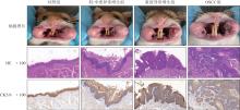
Fig 1
Construction of mucosal lesion models with different pathological states

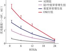
Fig 2
Quantitative analysis of fluorescence intensity in mucosal lesions of every group
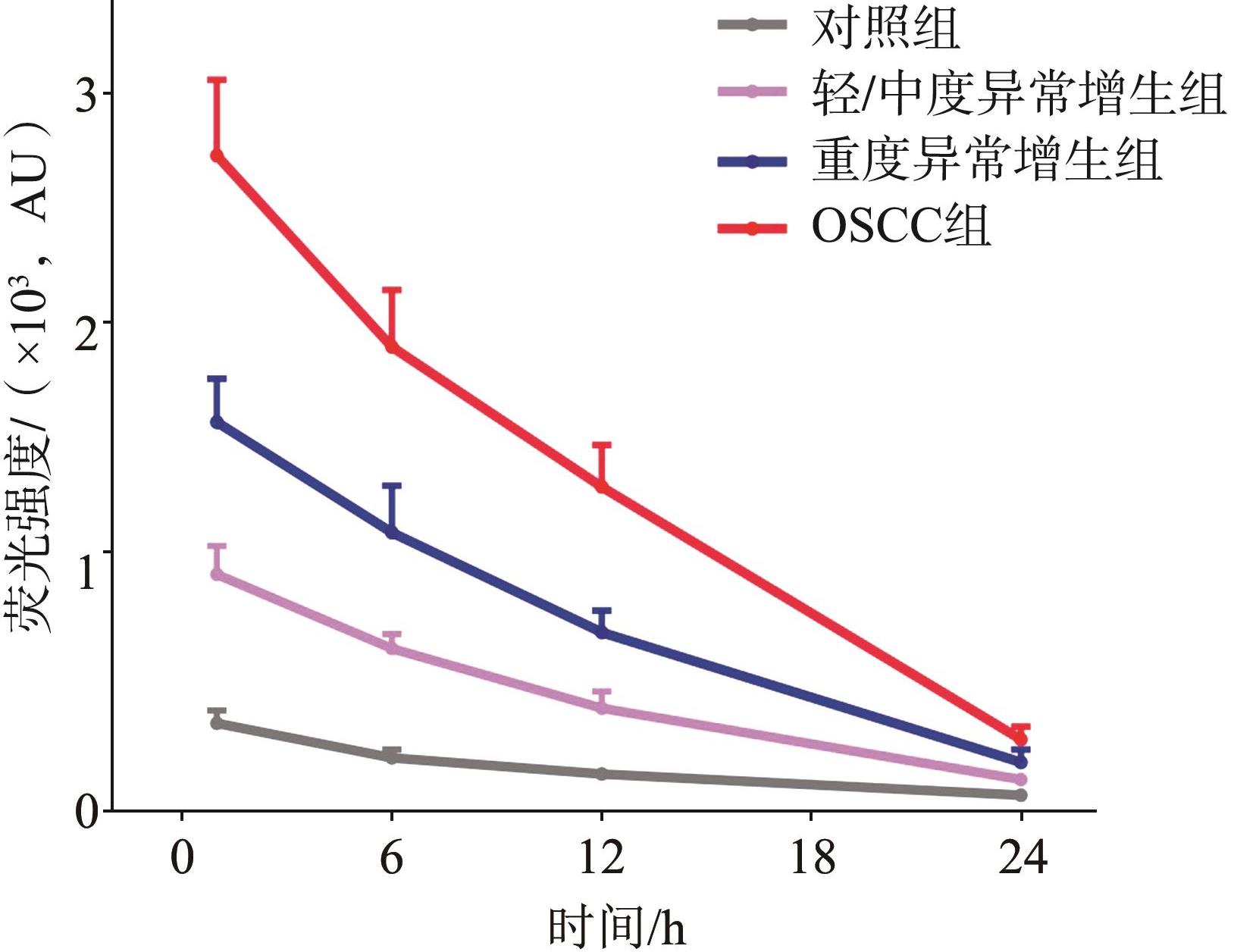
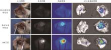
Fig 3
NIF imaging of mucosal lesions of every group

Tab 1
Quantitative analysis of ICG-NIF imaging, MVD and MLVD
| 组别 | MFI | SBR | MVD | MLVD |
|---|---|---|---|---|
| 对照组 | 65±9.16 | 14.4±2.97 | 7.8±2.59 | |
| 轻/中度异常增生组 | 123±12.26 | 1.89±0.32 | 26.4 ±5.32 | 6.4±1.64 |
| 重度异常增生组 | 214±21.12 | 3.27±0.46 | 73.2±11.14 | 5.6±1.32 |
| OSCC组 | 325±47.46 | 4.97±0.87 | 101.2±15.30 | 4.2±1.14 |
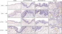
Fig 4
Immunohistochemical staining of blood vessels and lymphatic vessels in tissue slices of every group

Tab 2
Optical imaging in the diagnosis of OPMD malignant transformation
| 技术 | 检测深度/mm | 优势 | 不足 |
|---|---|---|---|
| 自体荧光成像 | 操作简便,阴性预测值较高 | 特异性较低 | |
| 窄带成像技术 | 0.17~0.20 | 显示上皮内血管,提高组织表面结构的对比度 | 穿透深度不足,角化物影响血管可见性 |
| 反射式共聚焦显微镜 | 0.2~03 | 高分辨率显示亚细胞结构 | 穿透深度不足,角化物影响观察 |
| 光学相干断层扫描技术 | 1.5~2 | 提供口腔组织微观结构和微血管的二维或三维成像 | 机械压力会影响,软组织的光学特征 |
| ICG-NIF成像 | 5~10 | 足够的穿透深度,可定量分析 | 需注射荧光显影剂 |
| 1 | Siegel RL, Miller KD, Fuchs HE, et al. Cancer statistics, 2021[J]. CA Cancer J Clin, 2021, 71(1): 7-33. |
| 2 | Warnakulasuriya S. Global epidemiology of oral and oropharyngeal cancer[J]. Oral Oncol, 2009, 45(4/5): 309-316. |
| 3 | Xia CF, Dong XS, Li H, et al. Cancer statistics in China and United States, 2022: profiles, trends, and determinants[J]. Chin Med J, 2022, 135(5): 584-590. |
| 4 | Parak U, Lopes Carvalho A, Roitberg F, et al. Effectiveness of screening for oral cancer and oral potentially malignant disorders (OPMD): a systematic review[J]. Prev Med Rep, 2022, 30: 101987. |
| 5 | Bouaoud J, Bossi P, Elkabets M, et al. Unmet needs and perspectives in oral cancer prevention[J]. Cancers, 2022, 14(7): 1815. |
| 6 | 梁甲武, 卫明慧, 刘青. 口腔潜在恶性病变癌变的早期无创检查方法[J]. 实用口腔医学杂志, 2018, 34(4): 557-560. |
| Liang JW, Wei MH, Liu Q. Early non-invasive examination methods for malignant transformation of potential oral lesions[J]. J Pract Stomatol, 2018, 34(4): 557-560. | |
| 7 | Xu Y, Deng X, Sun Y, et al. Optical imaging in the diagnosis of OPMDs malignant transformation[J]. J Dent Res, 2022, 101(7): 749-758. |
| 8 | Fang J, Islam W, Maeda H. Exploiting the dynamics of the EPR effect and strategies to improve the therapeutic effects of nanomedicines by using EPR effect enhancers[J]. Adv Drug Deliv Rev, 2020, 157: 142-160. |
| 9 | Shi Y, van der Meel R, Chen XY, et al. The EPR effect and beyond: strategies to improve tumor targeting and cancer nanomedicine treatment efficacy[J]. Theranostics, 2020, 10(17): 7921-7924. |
| 10 | Ikeda-Imafuku M, Wang LLW, Rodrigues D, et al. Stra-tegies to improve the EPR effect: a mechanistic perspective and clinical translation[J]. J Control Release, 2022, 345: 512-536. |
| 11 | Egloff-Juras C, Bezdetnaya L, Dolivet G, et al. NIR fluorescence-guided tumor surgery: new strategies for the use of indocyanine green[J]. Int J Nanomedicine, 2019, 14: 7823-7838. |
| 12 | Mieog JSD, Achterberg FB, Zlitni A, et al. Fundamentals and developments in fluorescence-guided cancer surgery[J]. Nat Rev Clin Oncol, 2022, 19(1): 9-22. |
| 13 | Iocca O, Sollecito TP, Alawi F, et al. Potentially malignant disorders of the oral cavity and oral dysplasia: a systematic review and Meta-analysis of malignant transformation rate by subtype[J]. Head Neck, 2020, 42(3): 539-555. |
| 14 | Mello FW, Miguel AFP, Dutra KL, et al. Prevalence of oral potentially malignant disorders: a systematic review and Meta-analysis[J]. J Oral Pathol Med, 2018, 47(7): 633-640. |
| 15 | 李晨曦, 施芷怡, 喻仲麟, 等. 口腔白斑病自体荧光成像结果与临床病理特征相关性分析[J]. 临床口腔医学杂志, 2023, 39(3): 153-157. |
| Li CX, Shi ZY, Yu ZL, et al. Correlation analysis between autofluorescence imaging results and clinicopathological characteristics of oral leukoplakia[J]. J Clin Stomatol, 2023, 39(3): 153-157. | |
| 16 | 王倩, 徐偲, 韩莹, 等. 甲苯胺蓝染色在口腔潜在恶性疾患及口腔鳞状细胞癌早期诊断中的临床应用价值研究[J]. 中国实用口腔科杂志, 2020, 13(12): 738-744. |
| Wang Q, Xu S, Han Y, et al. Clinical application value of toluidine blue staining in the early diagnosis of oral potential malignant disorders and oral squamous cell carcinoma[J]. Chin J Pract Stomatol, 2020, 13 (12): 738-744. | |
| 17 | 王映, 李志萍, 王浩然, 等. VELscope自体荧光检查在糜烂型口腔扁平苔藓诊断及恶变筛查中的应用研究[J]. 中国实用口腔科杂志, 2022, 15(2): 212-215. |
| Wang Y, Li ZP, Wang HR, et al. Application of VELscope autofluorescence examination in diagnosis and malignancy screening of erosive oral lichen planus[J]. Chin J Pract Stomatol, 2022, 15(2): 212-215. | |
| 18 | Lee YJ, Krishnan G, Nishio N, et al. Intraoperative fluorescence-guided surgery in head and neck squamous cell carcinoma[J]. Laryngoscope, 2021, 131(3): 529-534. |
| 19 | 王育新, 王志勇, 王永功, 等. 吲哚菁绿荧光成像技术在口腔鳞癌治疗中的应用: 中国专家共识[J]. 中国口腔颌面外科杂志, 2022, 20(1): 1-6. |
| Wang YX, Wang ZY, Wang YG, et al. Chinese expert consensus statement on the application of of indocyanine green (ICG) fluorescence imaging in the treatment of oral squamous cell carcinoma[J]. Chin J Oral Maxillofac Surg, 2022, 20(1): 1-6. | |
| 20 | 张誉, 季彤. 吲哚菁绿在口腔癌可视化治疗中的应用[J]. 口腔疾病防治, 2020, 28(2): 118-122. |
| Zhang Y, Ji T. Application of indocyanine green in visual treatment of oral cancer[J]. J Prev Treat Stomatol Dis, 2020, 28(2): 118-122. | |
| 21 | Pan J, Deng H, Hu S, et al. Real-time surveillance of surgical margins via ICG-based near-infrared fluorescence imaging in patients with OSCC[J]. World J Surg Oncol, 2020, 18(1): 96. |
| 22 | Wu ZH, Dong YC, Wang YX, et al. Clinical application of indocyanine green fluorescence navigation technology to determine the safe margin of advanced oral squamous cell carcinoma[J]. Gland Surg, 2022, 11(2): 352-357. |
| 23 | Vairaktaris E, Spyridonidou S, Papakosta V, et al. The hamster model of sequential oral oncogenesis[J]. Oral Oncol, 2008, 44(4): 315-324. |
| 24 | Wang ZD, Cormier RT. Golden Syrian hamster models for cancer research[J]. Cells, 2022, 11(15): 2395. |
| 25 | Xie DY, Wang YX, Wang ZY, et al. Kinetics analysis of indocyanine green based on a novel mouse model to distinguish between tumor and inflammation[J]. Anal Methods, 2019, 11(44): 5704-5710. |
| 26 | Pal R, Lwin TM, Krishnamoorthy M, et al. Fluorescence lifetime of injected indocyanine green as a universal marker of solid tumours in patients[J]. Nat Biomed Eng, 2023, 7(12): 1649-1666. |
| [1] | Yu Li, Zhou Tiejun, Wu Xiao, Lin Xinhong, Zhang Xiaoyan, Lai Yongxian, Liao Xinyue, Si Hang, Feng Yun, Jian Jie, Feng Yan. miR-302a-3p targeting lysosomal-associated membrane protein 5 inhibits the invasion and metastasis of oral squamous cell carcinoma [J]. West China Journal of Stomatology, 2025, 43(4): 547-558. |
| [2] | Cui Yingying, Ding Chuanyang, Peng Chaoran, Zhang Jianyun, Cai Xinjia, Li Tiejun. Progress in clinicopathological diagnosis of oral potentially malignant disorders [J]. West China Journal of Stomatology, 2025, 43(3): 314-324. |
| [3] | Ding Xiao, Chen Jiawen, Qu Pengyu, Sun Chenyu, Li Hongli, Hu Wenting, Fan Xin. miR-362-3p inhibited the invasion and metastasis of oral squamous cell carcinoma cells by targeting the regulation of pituitary tumor-transforming gene 1 [J]. West China Journal of Stomatology, 2024, 42(1): 46-55. |
| [4] | Cong Biqiao, Liu Xiaoping, Chen Jiawen, Li Hongli, Fan Xin. Effect of microRNA-663b on migration, invasion and epithelial‑mesenchymal transition of oral squamous cell carcinoma cells [J]. West China Journal of Stomatology, 2022, 40(4): 386-393. |
| [5] | Zhang Yuting, Liu Jiang, Zhao Hang, He Yang, Chen Qianming. Synthesis of 5-fluorouracil-lactoside derivatives and experimental study on their anti-oral squamous cell carcinoma activity [J]. West China Journal of Stomatology, 2022, 40(1): 32-38. |
| [6] | Gao Yongqiang, Shi Pengwei, Shi Wenkai, Liu Yiming. Expression and mechanism of long non-coding RNA HCG22 in oral squamous cell carcinoma [J]. West China Journal of Stomatology, 2021, 39(6): 658-666. |
| [7] | Zhang Shuaiyuan, Qin Shuo, Li Guanghui, Yi Yaqun, Fu Haojie, Gao Yajing, Sun Minglei.. Detection of peripheral blood circulating tumor cells in oral squamous cell carcinoma and its clinical significance [J]. West China Journal of Stomatology, 2021, 39(5): 591-597. |
| [8] | Zhou Haixia, Wang Luyao, Chen Shuai, Wang Dandan, Fang Zheng. circ_0005379 inhibits the progression of oral squamous cell carcinoma by regulating the miR-17-5p/acyl-CoA oxidase 1 axis [J]. West China Journal of Stomatology, 2021, 39(4): 425-433. |
| [9] | Wan Zixin, Zheng Zhijian, Huang Meichang, Chen Yu, Yao Lihong. Expression of Ki-67, Cyclin D1, P53, and P16 in patients with oral leukoplakia and leukoplakia cancerization with spicy diet in Chengdu [J]. West China Journal of Stomatology, 2021, 39(4): 434-440. |
| [10] | Lin Chengzhong, Liu Zheqi, Zhou Wenkai, Ji Tong, Cao Wei. Effect of the regulator of G-protein signaling 2 on the proliferation and invasion of oral squamous cell carcinoma cells and its molecular mechanism [J]. West China Journal of Stomatology, 2021, 39(3): 320-327. |
| [11] | Wu Baoqin, Li Chunhui, Zhang Menglian, Nie Minhai. microRNA-1 gene delivery mediated by exosomes suppresses CAL-27 cell proliferation [J]. West China Journal of Stomatology, 2021, 39(2): 136-142. |
| [12] | Xia Xiaoyang, Fang Fei, Liu Yan, Che Chao, Ke Jinjuan, Jiang Shengjun. Expression of cyclophilin A in oral squamous cell carcinoma and its effect on cell proliferation and invasion [J]. West China Journal of Stomatology, 2021, 39(2): 164-169. |
| [13] | Huang Sheng, Zhang Qiyuan, He Aie, Li Hongbo, Zhang Zhixing. Sex determining region Y-box 9 induced microtubule formation and epithelial⁃mesenchymal transition in human oral squamous cell carcinoma CAL27 cells [J]. West China Journal of Stomatology, 2021, 39(1): 74-80. |
| [14] | Zhao Ge, Li Changxue, Guo Chao, Zhu Hui. MicroRNA model that can predict the prognosis of oral squamous cell carcinoma based on bioinformatics analysis [J]. West China Journal of Stomatology, 2020, 38(6): 622-627. |
| [15] | Zhong Laiping. Standardized and individualized diagnosis and treatment of oral squamous cell carcinoma: opportunities and challenges [J]. West China Journal of Stomatology, 2020, 38(5): 484-488. |
| Viewed | ||||||
|
Full text |
|
|||||
|
Abstract |
|
|||||
 This work is licensed under a Creative Commons Attribution 3.0 License.
