West China Journal of Stomatology ›› 2025, Vol. 43 ›› Issue (6): 819-828.doi: 10.7518/hxkq.2025.2025044
• Basic Research • Previous Articles Next Articles
Gao Li1,2( ), Zhao Mingyue1,2, Yang Shun1, Wang Runan1, Cheng Jiajia1,2, Chen Guangsheng1,2
Received:2025-02-06
Revised:2025-06-02
Online:2025-12-01
Published:2025-11-27
Contact:
Gao Li
E-mail:467278759@qq.com
Supported by:CLC Number:
Gao Li, Zhao Mingyue, Yang Shun, Wang Runan, Cheng Jiajia, Chen Guangsheng. Preparation of polycaprolactone-polyethylene glycol-concentrated growth factor composite scaffolds and the effects on the biological properties of human periodontal ligament stem cells[J]. West China Journal of Stomatology, 2025, 43(6): 819-828.
Add to citation manager EndNote|Ris|BibTeX
Fig 1
Primary and subcultures of hPDLSCs under the microscope
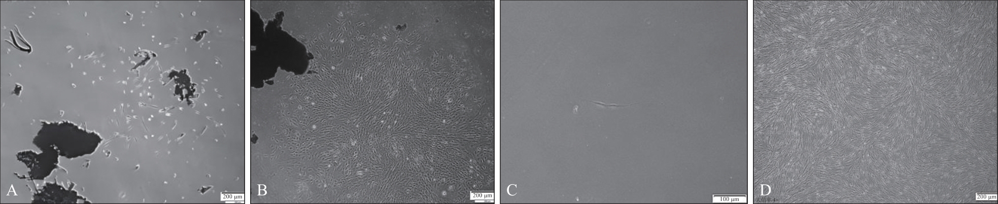
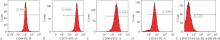
Fig 2
Expression of the relevant phenotypic molecules of hPDLSCs was determined by flow cytometry
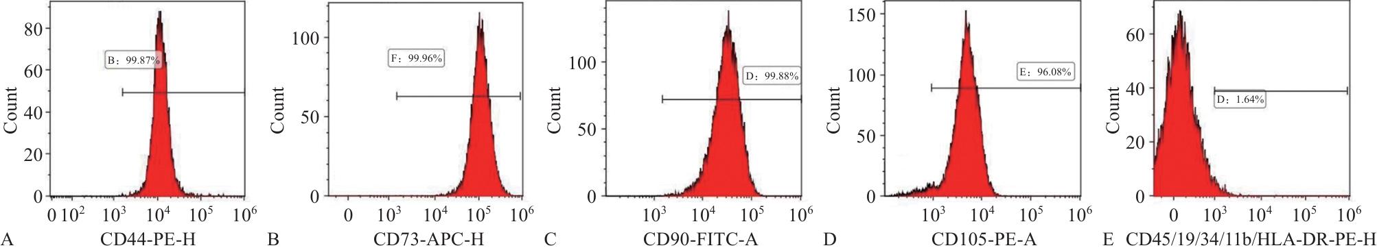
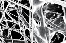
Fig 3
Structural features of the PCL-PEG scaffold and the PCL-PEG-CGF composite scaffold under SEM × 5 μm
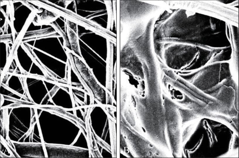
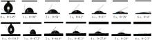
Fig 4
Hydrophilicity detection of the PCL-PEG scaffold and the PCL-PEG-CGF composite scaffold

Tab 1
Mechanical properties of the PCL-PEG scaffold and the PCL-PEG-CGF composite scaffold
| 项目 | PCL-PEG支架 | PCL-PEG-CGF复合支架 | P值 |
|---|---|---|---|
| 断裂拉伸强度 | 4.573 3±0.105 0 | 4.440 0±0.115 3 | 0.213 |
| 断裂延伸率 | 123.333 3±0.577 4 | 124.000 0±1.000 0 | 0.374 |
| 杨氏弹性模量 | 5.270 0±0.111 4 | 4.590 0±0.149 3 | 0.003* |
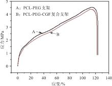
Fig 5
Stress-strain detection of the PCL-PEG scaffold and the PCL-PEG-CGF composite scaffold
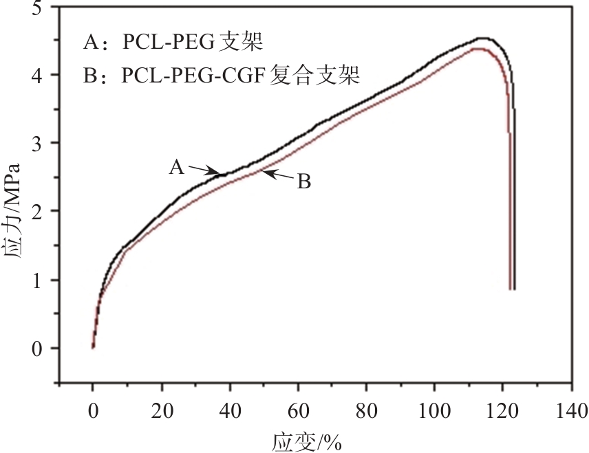
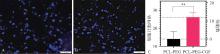
Fig 6
Nuclei of hPDLSCs stained with DAPI
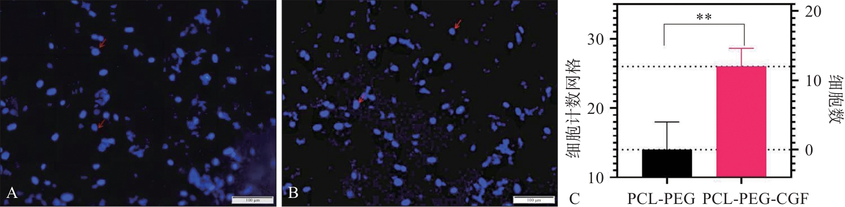
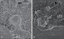
Fig 7
hPDLSCs growth on two scaffolds SEM × 10 μm
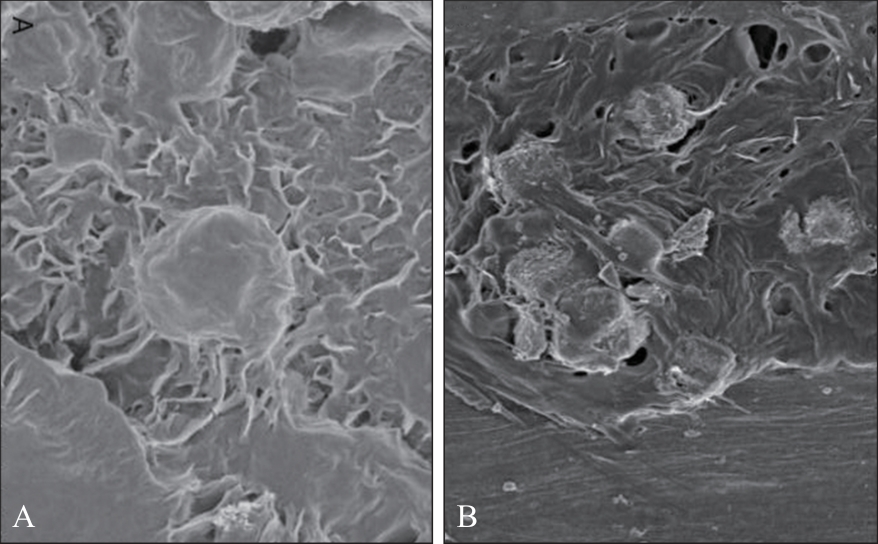
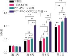
Fig 8
CCK-8 assay results of hPDLSCs proliferation in each group at 1, 3, 5, and 7 days

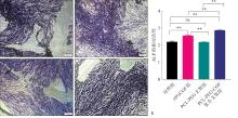
Fig 9
ALP staining and activity of hPDLSCs after 7 days of osteogenesis induction

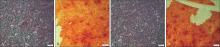
Fig 10
Alizarin Red S staining of hPDLSCs after 21-day osteogenic induction

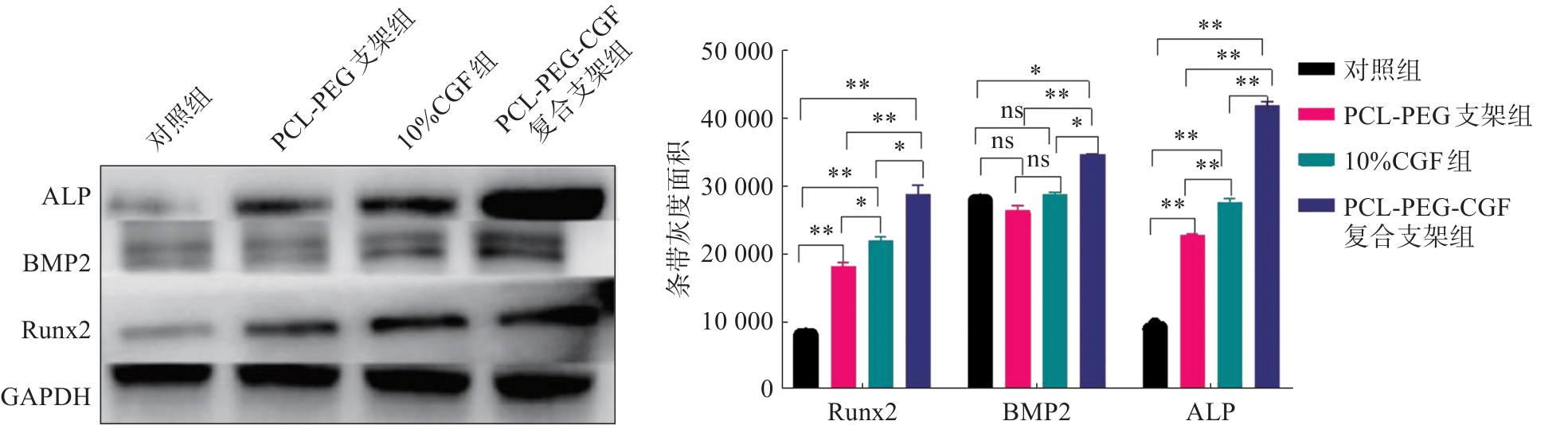
Fig 11
Relative quantification of hPDLSCs osteogenesis-related protein expression in each group
| [1] | Pigeot S, Klein T, Gullotta F, et al. Manufacturing of human tissues as off-the-shelf grafts programmed to induce regeneration[J]. Adv Mater, 2021, 33(43): e2103737. |
| [2] | Maia FR, Bastos AR, Oliveira JM, et al. Recent approa-ches towards bone tissue engineering[J]. Bone, 2022, 154: 116256. |
| [3] | Atala A. Bioengineered tissues for urogenital repair in children[J]. Pediatr Res, 2008, 63(5): 569-575. |
| [4] | Elghblawi E. Platelet-rich plasma, the ultimate secret for youthful skin elixir and hair growth triggering[J]. J Cosmet Dermatol, 2018, 17(3): 423-430. |
| [5] | Sarkar R, Gupta M. Platelet-rich plasma in melasma-A systematic review[J]. Dermatol Surg, 2022, 48(1): 131-134. |
| [6] | Sheikh Z, Hamdan N, Ikeda Y, et al. Natural graft tissues and synthetic biomaterials for periodontal and alveolar bone reconstructive applications: a review[J]. Biomater Res, 2017, 21(1): 9. |
| [7] | 王妮, 程佳佳, 高丽. 静电纺丝复合支架在骨组织工程中的应用[J]. 遵义医科大学学报, 2024, 47(2): 194-200. |
| Wang N, Cheng JJ, Gao L. Application of electrospinning composite scaffold in bone tissue engineering[J]. J Zunyi Med Univ, 2024, 47(2): 194-200. | |
| [8] | Pan L, Yang J, Xu L. Preparation and characterization of simvastatin-loaded PCL/PEG nanofiber membranes for drug sustained release[J]. Molecules, 2022, 27(21): 7158. |
| [9] | Koupaei N, Karkhaneh A, Daliri Joupari M. Preparation and characterization of (PCL-crosslinked-PEG)/hydroxyapatite as bone tissue engineering scaffolds[J]. J Biomed Mater Res A, 2015, 103(12): 3919-3926. |
| [10] | Dai T, Ma J, Ni S, et al. Attapulgite-doped electrospun PCL scaffolds for enhanced bone regeneration in rat cranium defects[J]. Biomater Adv, 2022, 133: 112656. |
| [11] | Mijiritsky E, Assaf HD, Peleg O, et al. Use of PRP, PRF and CGF in periodontal regeneration and facial rejuvenation-A narrative review[J]. Biology (Basel), 2021, 10(4): 317. |
| [12] | Honda H, Tamai N, Naka N, et al. Bone tissue enginee-ring with bone marrow-derived stromal cells integrated with concentrated growth factor in Rattus norvegicus calvaria defect model[J]. J Artif Organs, 2013, 16(3): 305-315. |
| [13] | Kashef-Saberi MS, Hayati Roodbari N, Parivar K, et al. Enhanced osteogenic differentiation of mesenchymal stem cells on electrospun polyethersulfone/poly (vinyl) alcohol/platelet rich plasma nanofibrous scaffolds[J]. ASAIO J, 2018, 64(5): e115-e122. |
| [14] | Pini M. Mechanical properties of the periodontal ligament: a systematic review[J]. J Biomech, 2019, 87: 1-11. |
| [15] | Ritsvall O, Albinsson S. Emerging role of YAP/TAZ in vascular mechanotransduction and disease[J]. Microcirculation, 2024, 31(4): e12838. |
| [16] | Inchingolo AD, Inchingolo AM, Malcangi G, et al. Effects of resveratrol, curcumin and quercetin supplementation on bone metabolism-A systematic review[J]. Nutrients. 2022, 14(17): 3519. |
| [17] | Liu Y, Cooper PR, Barralet JE, et al. Influence of calcium phosphate crystal assemblies on the proliferation and osteogenic gene expression of rat bone marrow stromal cells[J]. Biomaterials, 2007, 28(7): 1393-1403. |
| [18] | McMurray RJ, Gadegaard N, Tsimbouri PM, et al. Nanoscale surfaces for the long-term maintenance of mesenchymal stem cell phenotype and multipotency[J]. Nat Mater, 2011, 10(8): 637-644. |
| [19] | Bharti R, Anisha, Tikku AP, et al. Effect of platelet-rich fibrin and concentrated growth factor on the regenerative potential of human-induced pluripotent stem cells: a comparative analysis[J]. J Conserv Dent Endod, 2024, 27(9): 975-982. |
| [20] | Dipalma G, Inchingolo AM, Colonna V, et al. Autologous and heterologous minor and major bone regeneration with platelet-derived growth factors[J]. J Funct Biomater, 2025, 16(1): 16. |
| [21] | Arya PN, Saranya I, Selvamurugan N. RUNX2 regulation in osteoblast differentiation: a possible therapeutic function of the lncRNA and miRNA-mediated network[J]. Differentiation, 2024, 140: 100803. |
| [22] | Wu M, Wu S, Chen W, Li YP. The roles and regulatory mechanisms of TGF-β and BMP signaling in bone and cartilage development, homeostasis and disease[J]. Cell Res, 2024, 34(2): 101-123. |
| [23] | Beederman M, Lamplot JD, Nan G, et al. BMP signaling in mesenchymal stem cell differentiation and bone formation[J]. J Biomed Sci Eng, 2013, 6(8A): 32-52. |
| [24] | Bartold M, Gronthos S, Haynes D, et al. Mesenchymal stem cells and biologic factors leading to bone formation[J]. J Clin Periodontol, 2019, 46 (): 12-32. |
| Viewed | ||||||
|
Full text |
|
|||||
|
Abstract |
|
|||||
 This work is licensed under a Creative Commons Attribution 3.0 License.
