West China Journal of Stomatology ›› 2024, Vol. 42 ›› Issue (1): 37-45.doi: 10.7518/hxkq.2024.2023213
• Basic Research • Previous Articles Next Articles
Tang Xiaoxue( ), Zhou Zheng, Li Qiqi, Jiang Dandan
Received:2023-07-07
Revised:2023-12-20
Online:2024-02-01
Published:2024-01-12
Contact:
Tang Xiaoxue
E-mail:tangxiaoxue638@163.com
Supported by:CLC Number:
Tang Xiaoxue, Zhou Zheng, Li Qiqi, Jiang Dandan. Effects of sitagliptin activation of the stromal cell-derived factor-1/CXC chemokine receptor 4 signaling pathway on the proliferation, apoptosis, inflammation, and osteogenic differentiation of human periodontal ligament stem cells induced by lipopolysaccharide[J]. West China Journal of Stomatology, 2024, 42(1): 37-45.
Add to citation manager EndNote|Ris|BibTeX
Tab 1
Primer sequences for RT-qPCR
| 基因名称 | 正向引物(5’-3’) | 反向引物(5’-3’) |
|---|---|---|
| RUNX2 | CGAATGGCTAGCACGCTATTAA | GTCGCCATAACAGATTCATCCG |
| OCN | TCACACTCCTCGCCCTATT | GATGTGGTCAGCCAACTCG |
| OPN | TCCTAGCCCCACAGACCCTT | CACACTATCACCTCGGCCAT |
| SDF-1 | ATTCTCAACACTCCAAACTGTGC | CGGTATGAACTGAACTTGCAATG |
| CXCR4 | AGCTGTTGGTGACACGCGTGGTCTATG | GCACTACTGGTGGCCCTTGGAGTATGA |
| GAPDH | GCACCGTCAAGGCTGAGAAC | TGGTGAAGACGCCAGTGGA |
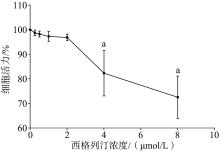
Fig 1
Effect of sitagliptin on the activity of hPDLSCs

Tab 2
Comparison of proliferative activity of hPDL-SCs in each group
| 组别 | 增殖活性 | ||
|---|---|---|---|
| 24 h | 48 h | 72 h | |
| 空白组 | 100.00±0.00 | 100.00±0.00 | 100.00±0.00 |
| 对照组 | 42.07±6.02a | 38.09±4.52a | 32.19±4.72a |
| 西格列汀低浓度组 | 60.13±8.11b | 53.38±6.90b | 48.25±5.96b |
| 西格列汀中浓度组 | 73.06±9.07bc | 67.94±8.57bc | 61.58±7.04bc |
| 西格列汀高浓度组 | 86.38±8.19bcd | 79.76±10.14bcd | 72.90±9.15bcd |
| 西格列汀高浓度+AMD3100组 | 51.74±7.08e | 45.21±6.35e | 41.55±6.03e |
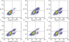
Fig 2
Apoptosis map of hPDLSCs in each group
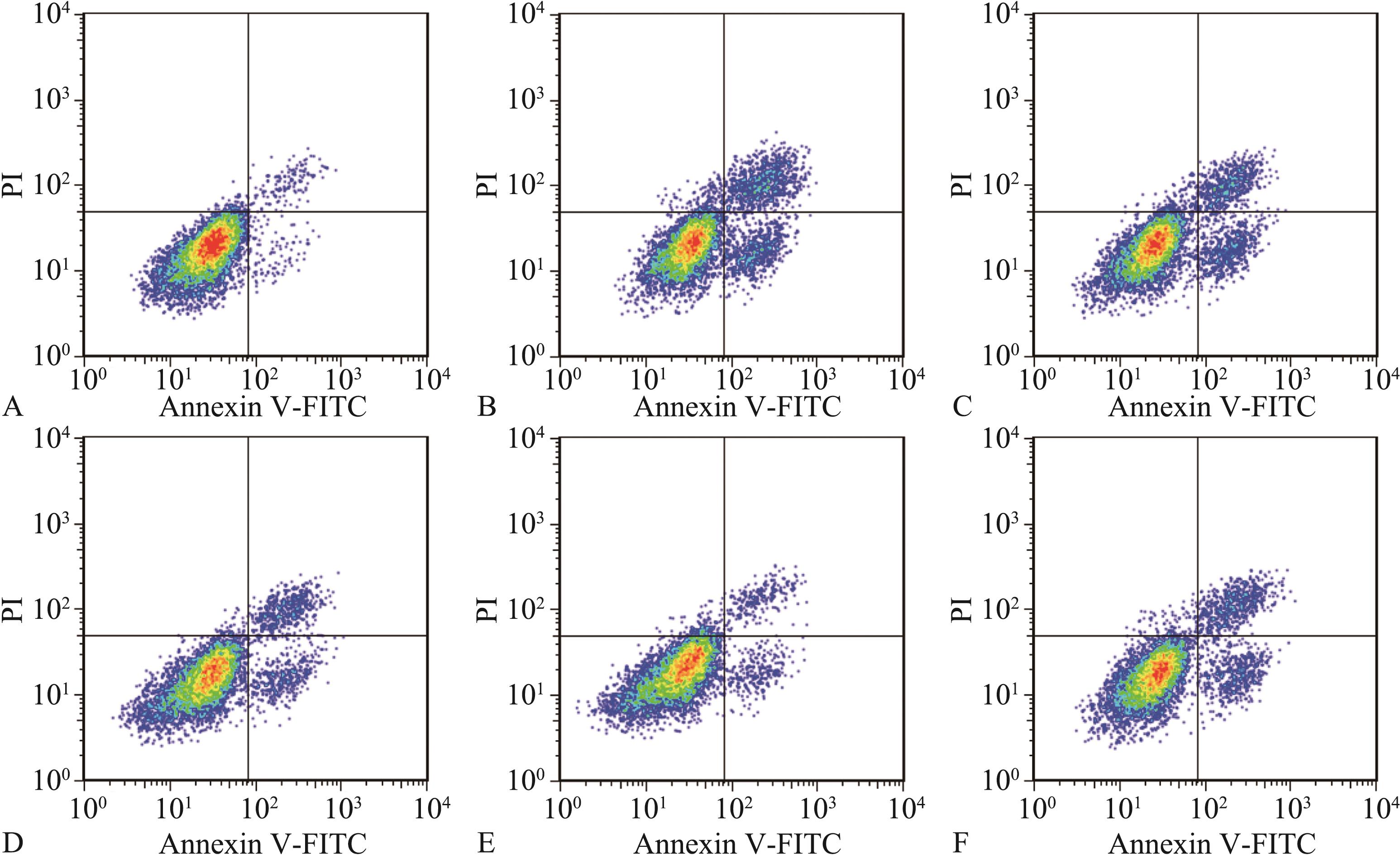
Fig 3
Formation of mineralized nodules in hPDLSCs of each group alizarin red staining × 200
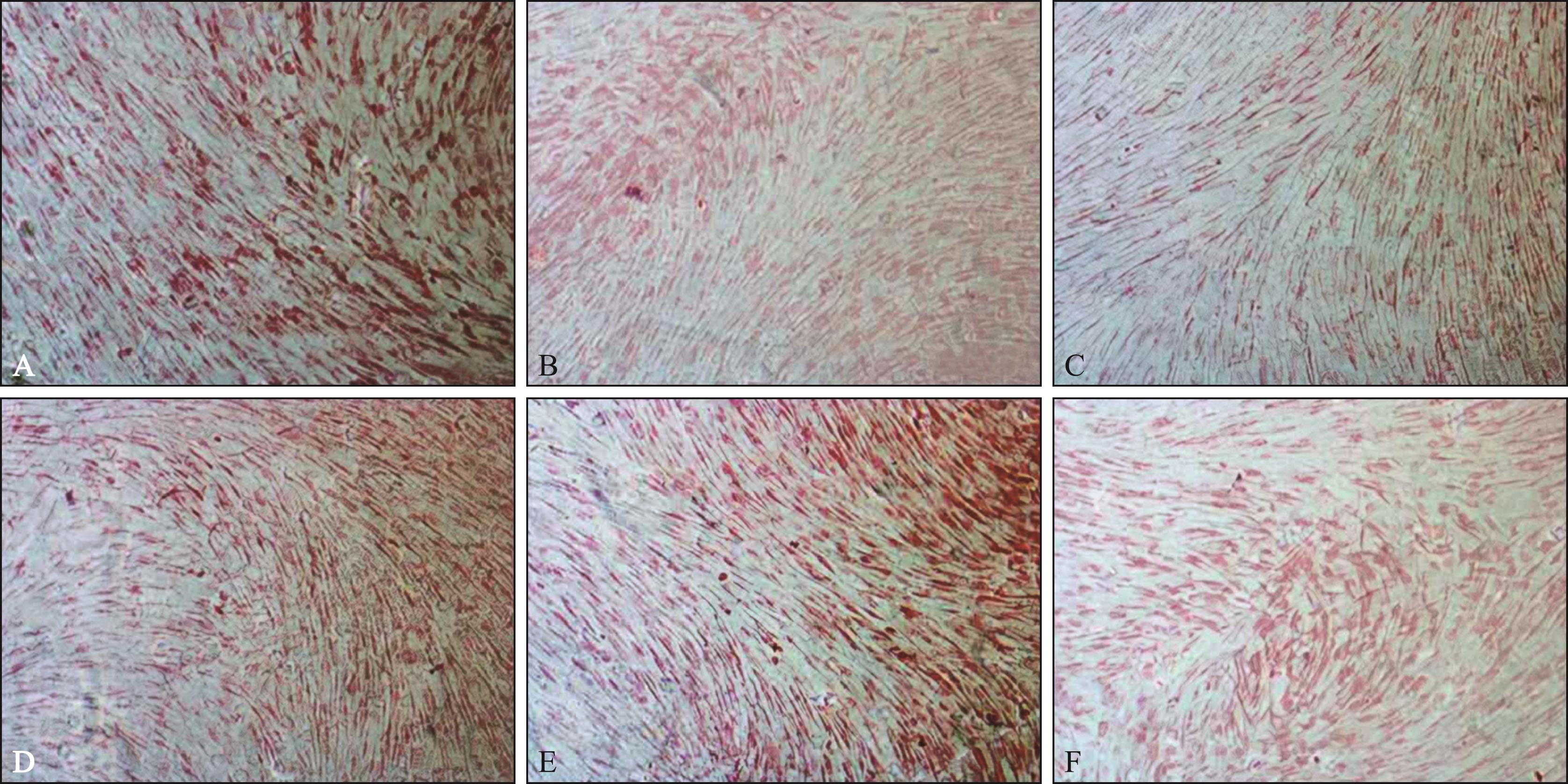
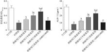
Fig 4
Comparison of staining intensity (left) and ALP activity (right) of hPDLSCs in each group


Fig 5
Comparison of TNF-α, IL-1β, and IL-6 levels in the culture supernatant of hPDLSCs in each group
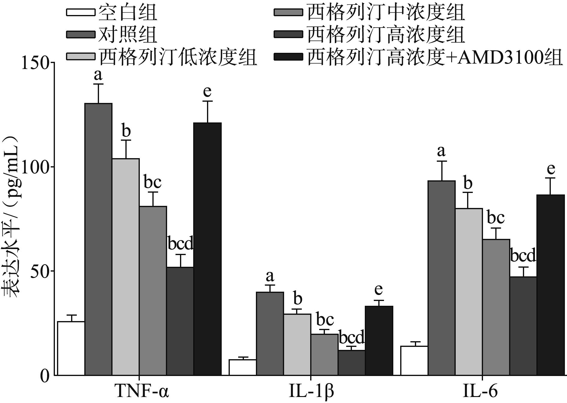
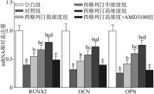
Fig 6
Comparison of mRNA expression levels of RUNX2, OCN, and OPN in hPDLSCs of each group


Fig 7
Western blot of SDF-1 and CXCR4 in hPDLSCs of each group
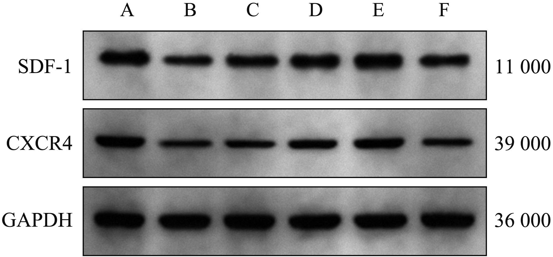
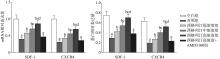
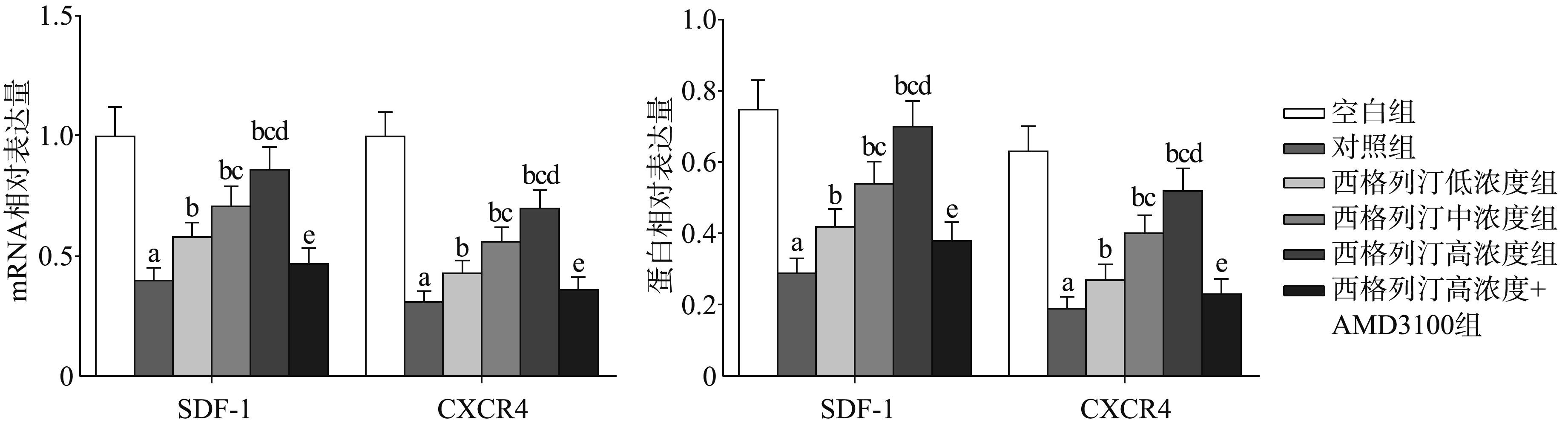
Fig 8
Comparison of mRNA (left) and protein (right) expression levels of SDF-1 and CXCR4 in hPDLSCs of each group
| 1 | Li W, Zheng Q, Xu M, et al. Association between circulating 25-hydroxyvitamin D metabolites and periodontitis: results from the NHANES 2009-2012 and Mendelian randomization study[J]. J Clin Periodontol, 2023, 50(2): 252-264. |
| 2 | Yoshino Y, Miyaji H, Nishida E, et al. Periodontal tissue regeneration by recombinant human collagen peptide granules applied with β-tricalcium phosphate fine particles[J]. J Oral Biosci, 2023, 65(1): 62-71. |
| 3 | Adachi O, Sugii H, Itoyama T, et al. Decorin promotes osteoblastic differentiation of human periodontal ligament stem cells[J]. Molecules, 2022, 27(23): 8224-8239. |
| 4 | Guo L, Li L. LIN28A alleviates inflammation, oxidative stress, osteogenic differentiation and mineralization in lipopolysaccharide (LPS)-treated human periodontal ligament stem cells[J]. Exp Ther Med, 2022, 23(6): 411-419. |
| 5 | Zhang H, Li X, Li J, et al. SDF-1 mediates mesenchymal stem cell recruitment and migration via the SDF-1/CXCR4 axis in bone defect[J]. J Bone Miner Metab, 2021, 39(2): 126-138. |
| 6 | 李胜鸿, 彭世元, 罗小玲, 等. 柚皮素通过基质细胞衍生因子1/趋化因子受体4信号轴对脂多糖作用下人牙周膜干细胞抗炎、成血管和成骨分化能力的影响[J]. 华西口腔医学杂志, 2023, 41(2): 175-184. |
| Li SH, Peng SY, Luo XL, et al. Effect of naringenin on the anti-inflammatory, vascularization, and osteogenesis differentiation of human periodontal ligament stem cells via the stromal cell-derived factor 1/C-X-C motif chemokine receptor 4 signaling axis stimulated by lipopolysaccharide[J]. West China J Stomatol, 2023, 41(2): 175-184. | |
| 7 | Lin FY, Shih CM, Huang CY, et al. Dipeptidyl peptidase-4 inhibitor decreases allograft vasculopathy via regula-ting the functions of endothelial progenitor cells in normoglycemic rats[J]. Cardiovasc Drugs Ther, 2021, 35(6): 1111-1127. |
| 8 | Yu G, Liu P, Shi Y, et al. Sitagliptin stimulates endothe-lial progenitor cells to induce endothelialization in aneurysm necks through the SDF-1/CXCR4/NRF2 signaling pathway[J]. Front Endocrinol (Lausanne), 2019, 10(1): 823-835. |
| 9 | Zhang Q, He L, Dong Y, et al. Sitagliptin ameliorates renal tubular injury in diabetic kidney disease via STAT3-dependent mitochondrial homeostasis through SDF-1α/CXCR4 pathway[J]. FASEB J, 2020, 34(6): 7500-7519. |
| 10 | Moraes RM, Lima GM, Oliveira FE, et al. Exenatide and sitagliptin decrease interleukin 1β, matrix metalloprotei-nase 9, and nitric oxide synthase 2 gene expression but does not reduce alveolar bone loss in rats with periodontitis[J]. J Periodontol, 2015, 86(11): 1287-1295. |
| 11 | Nie M, Li H, Liu P, et al. HMBOX1 attenuates LPS-induced periodontal ligament stem cell injury by inhibi-ting CXCL10 expression through the NF-κB signaling pathway[J]. Exp Ther Med, 2022, 23(3): 224-233. |
| 12 | 郭烨, 马庆云, 赵文丽, 等. GAS5靶向miR-222-3p对牙周膜干细胞成骨分化的影响机制[J]. 实用口腔医学杂志, 2021, 37(2): 271-274. |
| Guo Y, Ma QY, Zhao WL, et al. The effects of miR-222-3p targeted by GAS5 on osteogenic differentiation of pe-riodontal ligament stem cells[J]. J Pract Stomatol, 2021, 37(2): 271-274. | |
| 13 | 刘萍萍, 唐小莹, 袁小平. 基质细胞趋化因子-1对人牙周膜干细胞趋化因子受体——CXC亚家族受体4表达的影响研究[J]. 口腔医学研究, 2020, 36(1): 51-55. |
| Liu PP, Tang XY, Yuan XP. Effect of SDF-1 on CXCR4 expression of human periodontal stem cell chemokine receptor[J]. J Oral Sci Res, 2020, 36(1): 51-55. | |
| 14 | Li X, Wang X, Luan QX. Hyperresponsiveness of human gingival fibroblasts from patients with aggressive periodontitis against bacterial lipopolysaccharide[J]. Exp Ther Med, 2021, 21(5): 417-423. |
| 15 | Chen W, Su J, Cai S, et al. Cullin3 aggravates the inflammatory response of periodontal ligament stem cells via regulation of SHH signaling and Nrf2[J]. Bioengineered, 2021, 12(1): 3089-3100. |
| 16 | Chen J, Xu H, Xia K, et al. Resolvin E1 accelerates pulp repair by regulating inflammation and stimulating dentin regeneration in dental pulp stem cells[J]. Stem Cell Res Ther, 2021, 12(1): 75-88. |
| 17 | Kong L, Deng J, Zhou X, et al. Sitagliptin activates the p62-Keap1-Nrf2 signalling pathway to alleviate oxidati-ve stress and excessive autophagy in severe acute pancreatitis-related acute lung injury[J]. Cell Death Dis, 2021, 12(10): 928-938. |
| 18 | Zhao X, Huang P, Yuan J. Influence of glimepiride plus sitagliptin on treatment outcome, blood glucose, and oxidative stress in diabetic patients[J]. Am J Transl Res, 2022, 14(10): 7459-7466. |
| 19 | Zheng XY, Mao CY, Qiao H, et al. Plumbagin suppres-ses chronic periodontitis in rats via down-regulation of TNF-α, IL-1β and IL-6 expression[J]. Acta Pharmacol Sin, 2017, 38(8): 1150-1160. |
| 20 | 刘相, 康文燕, 商玲玲, 等. 西格列汀通过阻断核因子-κB信号通路抑制脂多糖诱导的人牙龈成纤维细胞炎症反应[J]. 华西口腔医学杂志, 2021, 39(2): 153-163. |
| Liu X, Kang WY, Shang LL, et al. Sitagliptin inhibits lipopolysaccharide-induced inflammatory response in human gingival fibroblasts by blocking nuclear factor-κB signaling pathway[J]. West China J Stomatol, 2021, 39(2): 153-163. | |
| 21 | 李颖辉, 齐芳芳, 韩行, 等. 不同浓度钙离子干预人牙周膜干细胞的增殖和成骨分化[J]. 中国组织工程研究, 2023, 27(19): 3005-3010. |
| Li YH, Qi FF, Han X, et al. Different concentrations of calcium ions interfere with the proliferation and osteoge-nic differentiation of human periodontal ligament stem cells[J]. Chin J Tissue Eng Res, 2023, 27(19): 3005-3010. | |
| 22 | Zhao A, Chung M, Yang Y, et al. The SDF-1/CXCR4 signaling pathway directs the migration of systemically transplanted bone marrow mesenchymal stem cells towards the lesion site in a rat model of spinal cord injury[J]. Curr Stem Cell Res Ther, 2023, 18(2): 216-230. |
| 23 | 刘玄林, 熊伟. 槲皮素调节SDF-1/CXCR4轴对下肢动脉硬化闭塞症大鼠的治疗作用[J]. 中国现代应用药学, 2023, 40(4): 455-460. |
| Liu XL, Xiong W. Therapeutic effect of quercetin on rats with arteriosclerosis occlusive disease of the lower extremities by regulating SDF-1/CXCR4 axis[J]. Chin J Modern Appl Pharm, 2023, 40(4): 455-460. |
| Viewed | ||||||
|
Full text |
|
|||||
|
Abstract |
|
|||||
 This work is licensed under a Creative Commons Attribution 3.0 License.
