West China Journal of Stomatology ›› 2019, Vol. 37 ›› Issue (2): 193-199.doi: 10.7518/hxkq.2019.02.012
Previous Articles Next Articles
Feng Liao1,Yao Liu2,Hanghang Liu2,Jian Hu1,Shuang Zhao3,Shimao Yang3( )
)
Received:2018-07-12
Revised:2018-12-19
Online:2019-04-01
Published:2019-04-28
Contact:
Shimao Yang
E-mail:yangshimao1986@163.com
Supported by:CLC Number:
Feng Liao,Yao Liu,Hanghang Liu,Jian Hu,Shuang Zhao,Shimao Yang. Effect of Angelica sinensis polysaccharide on the osteogenic differentiation of bone marrow mesenchymal stem cells of rats with high glucose levels[J]. West China Journal of Stomatology, 2019, 37(2): 193-199.
Add to citation manager EndNote|Ris|BibTeX
Tab 1
Sequences of primers
| 基因 | 引物序列(5’-3’) |
|---|---|
| Runx2 | F:GCGGACGAGGCAAGAGTT |
| R:TTGGTGCTGAGTTCAGGGAG | |
| Osx | F:CTGTGAAACCTCAAGTCCTATGGA |
| R:GCTCTGCAGTCAAGGGAGATG | |
| OCN | F:CAGGTGCAAAGCCCAGCGACT |
| R:AGGGGATCTGGGTAGGGGGCT | |
| Col-Ⅰ | F:CCACCTGCCTCTGGCTTCT |
| R:AGCTGTGGAGGAGGGTTTCA | |
| β-catenin | F:TGGCAACCAAGAAAGCAAG |
| R:CTGAACAAGAGTCCCAAGGAG | |
| CyclinD1 | F:CCCTCGGTGTCCTACTTCA |
| R:GTTTGTTCTCCTCCGCCTCT | |
| GAPDH | F:AGACCTTCAACACCCCAG |
| R:CACGATTTCCCTCTCAGC |
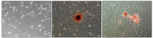
Fig.1
Rat BMSCs and osteogenic as well as adipogenic differentiation inverted microscope × 100
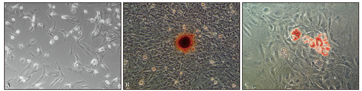
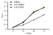
Fig.2
BMSCs proliferation (CCK-8) of every group
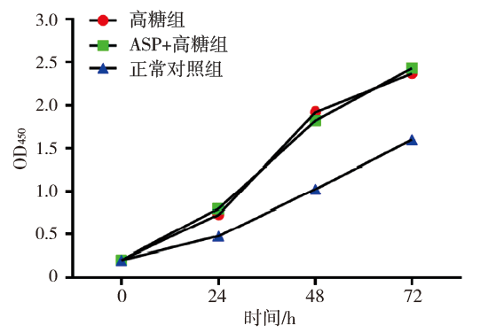
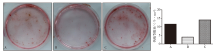
Fig.3
Alizarin red staining and semi-quantitative analysis of every group
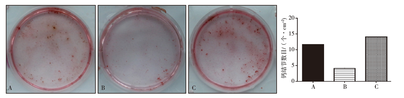
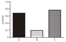
Fig.4
The ALP activity assay of every group

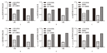
Fig.5
Relative mRNA levels of every group A:Runx2;B:Osx;C:OCN;D:Col-Ⅰ;E:CyclinD1;F:β-catenin。
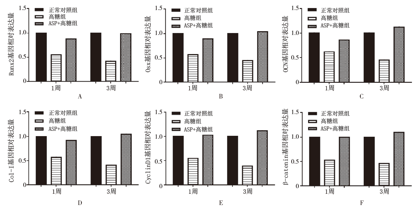
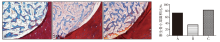
Fig.6
Masson staining and semi-quantitative analysis of every group

| [1] |
American Diabetes Association . Diagnosis and classification of diabetes mellitus[J]. Diabetes Care, 2014,37(Suppl 1):S81-S90.
doi: 10.2337/dc14-S081 URL |
| [2] | Goldenberg R, Punthakee Z . Definition, classification and diagnosis of diabetes, prediabetes and metabolic syndrome |
| [J]. Can J Diabetes , 2013,37(Suppl 1):S8-S11. | |
| [3] |
Spravchikov N, Sizyakov G, Gartsbein M , et al. Glucose effects on skin keratinocytes: implications for diabetes skin complications[J]. Diabetes, 2001,50(7):1627-1635.
doi: 10.2337/diabetes.50.7.1627 URL |
| [4] |
李昊, 周海伦, 王琪 , 等. 调节Lnk/干细胞因子-干细胞因子受体通路对糖尿病状态骨髓间充质干细胞成骨功能的影响[J]. 华西口腔医学杂志, 2015,33(6):633-637.
doi: 10.7518/hxkq.2015.06.017 |
|
Li H, Zhou HL, Wang Q , et al. Changes in bone marrow mesenchymal stem cells osteogenesis by the regulation of Lnk/stem cell factor-cKit signaling[J]. West Chin J Stoma-tol, 2015,33(6):633-637.
doi: 10.7518/hxkq.2015.06.017 |
|
| [5] |
Lim D, Kim Y . Anti-osteoporotic effects of angelica sinensis (oliv.) diels extract on ovariectomized rats and its oral toxi-city in rats[J]. Nutrients, 2014,6(10):4362-4372.
doi: 10.3390/nu6104362 URL |
| [6] |
Yang Q, Populo SM, Zhang JY , et al. Effect of Angelica sinensis on the proliferation of human bone cells[J]. Clin Chim Acta, 2002,324(1/2):89-97.
doi: 10.1016/S0009-8981(02)00210-3 URL |
| [7] |
Zhang S, He B, Ge JB , et al. Extraction, chemical analysis of Angelica sinensis polysaccharides and antioxidant activity of the polysaccharides in ischemia-reperfusion rats[J]. Int J Biol Macromol, 2010,47(4):546-550.
doi: 10.1016/j.ijbiomac.2010.07.012 URL |
| [8] | Zhang Y, Li MM, Zeng F , et al. Study to establish the role of JAK2 and SMAD1/5/8 pathways in the inhibition of hep-cidin by polysaccharides from Angelica sinensis[J]. J Eth-nopharmacol, 2012,144(2):433-440. |
| [9] |
Wang KP, Zeng F, Liu JN , et al. Inhibitory effect of poly-saccharides isolated from Angelica sinensis on hepcidin expression[J]. J Ethnopharmacol, 2011,134(3):944-948.
doi: 10.1016/j.jep.2011.02.015 URL |
| [10] |
Jin ML, Zhao K, Huang QS , et al. Isolation, structure and bioactivities of the polysaccharides from Angelica sinensis (Oliv.) Diels: A review[J]. Carbohydrate Polymers, 2012,89(3):713-722.
doi: 10.1016/j.carbpol.2012.04.049 URL |
| [11] |
Wang KP, Cao P, Wang HX , et al. Chronic administration of Angelica sinensis polysaccharide effectively improves fatty liver and glucose homeostasis in high-fat diet-fed mice[J]. Sci Rep, 2016,6(1):26229.
doi: 10.1038/srep26229 URL |
| [12] |
Srinivasan K, Viswanad B, Asrat L , et al. Combination of high-fat diet-fed and low-dose streptozotocin-treated rat: A model for type 2 diabetes and pharmacological screening[J]. Pharmacol Res, 2005,52(4):313-320.
doi: 10.1016/j.phrs.2005.05.004 URL |
| [13] | Balint E, Szabo P, Marshall CF , et al. Glucose-induced in-hibition of in vitro bone mineralization[J]. Bone, 2001,28 |
| ( 1):21-28. | |
| [14] |
Terada M, Inaba M, Yano Y , et al. Growth-inhibitory effect of a high glucose concentration on osteoblast-like cells[J]. Bone, 1998,22(1):17-23.
doi: 10.1016/S8756-3282(97)00220-2 URL |
| [15] |
Carnevale V, Romagnoli E , D’Erasmo E . Skeletal involve-ment in patients with diabetes mellitus[J]. Diabetes Metab Res Rev, 2004,20(3):196-204.
doi: 10.1002/(ISSN)1520-7560 URL |
| [16] |
Hofbauer LC, Brueck CC, Singh SK , et al. Osteoporosis in patients with diabetes mellitus[J]. J Bone miner Res, 2007,22(9):1317-1328.
doi: 10.1359/jbmr.070510 URL |
| [17] |
Colombo JS, Balani D, Sloan AJ , et al. Delayed osteoblast differentiation and altered inflammatory response around implants placed in incisor sockets of type 2 diabetic rats[J]. Clin Oral Implants Res, 2011,22(6):578-586.
doi: 10.1111/clr.2011.22.issue-6 URL |
| [18] |
Wongdee K, Charoenphandhu N . Osteoporosis in diabetes mellitus: Possible cellular and molecular mechanisms[J]. World J Diabetes, 2011,2(3):41-48.
doi: 10.4239/wjd.v2.i3.41 URL |
| [19] |
Fang SH, Jin YH, Zheng HX , et al. High glucose condition upregulated Txnip expression level in rat mesangial cells through ROS/MEK/MAPK pathway[J]. Mol Cell Biochem, 2011,347(1/2):175-182.
doi: 10.1007/s11010-010-0626-z URL |
| [20] |
Gopalakrishnan V, Vignesh RC, Arunakaran J , et al. Effects of glucose and its modulation by insulin and estradiol on BMSC differentiation into osteoblastic lineages[J]. Biochem Cell Biol, 2006,84(1):93-101.
doi: 10.1139/o05-163 URL |
| [21] |
Day TF, Guo XZ, Garrett-Beal L , et al. Wnt/β-catenin sig-naling in mesenchymal progenitors controls osteoblast and chondrocyte differentiation during vertebrate skeletogenesis[J]. Dev Cell, 2005,8(5):739-750.
doi: 10.1016/j.devcel.2005.03.016 URL |
| [22] |
Rodda SJ , McMahon AP. Distinct roles for Hedgehog and canonical Wnt signaling in specification, differentiation and maintenance of osteoblast progenitors[J]. Development, 2006,133(16):3231-3244.
doi: 10.1242/dev.02480 URL |
| Viewed | ||||||
|
Full text |
|
|||||
|
Abstract |
|
|||||
 This work is licensed under a Creative Commons Attribution 3.0 License.
