West China Journal of Stomatology ›› 2025, Vol. 43 ›› Issue (1): 76-83.doi: 10.7518/hxkq.2024.2024268
• Clinical Research • Previous Articles Next Articles
Shi Ruiwen1,2( ), Yang Hu1, Liu Yue1, Shi Yilin1, Zhang Shengben1, Liu Yu1, Song Feng1, Lan Jing1()
Received:2024-07-22
Revised:2024-09-03
Online:2025-02-01
Published:2025-01-22
Contact:
Lan Jing
E-mail:srw19980625@163.com;kqlj@sdu.edu.cn
CLC Number:
Shi Ruiwen, Yang Hu, Liu Yue, Shi Yilin, Zhang Shengben, Liu Yu, Song Feng, Lan Jing. L-shape technique with concentrated growth factor for horizontal bone defects in the maxillary anterior region: a clinical and radiographic study[J]. West China Journal of Stomatology, 2025, 43(1): 76-83.
Add to citation manager EndNote|Ris|BibTeX
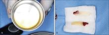
Fig 1
Preparation of CGF

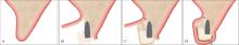
Fig 2
L-shape technique diagram
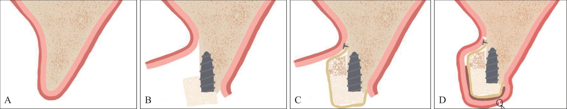

Fig 3
Preparation of DBBM-C
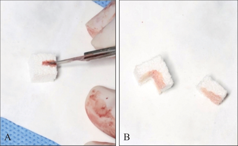

Fig 4
Surgical procedure of the test group
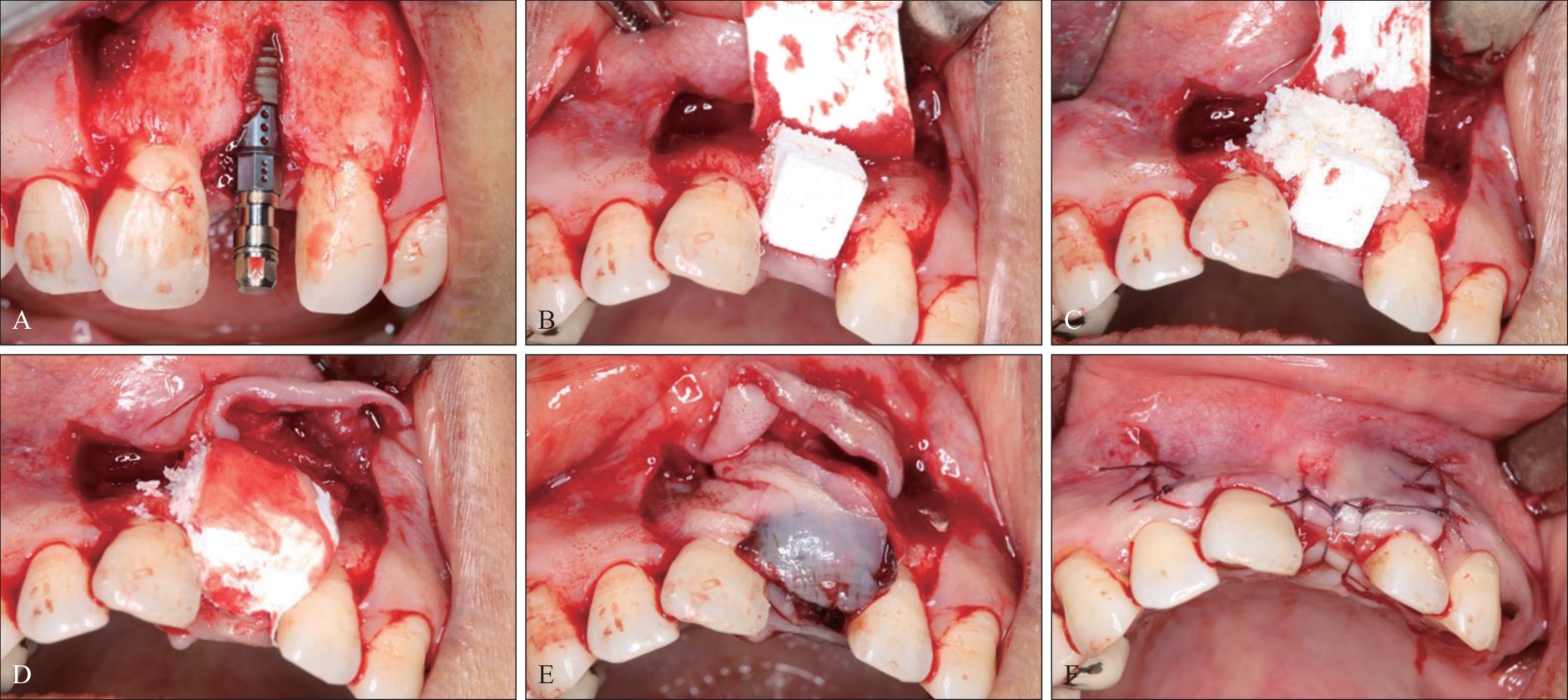

Fig 5
Surgical procedure of the control group
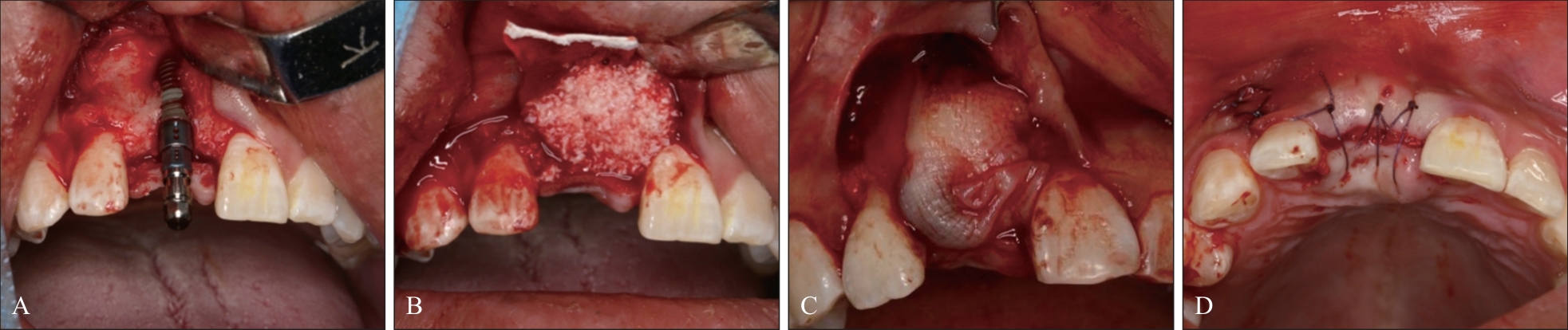
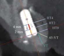
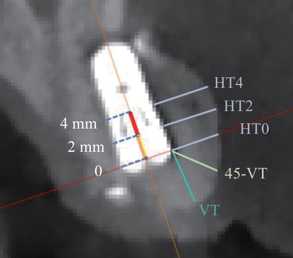
Fig 6
Measurement site diagram
Tab 1
General information of patients
| 项目 | 试验组 | 对照组 | P值 | |
|---|---|---|---|---|
| 年龄/岁 | 54.00±12.32 | 44.28±13.40 | 0.075 | |
| 性别 | 1.000 | |||
| 男 | 7(63.6%) | 9(64.3%) | ||
| 女 | 4(36.4%) | 5(35.7%) | ||
| 牙位 | 0.288 | |||
| 中切牙 | 8(72.7%) | 13(92.9%) | ||
| 侧切牙 | 3(27.3%) | 1(7.1%) | ||
| 种植体直径/mm | 1.000 | |||
| 3.3 | 8(72.7%) | 11(78.6%) | ||
| 4.1 | 3(27.3%) | 3(21.4%) | ||
| 种植体长度/mm | 0.090 | |||
| 10 | 10(90.9%) | 8(57.1%) | ||
| 12 | 1(9.1%) | 6(42.9%) | ||
| 吸烟 | 1.000 | |||
| 是 | 2(18.2%) | 3(21.4%) | ||
| 否 | 9(81.8%) | 11(78.6%) | ||
| 饮酒 | 0.350 | |||
| 是 | 4(36.4%) | 2(14.3%) | ||
| 否 | 7(63.6%) | 12(85.7%) | ||
| 糖尿病 | 0.288 | |||
| 是 | 3(27.3%) | 1(7.1%) | ||
| 否 | 8(72.7%) | 13(92.9%) | ||
| 愈合基台 | 0.002 | |||
| 使用 | 1(9.1%) | 10(71.4%) | ||
| 未使用 | 10(90.9%) | 4(28.6%) | ||
| 临时义齿 | 0.675 | |||
| 粘接式义齿 | 1(9.1%) | 1(7.1%) | ||
| 隐形义齿 | 2(18.2%) | 1(7.1%) | ||
| 未使用 | 8(72.7%) | 12(85.7%) | ||
| 垂直附加切口设计 | 0.288 | |||
| 单侧 | 3(27.3%) | 1(14.3%) | ||
| 双侧 | 8(72.7%) | 13(85.7%) | ||
Tab 2
Early discomfort and wound healing comparison of two groups
| 项目 | 试验组 | 对照组 | t值 | P值 |
|---|---|---|---|---|
| 肿胀持续时间/d | 6.00±3.10 | 6.86±1.79 | 0.869 | 0.394 |
| 止痛药量/片 | 3.36±3.61 | 3.14±2.14 | -0.191 | 0.851 |
| VAS评分 | 3.45±1.51 | 3.43±1.16 | -0.049 | 0.962 |
| 创口炎症评分 | 1.18±0.75 | 1.64±0.84 | 1.424 | 0.168 |
| EHI分级 | 1.73±0.65 | 1.79±0.58 | 0.238 | 0.814 |
Tab 3
Bone thickness comparison at T1 of two groups mm
| 测量项目 | 试验组 | 对照组 | t值 | P值 |
|---|---|---|---|---|
| HT0 | 1.96±0.61 | 1.49±0.99 | -1.373 | 0.121 |
| HT2 | 2.31±0.55 | 2.04±0.91 | -0.870 | 0.115 |
| HT4 | 2.62±0.74 | 2.34±0.92 | -0.811 | 0.459 |
| VT | 1.98±0.96 | 1.02±0.99 | -2.443 | 0.023* |
| 45-VT | 1.84±0.84 | 1.12±0.84 | -2.120 | 0.045* |
Tab 4
Bone thickness variation comparison from T0 to T1 of two groups
| 项目 | 试验组 | 对照组 | t值 | P值 |
|---|---|---|---|---|
| △HT0 | 2.78±1.11 | 1.63±1.23 | -2.414 | 0.024* |
| △HT2 | 2.60±1.01 | 1.51±1.11 | -2.556 | 0.018* |
| △HT4 | 2.64±1.21 | 1.50±1.26 | -2.305 | 0.031* |
| △VT | 1.54±0.91 | 1.20±1.50 | -0.661 | 0.515 |
| △45-VT | 2.14±0.95 | 1.35±1.24 | -1.742 | 0.095 |
Tab 5
The gray scale comparison of two groups
| 项目 | 试验组 | 对照组 | t值 | P值 |
|---|---|---|---|---|
| T0灰度值 | 782.31±212.18 | 926.83±136.88 | 2.065 | 0.050* |
| T1灰度值 | 1 411.02±322.31 | 1 232.25±412.68 | -1.216 | 0.236 |
| T1-T0灰度值变化量 | 628.71±320.55 | 305.42±372.59 | -2.33 | 0.029* |
| 1 | Jung RE, Brügger LV, Bienz SP, et al. Clinical and radiographical performance of implants placed with simultaneous guided bone regeneration using resorbable and nonresorbable membranes after 22-24 years, a prospective, controlled clinical trial[J]. Clin Oral Implants Res, 2021, 32(12): 1455-1465. |
| 2 | Mir-Mari J, Benic GI, Valmaseda-Castellón E, et al. Influence of wound closure on the volume stability of particulate and non-particulate GBR materials: an in vitro cone-beam computed tomographic examination. PartⅡ[J]. Clin Oral Implants Res, 2017, 28(6): 631-639. |
| 3 | McAllister BS, Haghighat K. Bone augmentation techniques[J]. J Periodontol, 2007, 78(3): 377-396. |
| 4 | Islam MT, Felfel RM, Abou Neel EA, et al. Bioactive calcium phosphate-based glasses and ceramics and their biomedical applications: a review[J]. J Tissue Eng, 2017, 8: 2041731417719170. |
| 5 | Benic GI, Eisner BM, Jung RE, et al. Hard tissue changes after guided bone regeneration of peri-implant defects comparing block versus particulate bone substitutes: 6-month results of a randomized controlled clinical trial[J]. Clin Oral Implants Res, 2019, 30(10): 1016-1026. |
| 6 | Troeltzsch M, Troeltzsch M, Kauffmann P, et al. Clinical efficacy of grafting materials in alveolar ridge augmentation: a systematic review[J]. J Craniomaxillofac Surg, 2016, 44(10): 1618-1629. |
| 7 | Wang D, Jin J, Qi W, et al. The two-dimensional size of peri-implant soft tissue in the anterior maxilla and some relevance: a 1-to 7-year cross-sectional study[J]. J Clin Periodontol, 2020, 47(4): 509-517. |
| 8 | Oh S, Chung SH, Han JY. Periodontal regenerative the-rapy in endo-periodontal lesions: a retrospective study over 5 years[J]. J Periodontal Implant Sci, 2019, 49(2): 90-104. |
| 9 | Silva CGB, Sapata VM, Llanos AH, et al. Peri-implant tissue changes at sites treated with alveolar ridge preservation in the aesthetic zone: twenty-two months follow-up of a randomized clinical trial[J]. J Clin Periodontol, 2022, 49(1): 39-47. |
| 10 | Thoma DS, Bienz SP, Lim HC, et al. Explorative randomized controlled study comparing soft tissue thickness, contour changes, and soft tissue handling of two ridge preservation techniques and spontaneous healing two months after tooth extraction[J]. Clin Oral Implants Res, 2020, 31(6): 565-574. |
| 11 | Gabay E, Katorza A, Zigdon-Giladi H, et al. Histological and dimensional changes of the alveolar ridge following tooth extraction when using collagen matrix and collagen-embedded xenogenic bone substitute: a randomized clinical trial[J]. Clin Implant Dent Relat Res, 2022, 24(3): 382-390. |
| 12 | Rohner D, Hailemariam S, Hammer B. Le Fort I osteotomies using Bio-Oss® collagen to promote bony union: a prospective clinical split-mouth study[J]. Int J Oral Maxillofac Surg, 2013, 42(5): 585-591. |
| 13 | Jung RE, Philipp A, Annen BM, et al. Radiographic evaluation of different techniques for ridge preservation after tooth extraction: a randomized controlled clinical trial[J]. J Clin Periodontol, 2013, 40(1): 90-98. |
| 14 | Arahira T, Todo M. Effects of proliferation and differentiation of mesenchymal stem cells on compressive mechanical behavior of collagen/β-TCP composite scaffold[J]. J Mech Behav Biomed Mater, 2014, 39: 218-230. |
| 15 | Zuercher AN, Mancini L, Naenni N, et al. The L-shape technique in guided bone regeneration with simulta-neous implant placement in the esthetic zone: a step-by-step protocol and a 2-14 year retrospective study[J]. J Esthet Restor Dent, 2023, 35(1): 197-205. |
| 16 | Ohnhaus EE, Adler R. Methodological problems in the measurement of pain: a comparison between the verbal rating scale and the visual analogue scale[J]. Pain, 1975, 1(4): 379-384. |
| 17 | Wachtel H, Schenk G, Böhm S, et al. Microsurgical access flap and enamel matrix derivative for the treatment of periodontal intrabony defects: a controlled clinical study[J]. J Clin Periodontol, 2003, 30(6): 496-504. |
| 18 | Jung EH, Jeong SN, Lee JH. Augmentation stability and early wound healing outcomes of guided bone regeneration in peri-implant dehiscence defects with L-and I-shaped soft block bone substitutes: a clinical and radiographic study[J]. Clin Oral Implants Res, 2021, 32(11): 1308-1317. |
| 19 | Wang HL, Boyapati L. “PASS” principles for predictable bone regeneration[J]. Implant Dent, 2006, 15(1): 8-17. |
| 20 | 朱悦萌, 贾克文, 焦俊杰, 等. 自体血小板浓缩物在角化黏膜增量中的临床应用[J]. 中国口腔种植学杂志, 2023, 28(4): 250-259. |
| Zhu YM, Jia KW, Jiao JJ, et al. Clinical applications of autologous platelet-rich concentrate in keratinized mucosal augmentation[J]. Chin J Oral Implant, 2023, 28(4): 250-259. | |
| 21 | Kao CH. Use of concentrate growth factors gel or membrane in chronic wound healing: description of 18 cases[J]. Int Wound J, 2020, 17(1): 158-166. |
| 22 | Chen Y, Cai Z, Zheng D, et al. Inlay osteotome sinus floor elevation with concentrated growth factor application and simultaneous short implant placement in severely atrophic maxilla[J]. Sci Rep, 2016, 6: 27348. |
| 23 | Elayah SA, Younis H, Cui H, et al. Alveolar ridge pre-servation in post-extraction sockets using concentrated growth factors: a split-mouth, randomized, controlled clinical trial[J]. Front Endocrinol (Lausanne), 2023, 14: 1163696. |
| 24 | 刘雨蒙, 袁长永. 浓缩生长因子软组织封闭功能在口腔种植中的应用[J]. 中国口腔种植学杂志, 2021, 26(6): 396-399. |
| Liu YM, Yuan CY. Application of concentrated growth factor in soft tissue sealing of oral implantation[J]. Chin J Oral Implant, 2021, 26(6): 396-399. | |
| 25 | Yu B, Wang Z. Effect of concentrated growth factors on beagle periodontal ligament stem cells in vitro [J]. Mol Med Rep, 2014, 9(1): 235-242. |
| [1] | Wu Hao, Cao Ning, Cao Liangwei, Yu Fei, Zhang Xu, Wei Shibo, Wei Hongwu, Guo Shuigen. Clinical efficacy of demineralized dentin matrix particles in immediate implantation for bone defects in posterior region: a 1 to 5-year follow-up study [J]. West China Journal of Stomatology, 2025, 43(4): 570-583. |
| [2] | Mo Minxuan, Pi Huaijin, Lin Youkai, Long Yifei, Fu Xiangqing, Duan Peipei. Analysis of the associations between maxillary anterior teeth and facial measurements in Han Chinese individuals with the most attractive smiles [J]. West China Journal of Stomatology, 2025, 43(4): 584-591. |
| [3] | Zhao Pengyu, Chen Gang, Cheng Yi, Wang Chao, Chen Dan, Huang Haitao. Clinical and histological evaluation of three-dimensional printing individualized titanium mesh for alveolar bone defect repair [J]. West China Journal of Stomatology, 2025, 43(4): 592-602. |
| [4] | Su Wenqi, Zhang Dandan, Cheng Yan, Cui Wenjie, Lei Lang, Li Houxuan. Guided bone regeneration therapy based on plaque control of peri-implantitis with follow-up at 7 years [J]. West China Journal of Stomatology, 2025, 43(1): 133-139. |
| [5] | Chen Liangwei, Han Jianmin, Guo Chuanbin. Research status and prospects of biodegradable magnesium-based metal-guided bone regeneration membranes [J]. West China Journal of Stomatology, 2024, 42(4): 415-425. |
| [6] | Zhu Zhanfeng, Yang Tingting, Chen Qinyi, Qiu Weien, Li Yongshan, Lin Yilan, Ban Yu. Concentrated growth factor and collagen as barrier materials in alveolar ridge preservation for posterior teeth: a prospective cohort study with one-year follow-up [J]. West China Journal of Stomatology, 2024, 42(3): 346-352. |
| [7] | Yu Yuanyuan, Zhong Shuaiqi, Sun Weilian, Lei Lihong.. Application of digital technology and platelet-rich fibrin technology in a novel regenerative treatment for posterior lingual furcation defect: a 6-year follow-up case report [J]. West China Journal of Stomatology, 2023, 41(5): 582-591. |
| [8] | Liu Yiming, Zhao Yun, Han Mei, Zhang Yuqiu, Mi Fanglin, Wang Bing. Preparation of functional poly-(lactic acid-co-glycolic acid)-based guided bone-regeneration membrane and its application in the reconstruction of mandibular defects in rats [J]. West China Journal of Stomatology, 2022, 40(5): 522-531. |
| [9] | Bolatihan Baligen, Lin Zhihui, Man Yi. Effect of Er:YAG laser combined with ethylenediamine tetra acetic acid on three-walled periodontal intrabony defects adjacent to implant sites [J]. West China Journal of Stomatology, 2021, 39(6): 718-723. |
| [10] | Fan Zhen, Liu Yue, Wang Zuolin. Tilted implantation technique for edentulous patients [J]. West China Journal of Stomatology, 2021, 39(4): 377-385. |
| [11] | Chen Luyi, Huang Min, Wu Jiaqi, Luo Jun. Guided bone regeneration-assisted orthodontic treatment for closing the space of missing central incisors [J]. West China Journal of Stomatology, 2021, 39(4): 482-488. |
| [12] | Hu Shuang, Li Chunmei, Zhang Shuaiyuan, Qin Shuo, Xie Chenlu, Niu Zhixing, Sun Minglei. Clinical value of oral repair membrane and β-tricalcium phosphate in the treatment of the postoperative bone defect of jaw cyst [J]. West China Journal of Stomatology, 2020, 38(5): 541-545. |
| [13] | Liu Shibo, Liu Xian. Review for different sources of exosomes in bone tissue engineering research [J]. West China Journal of Stomatology, 2020, 38(2): 193-197. |
| [14] | Tiantian Yu,Jin Liu,Junjing Yin,Xiangna Xu,Shengjie Yan,Jing Lan. Effects of concentrated growth factors on relieving postoperative reaction of guided bone regeneration in the esthetic zone [J]. West China Journal of Stomatology, 2019, 37(4): 398-402. |
| [15] | Yubin Cao,Chang Liu,Weilin Pan,Yuan Tu,Chunjie Li,Chengge Hua. Research progress on the modification of guided bone regeneration membranes [J]. West China Journal of Stomatology, 2019, 37(3): 325-329. |
| Viewed | ||||||
|
Full text |
|
|||||
|
Abstract |
|
|||||
 This work is licensed under a Creative Commons Attribution 3.0 License.
