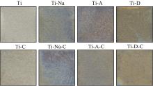
| 1 |
Antoci V, Chen AF, Parvizi J. Orthopedic implant use and infection[J]. Comprehen Biomater, 2017, 7: 133-151.
|
| 2 |
Jakubowicz J. Special issue: Ti-based biomaterials: synthesis, properties and applications[J]. Materials (Basel), 2020, 13(7): E1696.
|
| 3 |
韩宇. 抗菌肽GL13K涂层钛的抗腐蚀性能检测及其调节巨噬细胞促进成骨、成血管分化的基础研究[D]. 福州: 福建医科大学, 2021.
|
|
Han Y. Detection of corrosion resistance of titanium modified by antimicrobial peptide GL13K and regulation of macrophage behavior to promote osteogenic differentiation and angiogenic differentiation[D]. Fuzhou: Fujian Medical University, 2021.
|
| 4 |
Jemat A, Ghazali MJ, Razali M, et al. Surface modifications and their effects on titanium dental implants[J]. Biomed Res Int, 2015, 2015: 791725.
|
| 5 |
Nicholson JW. Titanium alloys for dental implants: a review[J]. Prosthesis, 2020, 2(2): 100-116.
|
| 6 |
Kurup A, Dhatrak P, Khasnis N. Surface modification techniques of titanium and titanium alloys for biomedical dental applications: a review[J]. Mater Today Proceed, 2021, 39: 84-90.
|
| 7 |
Liu CF, Chang KC, Sun YS, et al. Immobilizing type Ⅰcollagen via natural cross-linker genipin to enhance the osteogenic responses to titanium implant surface[J]. J Mater Res Technol, 2021, 15: 885-900.
|
| 8 |
Norris K, Mishukova OI, Zykwinska A, et al. Marine polysaccharide-collagen coatings on Ti6Al4V alloy formed by self-assembly[J]. Micromachines (Basel), 2019, 10(1): E68.
|
| 9 |
Van den Borre CE, Zigterman BGR, Mommaerts MY, et al. How surface coatings on titanium implants affect keratinized tissue: a systematic review[J]. J Biomed Mater Res B Appl Biomater, 2022, 110(7): 1713-1723.
|
| 10 |
Popova AD, Sheveyko AN, Kuptsov KA, et al. Osteoconductive, osteogenic, and antipathogenic plasma electrolytic oxidation coatings on titanium implants with BMP-2[J]. ACS Appl Mater Interfaces, 2023, 15(31): 37274-37289.
|
| 11 |
Wall I, Donos N, Carlqvist K, et al. Modified titanium surfaces promote accelerated osteogenic differentiation of mesenchymal stromal cells in vitro [J]. Bone, 2009, 45(1): 17-26.
|
| 12 |
Zhao Y, Sun Y, Hang R, et al. Biocompatible silane adhesion layer on titanium implants improves angiogenesis and osteogenesis[J]. Biomater Adv, 2022, 139: 213033.
|
| 13 |
Lee DJ, Tseng HC, Wong SW, et al. Dopaminergic effects on in vitro osteogenesis[J]. Bone Res, 2015, 3: 15020.
|
| 14 |
Yu Y, Li X, Li J, et al. Dopamine-assisted co-deposition of hydroxyapatite-functionalised nanoparticles of polydopamine on implant surfaces to promote osteogenesis in environments with high ROS levels[J]. Mater Sci Eng C Mater Biol Appl, 2021, 131: 112473.
|
| 15 |
Behrens C, Kauffmann P, von Hahn N, et al. Collagen-based osteogenic nanocoating of microrough titanium surfaces[J]. Int J Mol Sci, 2022, 23(14): 7803.
|
| 16 |
张文思, 朱文卿, 邱憬. 纯钛表面复合胶原蛋白凝胶涂层改性及其生物学性能初探[J]. 南京医科大学学报: 自然科学版, 2021, 41(9): 1329-1335.
|
|
Zhang WS, Zhu WQ, Qiu J. Modification of collagen gel coating on pure titanium surface and its biological properties: a preliminary study[J]. J Nanjing Med Univ (Nat Sci), 2021, 41(9): 1329-1335.
|
| 17 |
Zhu LY, Xie YY, Wen BT, et al. Porcine bone collagen peptides promote osteoblast proliferation and differentiation by activating the PI3K/Akt signaling pathway[J]. J Function Food, 2019, 64:103697.
|
| 18 |
Chen W, Li W, Xu K, et al. Functionalizing titanium surface with PAMAM dendrimer and human BMP2 gene via layer-by-layer assembly for enhanced osteogenesis[J]. J Biomed Mater Res A, 2018, 106(3): 706-717.
|
| 19 |
Han X, Gao W, Zhou Z, et al. Application of biomolecules modification strategies on PEEK and its composites for osteogenesis and antibacterial properties[J]. Colloids Surf B Biointerfaces, 2022, 215: 112492.
|
| 20 |
Mieszkowska A, Beaumont H, Martocq L, et al. Phenolic-enriched collagen fibrillar coatings on titanium alloy to promote osteogenic differentiation and reduce inflammation[J]. Int J Mol Sci, 2020, 21(17): E6406.
|
| 21 |
Babaei M, Bonakdar S, Nasernejad B. Selective biofunctionalization of 3D cell-imprinted PDMS with collagen immobilization for targeted cell attachment[J]. Sci Rep, 2022, 12(1): 12837.
|
| 22 |
Kim H, Lee YH, Kim NK, et al. Immobilization of collagen on the surface of a PEEK implant with monolayer nanopores[J]. Polymers (Basel), 2022, 14(9): 1633.
|
| 23 |
Li X, Liu X, Xing Y, et al. Erianin controls collagen-mediated retinal angiogenesis via the RhoA/ROCK1 signaling pathway induced by the alpha2/beta1 integrin-collagen interaction[J]. Invest Ophthalmol Vis Sci, 2022, 63(1): 27.
|
| 24 |
Manivasagam VK, Sabino RM, Kantam P, et al. Surface modification strategies to improve titanium hemocompatibility: a comprehensive review[J]. Mater Adv, 2021, 2(18): 5824-5842.
|
| 25 |
徐丽娜, 虞颖娟, 孙健. 低密度脂蛋白在钛金属表面的吸附行为分析[J]. 上海口腔医学, 2021, 30(1): 50-54.
|
|
Xu LN, Yu YJ, Sun J. Adsorption behavior of low density lipoprotein on titanium surface[J]. Shanghai J Stomatol, 2021, 30(1): 50-54.
|
| 26 |
Li H, Huang J, Wang Y, et al. Nanoscale modification of titanium implants improves behaviors of bone mesenchymal stem cells and osteogenesis in vivo [J]. Oxid Med Cell Longev, 2022, 2022: 2235335.
|
| 27 |
Yin J, Zhuang G, Zhu Y, et al. miR-615-3p inhibits the osteogenic differentiation of human lumbar ligamentum flavum cells via suppression of osteogenic regulators GDF5 and FOXO1[J]. Cell Biol Int, 2017, 41(7): 779-786.
|
| 28 |
Kim JY, Kim BI, Jue SS, et al. Localization of osteopontin and osterix in periodontal tissue during orthodontic tooth movement in rats[J]. Angle Orthod, 2012, 82(1): 107-114.
|
| 29 |
Hsueh YH, Cheng CY, Chien HW, et al. Synergistic effects of collagen and silver on the deposition characteristics, antibacterial ability, and cytocompatibility of a collagen/silver coating on titanium[J]. J Alloy Compound, 2020, 830: 154490.
|
| 30 |
Cho WT, Kim SY, Jung SI, et al. Effects of gamma radiation-induced crosslinking of collagen type Ⅰ coated dental titanium implants on osseointegration and bone regeneration[J]. Materials (Basel), 2021, 14(12): 3268.
|

 ), 黄艳玲, 赖颖真(