West China Journal of Stomatology ›› 2025, Vol. 43 ›› Issue (4): 592-602.doi: 10.7518/hxkq.2025.2024481
• Clinical Research • Previous Articles Next Articles
Zhao Pengyu1( ), Chen Gang1, Cheng Yi2, Wang Chao3, Chen Dan3, Huang Haitao1()
Received:2024-12-31
Revised:2025-03-04
Online:2025-08-01
Published:2025-08-29
Contact:
Huang Haitao
E-mail:1154652307@qq.com;hht945@hotmail.com
Supported by:CLC Number:
Zhao Pengyu, Chen Gang, Cheng Yi, Wang Chao, Chen Dan, Huang Haitao. Clinical and histological evaluation of three-dimensional printing individualized titanium mesh for alveolar bone defect repair[J]. West China Journal of Stomatology, 2025, 43(4): 592-602.
Add to citation manager EndNote|Ris|BibTeX
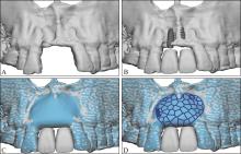
Fig 1
3D-PITM design and manufacturing process

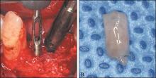
Fig 2
Acquisition of alveolar bone specimens

Tab 1
Basic information of patients
| 病例编号 | 性别 | 年龄/岁 | 缺牙位点 | 种植位点 | Terheyden分型 | 愈合期/月 |
|---|---|---|---|---|---|---|
| 1 | 男 | 48 | 12 | 12 | 3/4型 | 8 |
| 2 | 女 | 52 | 35、36、37 | 35 | 3/4型 | 9 |
| 37 | 3/4型 | |||||
| 3 | 男 | 52 | 21、22 | 21 | 3/4型 | 9 |
| 22 | 3/4型 | |||||
| 4 | 女 | 50 | 21、22 | 21 | 3/4型 | 8 |
| 22 | 3/4型 | |||||
| 5 | 男 | 38 | 21、22、23、24 | 22 | 3/4型 | 7 |
| 23 | 3/4型 | |||||
| 24 | 2/4型 | |||||
| 6 | 女 | 40 | 24、25、26、27 | 24 | 3/4型 | 11 |
| 26 | 3/4型 | |||||
| 27 | 3/4型 | |||||
| 7 | 女 | 49 | 36、37 | 36 | 3/4型 | 10 |
| 37 | 3/4型 | |||||
| 8 | 女 | 28 | 11、12、21、22 | 12 | 3/4型 | 11 |
| 22 | 3/4型 | |||||
| 9 | 男 | 37 | 11 | 11 | 3/4型 | 10 |
| 10 | 女 | 50 | 11、12 | 11 | 4/4型 | 10 |
| 12 | 3/4型 | |||||
| 11 | 女 | 57 | 11、21 | 11 | 3/4型 | 10 |
| 21 | 3/4型 | |||||
| 12 | 男 | 53 | 31、32、41、42 | 32 | 2/4型 | 9 |
| 42 | 2/4型 |
Tab 2
The occurrence of healing complications
| 病例编号 | 愈合并发症 | 备注 | |
|---|---|---|---|
| 创口裂开 | 3D-PITM暴露 | ||
| 1 | 术后2周创口裂开可吸收胶原膜暴露 | - | 术后4周内创口二次愈合 |
| 2 | - | - | - |
| 3 | - | - | - |
| 4 | - | - | - |
| 5 | - | - | - |
| 6 | 术后2周创口裂开可吸收胶原膜暴露 | - | 术后4周内创口二次愈合 |
| 7 | - | - | - |
| 8 | - | - | - |
| 9 | 术后2周创口裂开可吸收胶原膜暴露 | 术后3周暴露(早期暴露) | GBR手术失败 |
| 10 | - | 术后4月暴露(晚期暴露) | - |
| 11 | 术后10天创口裂开可吸收胶原膜暴露 | 术后4周暴露(早期暴露) | - |
| 12 | - | - | - |
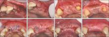
Fig 3
Soft tissue healing after GBR surgery

Tab 3
Imaging evaluation results of three-dimensio-nal bone augmentation volume
| 病例编号 | 计划骨增量体积/mm3 | 实际骨增量体积/mm3 | 实际成骨体积百分比/% |
|---|---|---|---|
| 1 | 175 | 289 | 165.14 |
| 2 | 573 | 321 | 56.02 |
| 3 | 401 | 380 | 94.76 |
| 4 | 107 | 95 | 88.79 |
| 5 | 583 | 526 | 90.22 |
| 6 | 757 | 696 | 91.94 |
| 7 | 1025 | 800 | 78.05 |
| 8 | 810 | 754 | 93.09 |
| 9 | 434 | 失败 | - |
| 10 | 1463 | 833 | 56.94 |
| 11 | 472 | 542 | 114.83 |
| 12 | 491 | 578 | 117.72 |
Tab 4
Histomorphometric results
| 标本编号 | 新生未矿化骨面积/mm2 | 新生骨总面积/mm2 | 视野内组织总面积/mm2 | 新生骨占比/% | 新生未矿化骨占比/% |
|---|---|---|---|---|---|
| 1 | 0.28 | 1.75 | 2.75 | 63.75 | 15.76 |
| 2 | 0.01 | 0.3 | 2.97 | 10.14 | 3.68 |
| 3 | 0.06 | 2.07 | 5.76 | 35.93 | 2.90 |
| 4 | 0.14 | 0.67 | 3.45 | 19.40 | 20.53 |
| 5 | 0.4 | 1.08 | 4.33 | 24.97 | 36.86 |
| 6 | 0.2 | 5.78 | 11.3 | 51.14 | 3.42 |
| 7 | 0.04 | 3.46 | 7.14 | 48.51 | 1.02 |
| 8 | 0.35 | 1.11 | 2.49 | 44.76 | 31.01 |
| 9 | 0.1 | 0.23 | 3.89 | 6.03 | 40.68 |
| 10 | 0.78 | 2.84 | 3.22 | 88.17 | 27.34 |
| 11 | 0.23 | 1.59 | 7.3 | 21.71 | 14.48 |
| 12 | 0.03 | 0.24 | 2.16 | 11.17 | 13.19 |
| 13 | 0.05 | 0.72 | 2.95 | 24.51 | 7.09 |
| 14 | 0.86 | 4.83 | 6.9 | 69.97 | 17.85 |
| 15 | 0.2 | 1.68 | 2.09 | 80.52 | 12.18 |
| 16 | 0.03 | 1.09 | 7.25 | 15.06 | 2.46 |
| 17 | 0.1 | 2.07 | 2.57 | 80.58 | 4.87 |
| 18 | 1.91 | 3.24 | 3.8 | 85.33 | 59.05 |
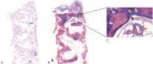
Fig 4
Alveolar bone specimens from the good healing group

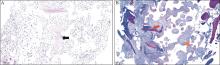
Fig 5
Alveolar bone specimens from 3D-PITM exposure group

Tab 5
Results of histomorphometric analysis of intergroup differences between the good healing group and the wound dehiscence group
| 骨组织类型 | 组别 | n | 中位数 | 四分位间距 | W值 | P值 |
|---|---|---|---|---|---|---|
| 新生骨占比 | 创口愈合良好 | 16 | 42.2% | 21.13%~72.61% | 21.0 | 0.549 |
| 创口裂开 | 2 | 29.9% | 22.49%~37.34% | |||
| 新生未矿化骨占比 | 创口愈合良好 | 16 | 13.8% | 4.57%~22.23% | 18.0 | 0.837 |
| 创口裂开 | 2 | 16.7% | 9.60%~23.87% |
Tab 6
Results of histomorphometric analysis of intergroup differences between the good healing group and the 3D-PITM exposure group
| 骨组织类型 | 组别 | n | 中位数 | 四分位间距 | W值 | P值 |
|---|---|---|---|---|---|---|
| 新生骨占比 | 创口愈合良好 | 16 | 42.2% | 21.13%~72.61% | 16.5 | 0.944 |
| 钛网暴露 | 2 | 47.8% | 31.43%~64.16% | |||
| 未矿化骨占比 | 创口愈合良好 | 16 | 13.8% | 4.57%~22.23% | 24.5 | 0.232 |
| 钛网暴露 | 2 | 7.3% | 4.89%~9.75% |
| [1] | Alotaibi FF, Rocchietta I, Buti J, et al. Comparative evidence of different surgical techniques for the management of vertical alveolar ridge defects in terms of complications and efficacy: a systematic review and network meta-analysis[J]. J Clin Periodontol, 2023, 50(11): 1487-1519. |
| [2] | Li SH, Zhao YX, Tian TR, et al. A minimally invasive method for titanium mesh fixation with resorbable sutures in guided bone regeneration: a retrospective study[J]. Clin Implant Dent Relat Res, 2023, 25(1): 87-98. |
| [3] | Yang W, Chen D, Wang C, et al. The effect of bone defect size on the 3D accuracy of alveolar bone augmentation performed with additively manufactured patient-specific titanium mesh[J]. BMC Oral Health, 2022, 22(1): 557. |
| [4] | Wang HL, Boyapati L. “PASS” principles for predicta-ble bone regeneration[J]. Implant Dent, 2006, 15(1): 8-17. |
| [5] | Xie Y, Li SH, Zhang TX, et al. Titanium mesh for bone augmentation in oral implantology: current application and progress[J]. Int J Oral Sci, 2020, 12(1): 37. |
| [6] | Cunha G, Carvalho PHA, Quirino LC, et al. Titanium mesh exposure after bone grafting: treatment approaches-a systematic review[J]. Craniomaxillofac Trauma Reconstr, 2022, 15(4): 397-405. |
| [7] | Briguglio F, Falcomatà D, Marconcini S, et al. The use of titanium mesh in guided bone regeneration: a systematic review[J]. Int J Dent, 2019, 2019: 9065423. |
| [8] | Ciocca L, Fantini M, De Crescenzio F, et al. Direct metal laser sintering (DMLS) of a customized titanium mesh for prosthetically guided bone regeneration of atrophic maxillary arches[J]. Med Biol Eng Comput, 2011, 49(11): 1347-1352. |
| [9] | Sumida T, Otawa N, Kamata YU, et al. Custom-made titanium devices as membranes for bone augmentation in implant treatment: clinical application and the comparison with conventional titanium mesh[J]. J Craniomaxillofac Surg, 2015, 43(10): 2183-2188. |
| [10] | Chiapasco M, Casentini P. Horizontal bone-augmentation procedures in implant dentistry: prosthetically guided regeneration[J]. Periodontol 2000, 2018, 77(1): 213-240. |
| [11] | Sagheb K, Schiegnitz E, Moergel M, et al. Clinical outcome of alveolar ridge augmentation with individualized CAD-CAM-produced titanium mesh[J]. Int J Implant Dent, 2017, 3(1): 36. |
| [12] | Cucchi A, Vignudelli E, Franceschi D, et al. Vertical and horizontal ridge augmentation using customized CAD/CAM titanium mesh with versus without resorbable membranes. A randomized clinical trial[J]. Clin Oral Implants Res, 2021, 32(12): 1411-1424. |
| [13] | Chiapasco M, Casentini P, Tommasato G, et al. Customized CAD/CAM titanium meshes for the guided bone regeneration of severe alveolar ridge defects: preliminary results of a retrospective clinical study in humans[J]. Clin Oral Implants Res, 2021, 32(4): 498-510. |
| [14] | Chrcanovic BR, Albrektsson T, Wennerberg A. Bone quality and quantity and dental implant failure: a systematic review and meta-analysis[J]. Int J Prosthodont, 2017, 30(3): 219-237. |
| [15] | Nicolielo LFP, van Dessel J, Jacobs R, et al. Relationship between trabecular bone architecture and early dental implant failure in the posterior region of the mandible[J]. Clin Oral Implants Res, 2020, 31(2): 153-161. |
| [16] | Li SH, Zhang TX, Zhou M, et al. A novel digital and visualized guided bone regeneration procedure and digital precise bone augmentation: a case series[J]. Clin Implant Dent Relat Res, 2021, 23(1): 19-30. |
| [17] | Pillai S, Upadhyay A, Khayambashi P, et al. Dental 3D-printing: transferring art from the laboratories to the clinics[J]. Polymers (Basel), 2021, 13(1): E157. |
| [18] | Fontana F, Maschera E, Rocchietta I, et al. Clinical classification of complications in guided bone regeneration procedures by means of a nonresorbable membrane[J]. Int J Periodontics Restorative Dent, 2011, 31(3): 265-273. |
| [19] | Simion M, Pistilli R, Vignudelli E, et al. Semi-occlusive CAD/CAM titanium mesh for guided bone regeneration: preliminary clinical and histological results[J]. Int J Oral Implantol (Berl), 2023, 16(4): 327-336. |
| [20] | Li L, Wang C, Li X, et al. Research on the dimensional accuracy of customized bone augmentation combined with 3D-printing individualized titanium mesh: a retrospective case series study[J]. Clin Implant Dent Relat Res, 2021, 23(1): 5-18. |
| [21] | Zhang G, Miao X, Lin H, et al. A tooth-supported titanium mesh bending and positioning module for alveolar bone augmentation and improving accuracy[J]. J Esthet Restor Dent, 2023, 35(4): 586-595. |
| [22] | Chen D, Zheng LL, Wang C, et al. Evaluation of surgical placement accuracy of customized CAD/CAM titanium mesh using screws-position-guided template: a retrospective comparative study[J]. Clin Implant Dent Relat Res, 2023, 25(3): 519-531. |
| [23] | 许来俊, 袁鹤, 叶青, 等. 负载纳米羟磷灰石的结冷胶修复大鼠下颌骨缺损的效果评价[J]. 上海口腔医学, 2022, 31(5): 449-453. |
| Xu LJ, Yuan H, Ye Q, et al. Repair of mandibular defects with. hydrogel loaded with nano-hydroxyapatite in rats[J]. Shanghai J Stomatol, 2022, 31(5): 449-453. | |
| [24] | 杜文瑜, 杨静文, 姜婷. 甲磺酸去铁胺促进大鼠颅骨临界骨缺损血管化骨再生的早期连续观察[J]. 北京大学学报(医学版), 2021, 53(6): 1171-1177. |
| Du WY, Yang JW, Jiang T. Early constant observation of the. effect of deferoxamine mesylate on improvement of vascularized bone regeneration in SD rat skull critical size defect model[J]. J Peking Uni (Health Sci), 2021, 53(6): 1171-1177. | |
| [25] | Cucchi A, Sartori M, Parrilli A, et al. Histological and histomorphometric analysis of bone tissue after guided bone regeneration with non-resorbable membranes vs resorbable membranes and titanium mesh[J]. Clin Implant Dent Relat Res, 2019, 21(4): 693-701. |
| [26] | Cucchi A, Bettini S, Fiorino A, et al. Histological and histomorphometric analysis of bone tissue using customized titanium meshes with or without resorbable membranes: a randomized clinical trial[J]. Clin Oral Implants Res, 2024, 35(1): 114-130. |
| [27] | MacBeth N, Trullenque-Eriksson A, Donos N, et al. Hard and soft tissue changes following alveolar ridge preservation: a systematic review[J]. Clin Oral Implants Res, 2017, 28(8): 982-1004. |
| [28] | Dellavia C, Canciani E, Pellegrini G, et al. Histological assessment of mandibular bone tissue after guided bone regeneration with customized computer-aided design/computer-assisted manufacture titanium mesh in humans: a cohort study[J]. Clin Implant Dent Relat Res, 2021, 23(4): 600-611. |
| [29] | Aludden H, Mordenfeld A, Dahlin C, et al. Histological and histomorphometrical outcome after lateral guided bone regeneration augmentation of the mandible with different ratios of deproteinized bovine bone mineral and autogenous bone. A preclinical in vivo study[J]. Clin Oral Implants Res, 2020, 31(10): 1025-1036. |
| [30] | Gallo P, Díaz-Báez D, Perdomo S, et al. Comparative analysis of two biomaterials mixed with autogenous bone graft for vertical ridge augmentation: a histomorphometric study in humans[J]. Clin Implant Dent Relat Res, 2022, 24(5): 709-719. |
| [31] | Corinaldesi G, Pieri F, Marchetti C, et al. Histologic and histomorphometric evaluation of alveolar ridge augmentation using bone grafts and titanium micromesh in humans[J]. J Periodontol, 2007, 78(8): 1477-1484. |
| [32] | Zazou N, Diab N, Bahaa S, et al. Clinical comparison of different flap advancement techniques to periosteal releasing incision in guided bone regeneration: a randomized controlled trial[J]. Clin Implant Dent Relat Res, 2021, 23(1): 107-116. |
| [33] | Bertran Faus A, Cordero Bayo J, Velasco-Ortega E, et al. Customized titanium mesh for guided bone regeneration with autologous bone and xenograft[J]. Materials (Basel), 2022, 15(18): 6271. |
| [34] | Galindo-Moreno P, Moreno-Riestra I, Avila G, et al. Effect of anorganic bovine bone to autogenous cortical bone ratio upon bone remodeling patterns following maxillary sinus augmentation[J]. Clin Oral Implants Res, 2011, 22(8): 857-864. |
| [35] | Leblebicioglu B, Tatakis DN. Complications following alveolar ridge augmentation procedures[J]. Periodontol 2000, 2023, 93(1): 221-235. |
| [36] | Poli PP, Beretta M, Maiorana C, et al. Therapeutic strategies in the management of nonresorbable membrane and titanium mesh exposures following alveolar bone augmentation: a systematic scoping review[J]. Int J Oral Maxillofac Implants, 2022, 37(2): 250-269. |
| [37] | Lim G, Lin GH, Monje A, et al. Wound healing complications following guided bone regeneration for ridge augmentation: a systematic review and meta-analysis[J]. Int J Oral Maxillofac Implants, 2018, 33(1): 41-50. |
| [38] | Lizio G, Mazzone N, Corinaldesi G, et al. Reconstruction of extended and morphologically varied alveolar ridge defects with the titanium mesh technique: clinical and dental implants outcomes[J]. Int J Periodontics Restorative Dent, 2016, 36(5): 689-697. |
| [39] | Ciocca L, Lizio G, Baldissara P, et al. Prosthetically CAD-CAM-guided bone augmentation of atrophic jaws using customized titanium mesh: preliminary results of an open prospective study[J]. J Oral Implantol, 2018, 44(2): 131-137. |
| [40] | Zhou L, Su Y, Wang J, et al. Effect of exposure rates with customized versus conventional titanium mesh on guided bone regeneration: systematic review and meta-analysis[J]. J Oral Implantol, 2022, 48(4): 339-346. |
| [41] | Cucchi A, Vignudelli E, Napolitano A, et al. Evaluation of complication rates and vertical bone gain after guided bone regeneration with non-resorbable membranes versus titanium meshes and resorbable membranes. A randomized clinical trial[J]. Clin Implant Dent Relat Res, 2017, 19(5): 821-832. |
| [42] | Nan X, Wang C, Li LZ, et al. Application of three-dimensional printing individualized titanium mesh in alveolar bone defects with different Terheyden classifications: a retrospective case series study[J]. Clin Oral Implants Res, 2023, 34(6): 639-650. |
| [43] | Machtei EE. The effect of membrane exposure on the outcome of regenerative procedures in humans: a meta-analysis[J]. J Periodontol, 2001, 72(4): 512-516. |
| [44] | Atef M, Tarek A, Shaheen M, et al. Horizontal ridge augmentation using native collagen membrane vs titanium mesh in atrophic maxillary ridges: randomized clinical trial[J]. Clin Implant Dent Relat Res, 2020, 22(2): 156-166. |
| [45] | Hofferber CE, Beck JC, Liacouras PC, et al. Volumetric changes in edentulous alveolar ridge sites utilizing guided bone regeneration and a custom titanium ridge augmentation matrix (CTRAM): a case series study[J]. Int J Implant Dent, 2020, 6(1): 83. |
| [46] | Hartmann A, Seiler M. Minimizing risk of customized titanium mesh exposures-a retrospective analysis[J]. BMC Oral Health, 2020, 20(1): 36. |
| [47] | Rakhmatia YD, Ayukawa Y, Furuhashi A, et al. Microcomputed tomographic and histomorphometric analyses of novel titanium mesh membranes for guided bone regeneration: a study in rat calvarial defects[J]. Int J Oral Maxillofac Implants, 2014, 29(4): 826-835. |
| [48] | Lee WZ, Ong MMA, Yeo ABK. Gingival profiles in a select Asian cohort: a pilot study[J]. J Invest Clin Dent, 2018, 9(1). DOI:10.1111/jicd.12269 . |
| [49] | Hartmann A, Hildebrandt H, Schmohl JU, et al. Evaluation of risk parameters in bone regeneration using a customized titanium mesh: results of a clinical study[J]. Implant Dent, 2019, 28(6): 543-550. |
| [50] | Frost NA, Mealey BL, Jones AA, et al. Periodontal biotype: gingival thickness as it relates to probe visibility and buccal plate thickness[J]. J Periodontol, 2015, 86(10): 1141-1149. |
| [51] | Wang CX, Rong QG, Zhu N, et al. Finite element analysis of stress in oral mucosa and titanium mesh interface[J]. BMC Oral Health, 2023, 23(1): 25. |
| [52] | Lin GH, Chan HL, Wang HL. The significance of keratinized mucosa on implant health: a systematic review[J]. J Periodontol, 2013, 84(12): 1755-1767. |
| [53] | Miyamoto I, Funaki K, Yamauchi K, et al. Alveolar ridge reconstruction with titanium mesh and autogenous particulate bone graft: computed tomography-based evaluations of augmented bone quality and quantity[J]. Clin Implant Dent Relat Res, 2012, 14(2): 304-311. |
| [54] | Bahaa S, Diab N, Zazou N, et al. Evaluation of bone gain in horizontal ridge augmentation using titanium mesh in combination with different flap advancement techniques: a randomized clinical trial[J]. Int J Oral Ma-xillofac Surg, 2023, 52(3): 379-387. |
| [55] | Torres J, Tamimi F, Alkhraisat MH, et al. Platelet-rich plasma may prevent titanium-mesh exposure in alveolar ridge augmentation with anorganic bovine bone[J]. J Clin Periodontol, 2010, 37(10): 943-951. |
| [56] | Kaner D, Zhao H, Arnold W, et al. Pre-augmentation soft tissue expansion improves scaffold-based vertical bone regeneration-a randomized study in dogs[J]. Clin Oral Implants Res, 2017, 28(6): 640-647. |
| [57] | Kablan F, Laster Z. The use of free fat tissue transfer from the buccal fat pad to obtain and maintain primary closure and to improve soft tissue thickness at bone-augmented sites: technique presentation and report of case series[J]. Int J Oral Maxillofac Implants, 2014, 29(2): e220-e231. |
| [1] | Su Wenqi, Zhang Dandan, Cheng Yan, Cui Wenjie, Lei Lang, Li Houxuan. Guided bone regeneration therapy based on plaque control of peri-implantitis with follow-up at 7 years [J]. West China Journal of Stomatology, 2025, 43(1): 133-139. |
| [2] | Shi Ruiwen, Yang Hu, Liu Yue, Shi Yilin, Zhang Shengben, Liu Yu, Song Feng, Lan Jing. L-shape technique with concentrated growth factor for horizontal bone defects in the maxillary anterior region: a clinical and radiographic study [J]. West China Journal of Stomatology, 2025, 43(1): 76-83. |
| [3] | Chen Liangwei, Han Jianmin, Guo Chuanbin. Research status and prospects of biodegradable magnesium-based metal-guided bone regeneration membranes [J]. West China Journal of Stomatology, 2024, 42(4): 415-425. |
| [4] | Liu Yiming, Zhao Yun, Han Mei, Zhang Yuqiu, Mi Fanglin, Wang Bing. Preparation of functional poly-(lactic acid-co-glycolic acid)-based guided bone-regeneration membrane and its application in the reconstruction of mandibular defects in rats [J]. West China Journal of Stomatology, 2022, 40(5): 522-531. |
| [5] | Chen Luyi, Huang Min, Wu Jiaqi, Luo Jun. Guided bone regeneration-assisted orthodontic treatment for closing the space of missing central incisors [J]. West China Journal of Stomatology, 2021, 39(4): 482-488. |
| [6] | Tiantian Yu,Jin Liu,Junjing Yin,Xiangna Xu,Shengjie Yan,Jing Lan. Effects of concentrated growth factors on relieving postoperative reaction of guided bone regeneration in the esthetic zone [J]. West China Journal of Stomatology, 2019, 37(4): 398-402. |
| [7] | Yubin Cao,Chang Liu,Weilin Pan,Yuan Tu,Chunjie Li,Chengge Hua. Research progress on the modification of guided bone regeneration membranes [J]. West China Journal of Stomatology, 2019, 37(3): 325-329. |
| [8] | Deng Xia, Bai Shi. Biomineralization of electrospun polycaprolactone-guided bone regeneration membrane [J]. West China Journal of Stomatology, 2016, 34(6): 570-574. |
| [9] | Liu Man, Zhang Qiang, Zhou Liwei, Mo Anchun, Li Xiaoyu, Li Jidong. Research on the micro structure of antibacterial nanocomposite membrane and it’s biocompatibility as a guided bone regeneration membrane [J]. West China Journal of Stomatology, 2013, 31(2): 127-130. |
| [10] | Jiang Xinyi1, Yang Ji2, Chai Zhaowu3, Song Jinlin1, Deng Feng1, Wang Zhibiao4.. Low intensity pulsed ultrasound irradiating combined with guided bone regeneration for promoting the repair effect of periodontal bone defect [J]. West China Journal of Stomatology, 2012, 30(5): 487-492. |
| [11] | ZUO Yanping1, WANG Yong-yue2, ZHOU Ying3, FU Yun-ting4. Bone histomorphometry study in agglutination of femoral condyle of dog after being compressed [J]. West China Journal of Stomatology, 2009, 27(01): 104-106. |
| [12] | FEI Wei, YANG Xiao-min, LI Zheng, YIN Ming-ping, SHEN Zhi-hao, LIAO Chu-hang. Experimental study of the bioresorbable collagen membrane used for guided bone regeneration around dental implants [J]. West China Journal of Stomatology, 2008, 26(05): 494-498. |
| [13] | BAI Shi1, MO An- chun2, XIAN Su- qin2, ZUO Yi3, LI Yu- bao3, XU Wei2. Char acter ization and antibacter ial effect of Ag- nHA- nTiO2/polyamide 66 nanocomposite membr ane on oral bacter ia [J]. West China Journal of Stomatology, 2008, 26(04): 358-361. |
| [14] | LIU Man1,2, WANG Shao- an3, MO An- chun3, MENG Yao4, HU Jie5, LI Xiao- yu1. Study on the microstructur e of acellular dermal matr ix and its biocompatibility with MG63 osteoblast - like cells [J]. West China Journal of Stomatology, 2008, 26(02): 129-132. |
| [15] | Wang Zhiyong, Shi Bing, Lu Dawei, Song Qinggao. A Histological Study on Healing Process of Palatal Wound withDenuded Bone Restored with Transplanted Buccal or Palatal Mucosa [J]. West China Journal of Stomatology, 2002, 20(05): 326-329. |
| Viewed | ||||||
|
Full text |
|
|||||
|
Abstract |
|
|||||
 This work is licensed under a Creative Commons Attribution 3.0 License.
