West China Journal of Stomatology ›› 2025, Vol. 43 ›› Issue (5): 679-688.doi: 10.7518/hxkq.2025.2024449
• Clinical Research • Previous Articles Next Articles
Chen Shuangzhen1( ), Zhang Xianyue2, Jia Xiaofeng1, Xia Rong1(), Jiang Fan2()
Received:2024-12-12
Revised:2025-04-16
Online:2025-10-01
Published:2025-10-21
Contact:
Xia Rong,Jiang Fan
E-mail:doubletown@outlook.com;xiarongqh@aliyun.com;ahultrasound2005@126.com
Supported by:CLC Number:
Chen Shuangzhen, Zhang Xianyue, Jia Xiaofeng, Xia Rong, Jiang Fan. Effect of trapezoidal and modified triangular flaps on mucosal blood supply and osteogenesis after guided bone regeneration[J]. West China Journal of Stomatology, 2025, 43(5): 679-688.
Add to citation manager EndNote|Ris|BibTeX

Fig 1
Surgical procedure of the trapezoidal flap group
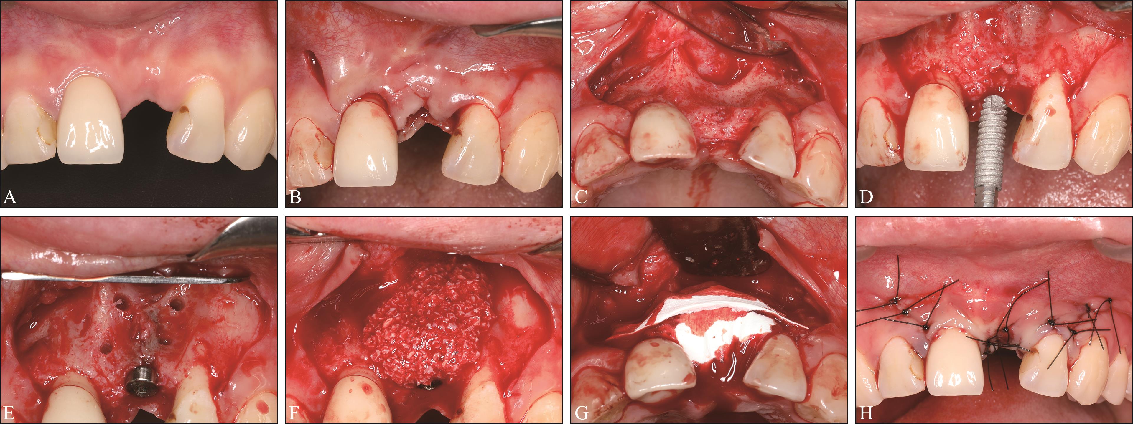
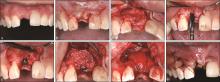
Fig 2
Surgical procedure of the modified triangular flap group

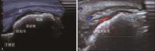
Fig 3
Ultrasound image
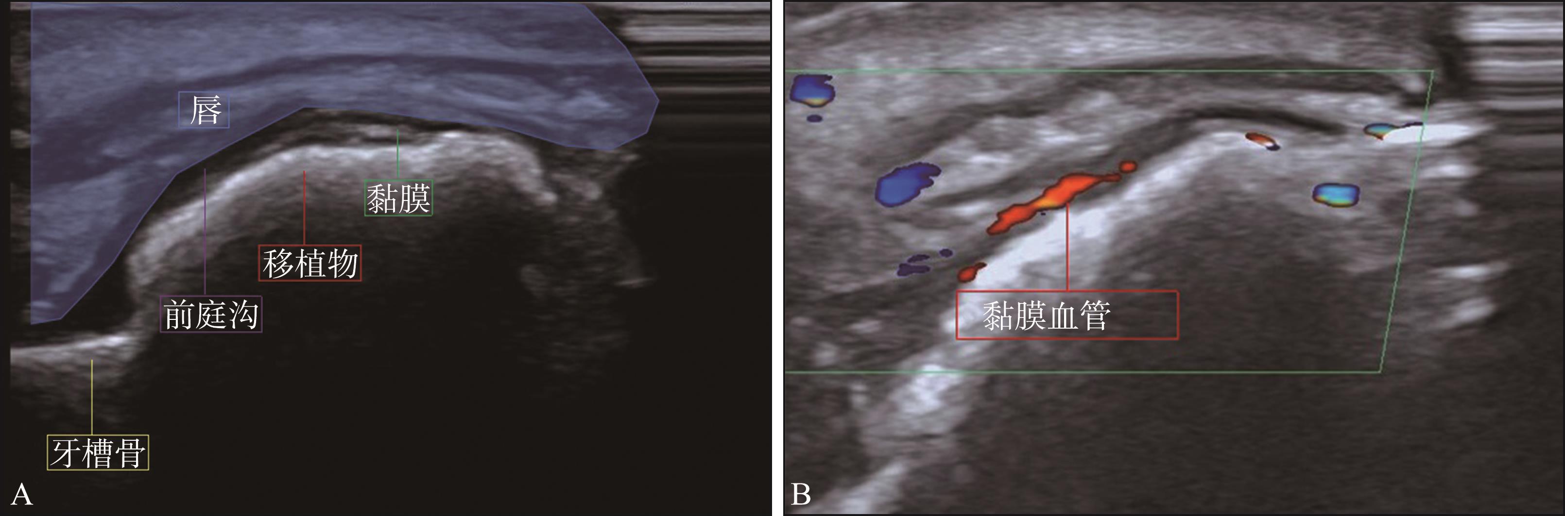
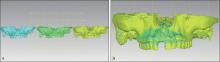
Fig 4
Contour change analysis
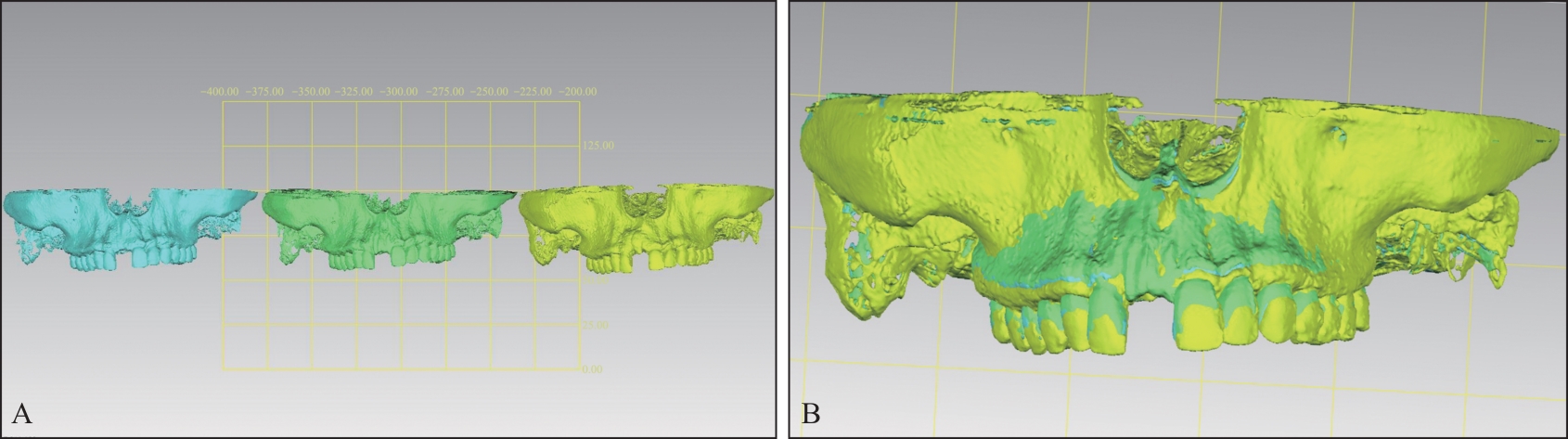
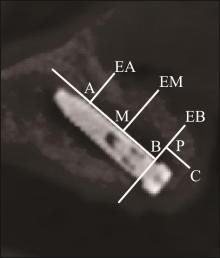
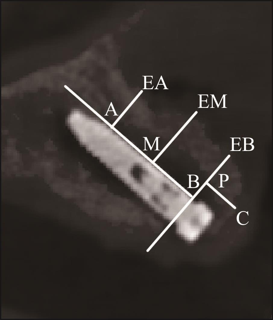
Fig 5
CBCT marker points and measurements
Tab 1
Characteristics of patients
| 特征 | 梯形瓣组 | 角形瓣组 | P值 | |
|---|---|---|---|---|
| 年龄/岁 | 36.1±14.1 | 34.8±13.0 | 0.828 | |
| 性别 | 男 | 4(36.4%) | 6(54.5%) | 0.392 |
| 女 | 7(63.6%) | 5(45.5%) | ||
| 缺失位置 | 中切牙 | 7(63.6%) | 7(63.6%) | 0.565 |
| 侧切牙 | 3(27.3%) | 4(36.4%) | ||
| 尖牙 | 1(9.1%) | 0(0%) | ||
| 牙龈生物型 | 厚龈生物型 | 4(36.4%) | 3(27.3%) | 0.647 |
| 薄龈生物型 | 7(63.6%) | 8(72.7%) | ||
| 缺牙原因 | 外伤 | 5(54.5%) | 6(54.5%) | 0.700 |
| 先天缺失 | 3(27.3%) | 1(9.1%) | ||
| 龋病 | 1(9.1%) | 2(18.2%) | ||
| 根尖周疾病 | 2(18.2%) | 2(18.2%) | ||
| 缺牙时间/年 | 3.7±6.4 | 4.4±7.8 | 0.809 | |
| 种植体尺寸 | 3.4 mm×12 mm | 9(81.8%) | 9(81.8%) | 1.000 |
| 3.4 mm×14 mm | 2(18.2%) | 2(18.2%) | ||
| 唇侧骨壁厚度/mm | 0.41±0.32 | 0.55±0.22 | 0.345 | |
Tab 2
Baseline values of vascular parameters
| 指标 | 总体 | 缺失牙 | 参照牙 | 平均差值 | 95%置信区间 |
|---|---|---|---|---|---|
| Vm/(cm/s) | 4.42±2.43 | 3.14±1.00 | 5.71±2.77 | -2.57±2.78* | 1.30,3.84 |
| Vmax/(cm/s) | 7.22±3.61 | 5.02±1.52 | 9.41±3.79 | -4.39±3.88* | 2.64,6.15 |
| S/D | 2.60±0.73 | 2.43±0.40 | 2.77±0.93 | -0.34±0.77 | -0.09,0.78 |
| PI | 1.02±0.37 | 0.96±0.23 | 1.09±0.46 | -0.13±0.34 | -0.09,0.36 |
| RI | 0.59±0.08 | 0.58±0.06 | 0.61±0.08 | -0.03±0.08 | -0.01,0.08 |
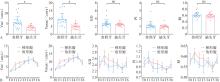
Fig 6
Vascular parameters statistical charts
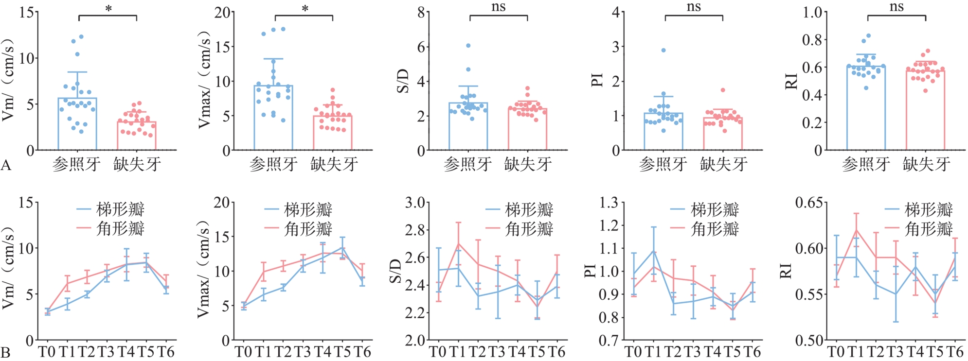
Tab 3
Vascular parameters of human gingival after flap surgery
| 组别 | 时间 | Vm/(cm/s) | Vmax/(cm/s) | S/D | PI | RI |
|---|---|---|---|---|---|---|
| 梯形瓣 | T0 | 3.08±1.16 | 4.93±1.77 | 2.51±0.53 | 0.99±0.30 | 0.59±0.08 |
| T1 | 3.92±2.10 | 6.60±3.04 | 2.52±0.43 | 1.09±0.34 | 0.59±0.07 | |
| T2 | 4.97±1.20 | 7.63±1.62 | 2.32±0.31 | 0.86±0.16 | 0.56±0.05 | |
| T3 | 6.96±2.10 | 10.74±2.95 | 2.35±0.64 | 0.87±0.25 | 0.55±0.10 | |
| T4 | 8.19±5.79 | 11.93±7.32 | 2.40±0.24 | 0.89±0.13 | 0.58±0.05 | |
| T5 | 8.41±3.43 | 13.41±4.99 | 2.29±0.46 | 0.85±0.18 | 0.55±0.07 | |
| T6 | 5.44±1.34 | 8.49±2.08 | 2.39±0.28 | 0.91±0.14 | 0.58±0.05 | |
| 角形瓣 | T0 | 3.19±0.87 | 5.12±1.31 | 2.35±0.23 | 0.93±0.13 | 0.57±0.04 |
| T1 | 6.16±2.82 | 9.91±4.51 | 2.70±0.51 | 1.02±0.21 | 0.62±0.06 | |
| T2 | 6.87±2.36 | 10.74±2.53 | 2.55±0.59 | 0.97±0.27 | 0.59±0.09 | |
| T3 | 7.56±2.22 | 11.57±2.67 | 2.50±0.36 | 0.96±0.21 | 0.59±0.06 | |
| T4 | 8.26±2.74 | 12.61±4.16 | 2.43±0.50 | 0.91±0.24 | 0.57±0.07 | |
| T5 | 8.44±1.70 | 12.53±2.68 | 2.24±0.26 | 0.83±0.13 | 0.54±0.05 | |
| T6 | 6.39±2.35 | 10.04±3.38 | 2.50±0.39 | 0.96±0.17 | 0.59±0.07 |
Tab 4
Alveolar bone parameter changes
| 测量参数及时间点 | 梯形瓣组 | 角形瓣组 | P值 | |
|---|---|---|---|---|
| A-EA/mm | T1 | 3.4±0.8 | 3.2±0.9 | 0.850 |
| T7 | 2.9±0.7 | 3.0±0.8 | 0.468 | |
| T7-T1 | -0.6±0.8 | -0.2±0.6 | 0.347 | |
| M-EM/mm | T1 | 4.4±0.8 | 4.1±0.8 | 0.390 |
| T7 | 3.6±0.3 | 3.8±0.9 | 0.587 | |
| T7-T1 | -0.9±0.6 | -0.3±0.6 | 0.075 | |
| B-EB/mm | T1 | 3.6±0.9 | 3.6±0.6 | 0.618 |
| T7 | 2.3±0.7 | 2.6±1.0 | 0.645 | |
| T7-T1 | -1.3±0.9 | -0.9±0.7 | 0.303 | |
| C-P/mm | T1 | 3.8±1.5 | 3.3±1.2 | 0.449 |
| T7 | 1.9±0.5 | 1.9±0.6 | 1.000 | |
| T7-T1 | -1.9±1.4 | -1.4±1.3 | 0.460 | |
| 体积/mm3 | T1-T0 | 448±48 | 418±120 | 0.521 |
| T7-T0 | 312±70 | 304±141 | 0.884 | |
| T7-T1 | -136±78 | -114±85 | 0.601 | |
| [1] | Ramasamy SK, Kusumbe AP, Schiller M, et al. Blood flow controls bone vascular function and osteogenesis[J]. Nat Commun, 2016, 7: 13601. |
| [2] | Huang L, Zou R, He J, et al. Comparing osteogenic effects between concentrated growth factors and the acellular dermal matrix[J]. Braz Oral Res, 2018, 32: e29. |
| [3] | Feng SW, Su YH, Lin YK, et al. Small blood stem cells for enhancing early osseointegration formation on dental implants: a human phaseⅠsafety study[J]. Stem Cell Res Ther, 2021, 12(1): 380. |
| [4] | Braut V, Bornstein MM, Belser U, et al. Thickness of the anterior maxillary facial bone wall—a retrospective radiographic study using cone beam computed tomography[J]. Int J Periodontics Restorative Dent, 2011, 31(2): 125-131. |
| [5] | Scarano A, Lorusso F, Arcangelo M, et al. Lateral sinus floor elevation performed with trapezoidal and modified triangular flap designs: a randomized pilot study of post-operative pain using thermal infrared imaging[J]. Int J Environ Res Public Health, 2018, 15(6): 1277. |
| [6] | Koymen R, Karacayli U, Gocmen-Mas N, et al. Flap and incision design in implant surgery: clinical and anatomical study[J]. Surg Radiol Anat, 2009, 31(4): 301-306. |
| [7] | Bhide A, Acharya G, Baschat A, et al. ISUOG Practice Guidelines (updated): use of Doppler velocimetry in obstetrics[J]. Ultrasound Obstet Gynecol, 2021, 58(2): 331-339. |
| [8] | Jiang L, Zhang D, Chen YN, et al. The value of conventional ultrasound combined with superb microvascular imaging and color Doppler flow imaging in the diagnosis of thyroid malignant nodules: a systematic review and meta-analysis[J]. Front Endocrinol (Lausanne), 2023, 14: 1182259. |
| [9] | Yu TF, He W, Gan CG, et al. Deep learning applied to two-dimensional color Doppler flow imaging ultrasound images significantly improves diagnostic performance in the classification of breast masses: a multicenter study[J]. Chin Med J (Engl), 2021, 134(4): 415-424. |
| [10] | Zarzecki M, Obuchowska I, Ustymowicz A, et al. Glaucoma surgery and ocular blood flow in colour Doppler imaging: is there a link[J]. Clin Ophthalmol, 2024, 18: 49-60. |
| [11] | Xue F, Wu BZ, Zhang R, et al. Analyses of gingival papilla blood flow via color doppler flow imaging and micro-flow imaging in patients with advanced periodontitis: a clinical pilot study[J]. Eur J Med Res, 2024, 29(1): 527. |
| [12] | Wu C, Liu X, Zhang H, et al. Response of human perio-dontal ligament to orthodontic force using superb microvascular imaging[J]. Am J Orthod Dentofacial Orthop, 2022, 162(5): e257-e266. |
| [13] | Abu Alhaija ES, Taha NA. A comparative study of initial changes in pulpal blood flow between conventional and self-ligating fixed orthodontic brackets during leveling and alignment stage[J]. Clin Oral Investig, 2021, 25(3): 971-981. |
| [14] | Urban IA, Lozada JL, Wessing B, et al. Vertical bone grafting and periosteal vertical mattress suture for the fi-xation of resorbable membranes and stabilization of particulate grafts in horizontal guided bone regeneration to achieve more predictable results: a technical report[J]. Int J Periodontics Restorative Dent, 2016, 36(2): 153-159. |
| [15] | Guo R, Yu Q, Lin Y, et al. Pulp blood flow changes in maxillary and mandibular anterior teeth after orthodontic retraction: a prospective study[J]. BMC Oral Health, 2022, 22(1): 508. |
| [16] | Retzepi M, Tonetti M, Donos N. Gingival blood flow changes following periodontal access flap surgery using laser Doppler flowmetry[J]. J Clin Periodontol, 2007, 34(5): 437-443. |
| [17] | Kijsamanmith K, Vongsavan N, Matthews B. Pulpal blood flow recorded from exposed dentine with a laser Doppler flow meter using red or infrared light[J]. Arch Oral Biol, 2018, 87: 163-167. |
| [18] | Rendell MS, Johnson ML, Smith D, et al. Skin blood flow response in the rat model of wound healing: expression of vasoactive factors[J]. J Surg Res, 2002, 107(1): 18-26. |
| [19] | Ahmed MV, Rastogi S, Baad RK, et al. Comparative study between two flaps-trapezoidal flap (TZF) and ocshenbein-leubke flap (OLF) in periapical surgeries[J]. J Maxillofac Oral Surg, 2013, 12(4): 440-446. |
| [20] | Schenk RK, Buser D, Hardwick WR, et al. Healing pattern of bone regeneration in membrane-protected defects: a histologic study in the canine mandible[J]. Int J Oral Maxillofac Implants, 1994, 9(1): 13-29. |
| [21] | Nobuto T, Suwa F, Kono T, et al. Microvascular response in the periosteum following mucoperiosteal flap surgery in dogs: 3-dimensional observation of an angiogenic process[J]. J Periodontol, 2005, 76(8): 1339-1345. |
| [22] | Pazzaglia UE. Periosteal and endosteal reaction to rea-ming and nailing: the possible role of revascularization on the endosteal anchorage of cementless stems[J]. Biomaterials, 1996, 17(10): 1009-1014. |
| [23] | Donos N, Akcali A, Padhye N, et al. Bone regeneration in implant dentistry: Which are the factors affecting the clinical outcome[J]. Periodontol 2000, 2023, 93(1): 26-55. |
| [24] | Marenzi G, Riccitiello F, Tia M, et al. Influence of leukocyte- and platelet-rich fibrin (L-PRF) in the healing of simple postextraction sockets: a split-mouth study[J]. Biomed Res Int, 2015, 2015: 369273. |
| [25] | Botticelli D, Berglundh T, Lindhe J. Hard-tissue alterations following immediate implant placement in extraction sites[J]. J Clin Periodontol, 2004, 31(10): 820-828. |
| [26] | Sanz M, Cecchinato D, Ferrus J, et al. A prospective, randomized-controlled clinical trial to evaluate bone pre-servation using implants with different geometry placed into extraction sockets in the maxilla[J]. Clin Oral Implants Res, 2010, 21(1): 13-21. |
| [27] | Grassi FR, Grassi R, Rapone B, et al. Dimensional chan-ges of buccal bone plate in immediate implants inserted through open flap, open flap and bone grafting and flapless techniques: a cone-beam computed tomography randomized controlled clinical trial[J]. Clin Oral Implants Res, 2019, 30(12): 1155-1164. |
| [28] | Degidi M, Daprile G, Nardi D, et al. Buccal bone plate in immediately placed and restored implant with Bio-Oss® collagen graft: a 1-year follow-up study[J]. Clin Oral Implants Res, 2013, 24(11): 1201-1205. |
| [29] | Fu JH, Yeh CY, Chan HL, et al. Tissue biotype and its relation to the underlying bone morphology[J]. J Periodontol, 2010, 81(4): 569-574. |
| [30] | Nagaraj KR, Savadi RC, Savadi AR, et al. Gingival biotype—Prosthodontic perspective[J]. J Indian Prosthodont Soc, 2010, 10(1): 27-30. |
| [31] | Barone A, Alfonsi F, Derchi G, et al. The effect of insertion torque on the clinical outcome of single implants: a randomized clinical trial[J]. Clin Implant Dent Relat Res, 2016, 18(3): 588-600. |
| [32] | Monje A, Roccuzzo A, Buser D, et al. Influence of buccal bone wall thickness on the peri-implant hard and soft tissue dimensional changes: a systematic review[J]. Clin Oral Implants Res, 2023, 34(3): 157-176. |
| [1] | Zhao Pengyu, Chen Gang, Cheng Yi, Wang Chao, Chen Dan, Huang Haitao. Clinical and histological evaluation of three-dimensional printing individualized titanium mesh for alveolar bone defect repair [J]. West China Journal of Stomatology, 2025, 43(4): 592-602. |
| [2] | Su Wenqi, Zhang Dandan, Cheng Yan, Cui Wenjie, Lei Lang, Li Houxuan. Guided bone regeneration therapy based on plaque control of peri-implantitis with follow-up at 7 years [J]. West China Journal of Stomatology, 2025, 43(1): 133-139. |
| [3] | Shi Ruiwen, Yang Hu, Liu Yue, Shi Yilin, Zhang Shengben, Liu Yu, Song Feng, Lan Jing. L-shape technique with concentrated growth factor for horizontal bone defects in the maxillary anterior region: a clinical and radiographic study [J]. West China Journal of Stomatology, 2025, 43(1): 76-83. |
| [4] | Chen Liangwei, Han Jianmin, Guo Chuanbin. Research status and prospects of biodegradable magnesium-based metal-guided bone regeneration membranes [J]. West China Journal of Stomatology, 2024, 42(4): 415-425. |
| [5] | Liu Yiming, Zhao Yun, Han Mei, Zhang Yuqiu, Mi Fanglin, Wang Bing. Preparation of functional poly-(lactic acid-co-glycolic acid)-based guided bone-regeneration membrane and its application in the reconstruction of mandibular defects in rats [J]. West China Journal of Stomatology, 2022, 40(5): 522-531. |
| [6] | Chen Luyi, Huang Min, Wu Jiaqi, Luo Jun. Guided bone regeneration-assisted orthodontic treatment for closing the space of missing central incisors [J]. West China Journal of Stomatology, 2021, 39(4): 482-488. |
| [7] | Tiantian Yu,Jin Liu,Junjing Yin,Xiangna Xu,Shengjie Yan,Jing Lan. Effects of concentrated growth factors on relieving postoperative reaction of guided bone regeneration in the esthetic zone [J]. West China Journal of Stomatology, 2019, 37(4): 398-402. |
| [8] | Yubin Cao,Chang Liu,Weilin Pan,Yuan Tu,Chunjie Li,Chengge Hua. Research progress on the modification of guided bone regeneration membranes [J]. West China Journal of Stomatology, 2019, 37(3): 325-329. |
| [9] | Deng Xia, Bai Shi. Biomineralization of electrospun polycaprolactone-guided bone regeneration membrane [J]. West China Journal of Stomatology, 2016, 34(6): 570-574. |
| [10] | Liu Man, Zhang Qiang, Zhou Liwei, Mo Anchun, Li Xiaoyu, Li Jidong. Research on the micro structure of antibacterial nanocomposite membrane and it’s biocompatibility as a guided bone regeneration membrane [J]. West China Journal of Stomatology, 2013, 31(2): 127-130. |
| [11] | Jiang Xinyi1, Yang Ji2, Chai Zhaowu3, Song Jinlin1, Deng Feng1, Wang Zhibiao4.. Low intensity pulsed ultrasound irradiating combined with guided bone regeneration for promoting the repair effect of periodontal bone defect [J]. West China Journal of Stomatology, 2012, 30(5): 487-492. |
| [12] | HUANG Xiao-feng1, ZENG Xiang -long2. Changes of blood flow volume in dog’s mental artery after mandible osteocompression [J]. West China Journal of Stomatology, 2010, 28(06): 664-667. |
| [13] | FEI Wei, YANG Xiao-min, LI Zheng, YIN Ming-ping, SHEN Zhi-hao, LIAO Chu-hang. Experimental study of the bioresorbable collagen membrane used for guided bone regeneration around dental implants [J]. West China Journal of Stomatology, 2008, 26(05): 494-498. |
| [14] | BAI Shi1, MO An- chun2, XIAN Su- qin2, ZUO Yi3, LI Yu- bao3, XU Wei2. Char acter ization and antibacter ial effect of Ag- nHA- nTiO2/polyamide 66 nanocomposite membr ane on oral bacter ia [J]. West China Journal of Stomatology, 2008, 26(04): 358-361. |
| [15] | LIU Man1,2, WANG Shao- an3, MO An- chun3, MENG Yao4, HU Jie5, LI Xiao- yu1. Study on the microstructur e of acellular dermal matr ix and its biocompatibility with MG63 osteoblast - like cells [J]. West China Journal of Stomatology, 2008, 26(02): 129-132. |
| Viewed | ||||||
|
Full text |
|
|||||
|
Abstract |
|
|||||
 This work is licensed under a Creative Commons Attribution 3.0 License.
