West China Journal of Stomatology ›› 2025, Vol. 43 ›› Issue (1): 114-125.doi: 10.7518/hxkq.2024.2024169
Previous Articles Next Articles
Chu Tianhao1,2( ), Zhang Xueying1, Wang Haocheng1, Ma Haojie1, Liu Yuanyuan1,2()
Received:2024-05-02
Revised:2024-08-05
Online:2025-02-01
Published:2025-01-22
Contact:
Liu Yuanyuan
E-mail:1072615131@qq.com;liuyuan113@sina.com
Supported by:CLC Number:
Chu Tianhao, Zhang Xueying, Wang Haocheng, Ma Haojie, Liu Yuanyuan. Three-dimensional finite element feature analysis of the mandible and morphology and position of temporomandibular joint in patients with unilateral and bilateral molar scissor bite[J]. West China Journal of Stomatology, 2025, 43(1): 114-125.
Add to citation manager EndNote|Ris|BibTeX
Tab 1
TMJ measurements and meanings
| 名称 | 英文 | 测量项目含义 |
|---|---|---|
| 关节窝宽度 | width of the fossa,WF | 在垂直于髁突长轴的矫正斜矢状位下,连接关节窝后壁最下点和关节结节最下点,此连线位于关节窝内的部分为关节窝宽度 |
| 关节窝高度 | height of the fossa,HF | 在垂直于髁突长轴的矫正斜矢状位下,连接关节窝后壁最下点和关节结节最下点,关节窝最上点到此连线的垂直距离为关节窝高度 |
| 关节结节倾斜度 | articular eminence inclination,AEI | 关节结节最下点与关节窝最上点连线与关节结节最下点与关节窝后壁最下点连线的交角 |
| 髁突长轴 | long axis of the condyle,LAC | 在髁突面积最大的横断面上测量髁突的最大内外径 |
| 髁突短轴 | minor axis of the condyle,MAC | 在髁突面积最大的横断面上测量髁突的最大前后径 |
| 髁突水平角 | horizontal angle of the condyle,HAC | 髁突长轴与冠状水平线间的夹角 |
| 关节前间隙 | anterior joint space,AJS | 过关节窝顶点向髁突前斜面作切线,切点与关节结节后斜面的最短距离 |
| 关节上间隙 | superior joint space,SJS | 关节窝顶点与髁突顶点连线的距离 |
| 关节后间隙 | posterior joint space,PJS | 过关节窝顶点向髁突后斜面作切线,切点与关节窝后壁的最短距离 |
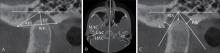
Fig 1
TMJ measurements
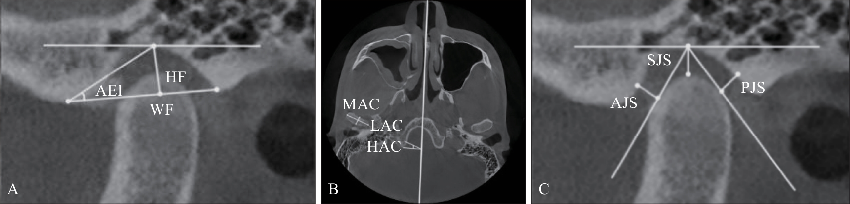
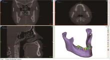
Fig 2
Apply Mimics Research 21.0 to preliminarily extract the mandible model
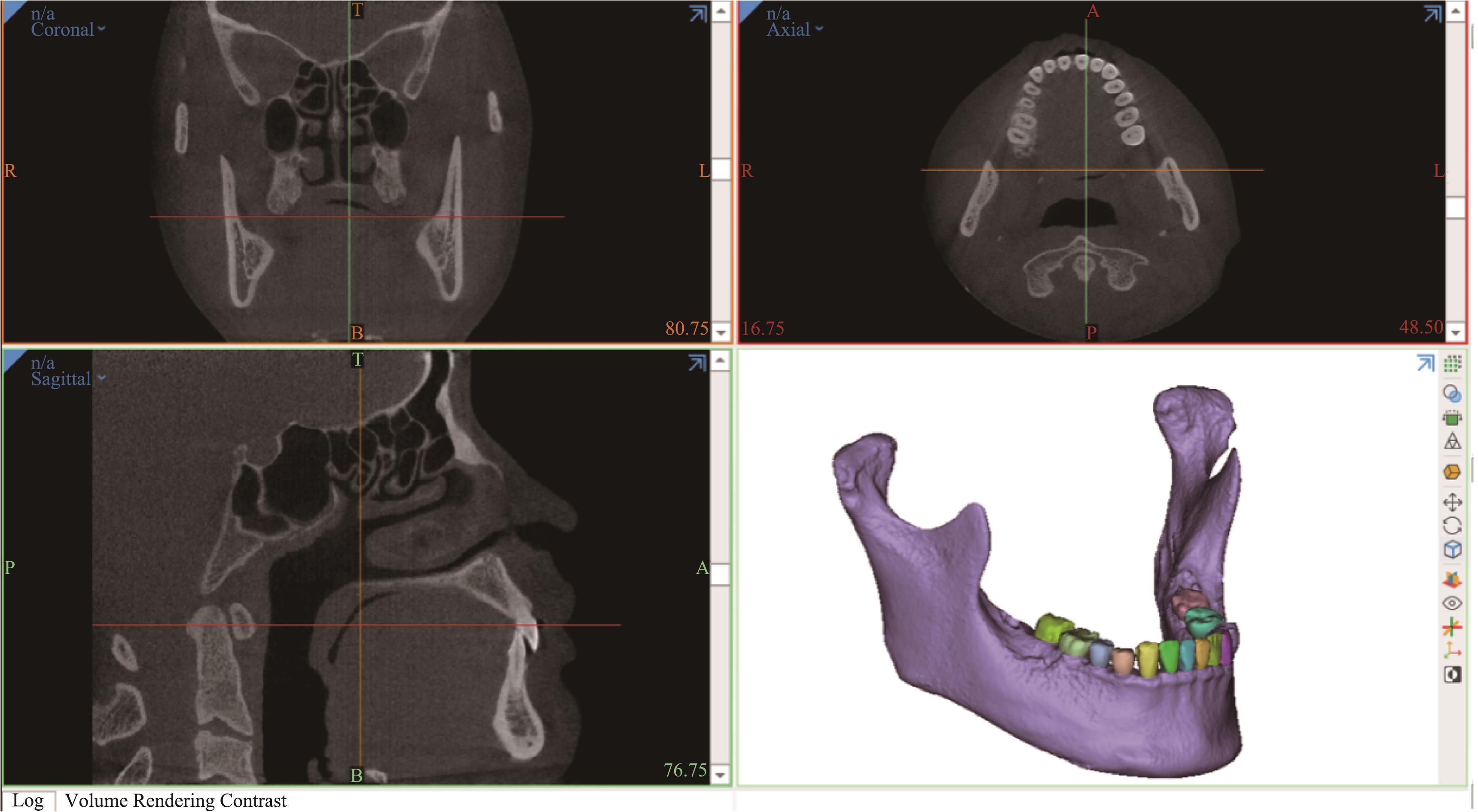
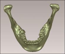
Fig 3
Apply Geomagic Wrap 2017 to further smooth the model
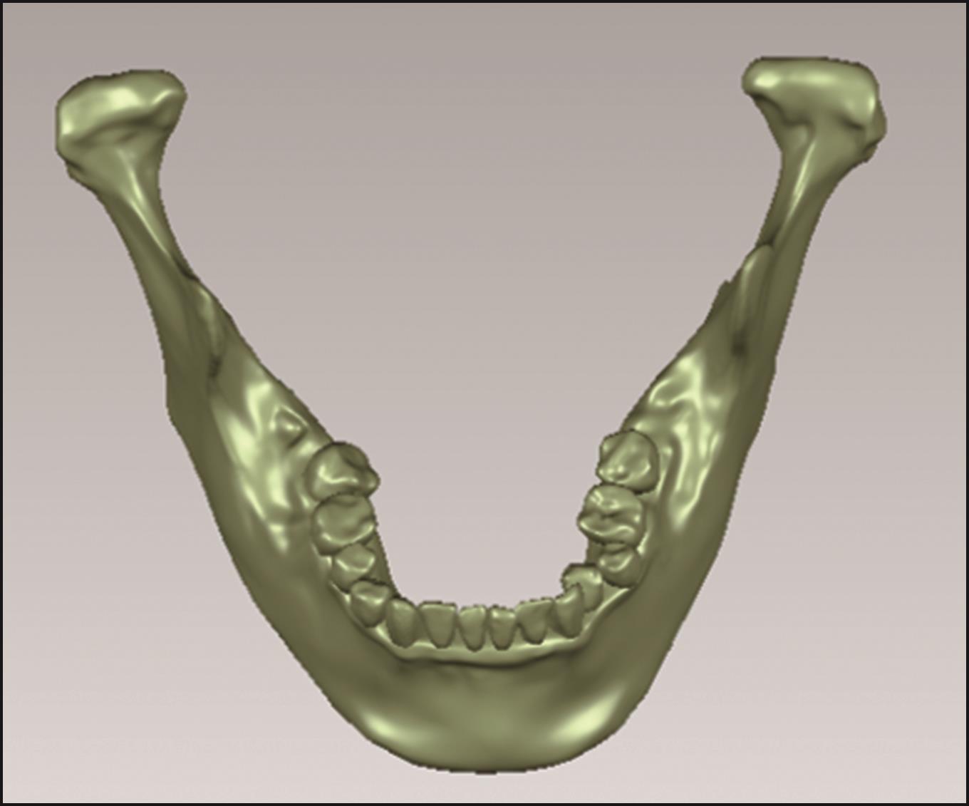
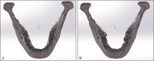
Fig 4
Apply SolidWorks 2024 to assemble tooth and mandible
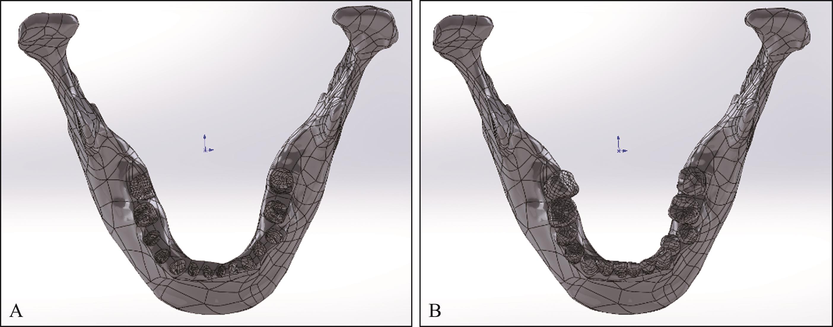
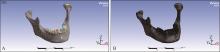
Fig 5
Use Ansys Workbench 2022 R1 imports and meshes models
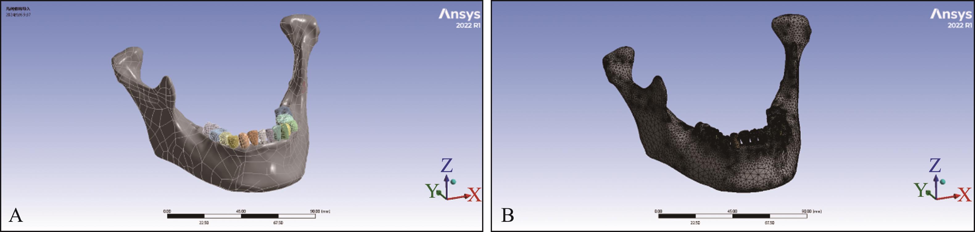
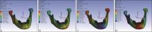
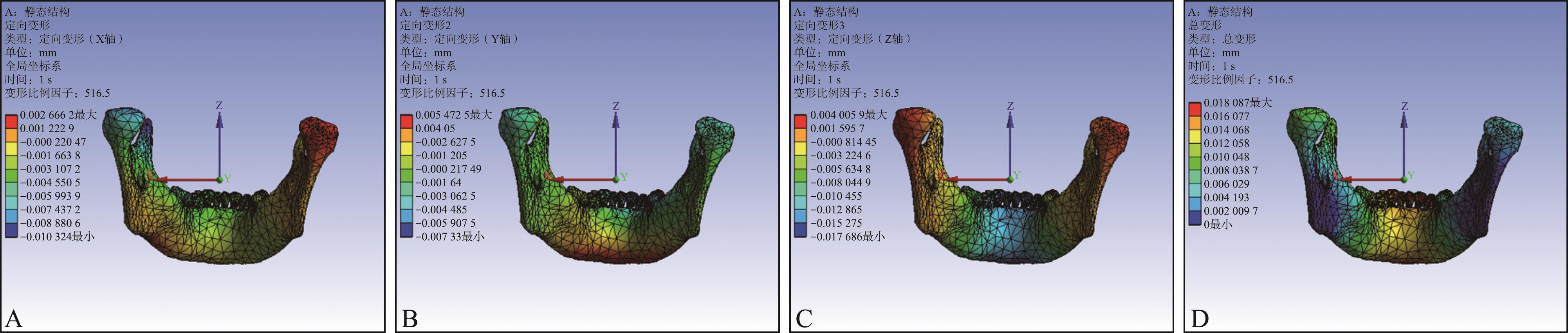
Fig 6
Directional deformation results of the model under stress load (frontal view)

Fig 7
Directional deformation results of the model under stress load (rear view)
Tab 2
Angle’s classification of patients in the unilate-ral group and the bilateral group
| 组别 | 安氏Ⅰ类 | 安氏Ⅱ类 | 安氏Ⅲ类 | 其他 |
|---|---|---|---|---|
| 单侧组 | 7/70.0 | 2/20.0 | 0/0.0 | 1/10.0 |
| 双侧组 | 5/50.0 | 3/30.0 | 1/10.0 | 1/10.0. |
Tab 3
Osseous classification of patients in the unilate-ral group, the bilateral group and the control group
| 组别 | 骨性Ⅰ类 | 骨性Ⅱ类 | 骨性Ⅲ类 |
|---|---|---|---|
| 单侧组 | 7/70.0 | 3/30.0 | 0/0.0 |
| 双侧组 | 4/40.0 | 6/60.0 | 0/0.0 |
| 对照组 | 9/45.0 | 11/55.0 | 0/0.0 |
Tab 4
Comparison of bilateral joint morphology and position measurements in the control group
| 项目 | 左侧 | 右侧 | P值 |
|---|---|---|---|
| 关节上间隙/mm | 3.91±1.12 | 3.92±0.92 | 0.975 |
| 关节前间隙/mm | 2.61±0.96 | 2.95±1.50 | 0.435 |
| 关节后间隙/mm | 1.97±0.39 | 2.09±0.43 | 0.639 |
| 髁突长轴/mm | 20.94±2.11 | 20.97±2.17 | 0.943 |
| 髁突短轴/mm | 8.66±1.01 | 8.35±0.76 | 0.111 |
| 关节窝高度/mm | 5.82±1.43 | 5.57±0.95 | 0.503 |
| 关节窝宽度/mm | 15.29±1.86 | 16.47±0.80 | 0.180 |
| 关节结节斜度/° | 35.86±7.37 | 35.57±10.78 | 0.948 |
| 髁突水平角/° | 15.23±4.95 | 17.94±6.36 | 0.374 |
Tab 5
Comparison of bilateral joint morphology and position in the unilateral group
| 项目 | 锁 侧 侧 | 正常侧 | P值 |
|---|---|---|---|
| 关节上间隙/mm | 2.48±0.44 | 2.64±0.20 | 0.234 |
| 关节前间隙/mm | 2.48±0.85 | 2.24±0.61 | 0.603 |
| 关节后间隙/mm | 1.80±0.44 | 1.76±0.38 | 0.852 |
| 髁突长轴/mm | 18.76±2.11 | 18.55±3.57 | 0.807 |
| 髁突短轴/mm | 8.02±1.43 | 7.99±1.49 | 0.932 |
| 关节窝高度/mm | 6.33±1.08 | 5.85±1.08 | 0.157 |
| 关节窝宽度/mm | 16.73±1.73 | 16.74±2.79 | 0.984 |
| 关节结节斜度/° | 35.60±6.48 | 36.02±7.80 | 0.872 |
| 髁突水平角/° | 23.32±7.93 | 21.71±7.66 | 0.421 |
Tab 6
Comparison of bilateral joint morphology and position in the bilateral group
| 项目 | 左侧 | 右侧 | P值 |
|---|---|---|---|
| 关节上间隙/mm | 2.94±0.74 | 2.66±0.78 | 0.344 |
| 关节前间隙/mm | 2.44±0.68 | 2.64±1.12 | 0.720 |
| 关节后间隙/mm | 2.04±0.45 | 1.73±0.33 | 0.239 |
| 髁突长轴/mm | 16.71±1.43 | 19.28±2.26 | 0.419 |
| 髁突短轴/mm | 6.83±1.11 | 6.64±1.25 | 0.675 |
| 关节窝高度/mm | 6.10±0.92 | 6.57±0.42 | 0.305 |
| 关节窝宽度/mm | 15.76±2.25 | 15.91±1.38 | 0.871 |
| 关节结节斜度/° | 34.78±2.93 | 36.79±2.62 | 0.292 |
| 髁突水平角/° | 25.44±7.40 | 25.71±8.67 | 0.888 |
Tab 7
Comparison of the morphology and position of the left joint in the control group and the scissor bite side of the unilateral group and the left joint in the bilateral group
| 项目 | 对照组左侧 | 单侧组锁 侧 侧 | 双侧组左侧 | F值/H值* | P值 |
|---|---|---|---|---|---|
| 关节上间隙/mm | 3.91±1.12 | 2.48±0.44a | 2.94±0.74 | 6.297* | 0.031* |
| 关节前间隙/mm | 2.61±0.96 | 2.48±0.85 | 2.44±0.68 | 0.306* | 0.858* |
| 关节后间隙/mm | 1.97±0.39 | 1.80±0.44 | 2.04±0.45 | 0.628 | 0.544 |
| 髁突长轴/mm | 20.94±2.11 | 18.76±2.11a,c | 16.71±1.43b | 7.777 | 0.003 |
| 髁突短轴/mm | 8.66±1.01 | 8.02±1.43 | 6.83±1.11b | 3.725 | 0.043 |
| 关节窝高度/mm | 5.82±1.43 | 6.33±1.08 | 6.10±0.92 | 0.354 | 0.706 |
| 关节窝宽度/mm | 15.29±1.86 | 16.73±1.73 | 15.76±2.25 | 1.071 | 0.363 |
| 关节结节斜度/° | 35.86±7.37 | 35.60±6.48 | 34.78±2.93 | 0.062 | 0.940 |
| 髁突水平角/° | 15.23±4.95 | 23.32±7.93a | 25.44±7.40b | 3.706 | 0.044 |
Tab 8
Comparison of the shape and position of the right joint in the control group, the normal side in the unilateral group and the right joint in the bilateral group
| 项目 | 对照组左侧 | 单侧组锁 侧 侧 | 双侧组左侧 | F值/H值* | P值 |
|---|---|---|---|---|---|
| 关节上间隙/mm | 3.92±0.92 | 2.64±0.20a | 2.66±0.78b | 9.2185* | 0.010* |
| 关节前间隙/mm | 2.95±1.50 | 2.24±0.61 | 2.64±1.12 | 1.488 | 0.251 |
| 关节后间隙/mm | 2.09±0.43 | 1.76±0.38 | 1.73±0.33 | 1.749 | 0.201 |
| 髁突长轴/mm | 20.97±2.17 | 18.55±3.57 | 19.28±2.26b | 4.319 | 0.028 |
| 髁突短轴/mm | 8.35±0.76 | 7.99±1.49c | 6.64±1.25 | 3.458 | 0.052 |
| 关节窝高度/mm | 5.57±0.95 | 5.85±1.08 | 6.57±0.42 | 2.240 | 0.134 |
| 关节窝宽度/mm | 16.47±0.80 | 16.74±2.79 | 15.91±1.38 | 0.480* | 0.787* |
| 关节结节斜度/° | 35.57±10.78 | 36.02±7.80 | 36.79±2.62 | 0.043 | 0.958 |
| 髁突水平角/° | 17.94±6.36 | 21.71±7.66 | 25.71±8.67 | 1.656 | 0.217 |
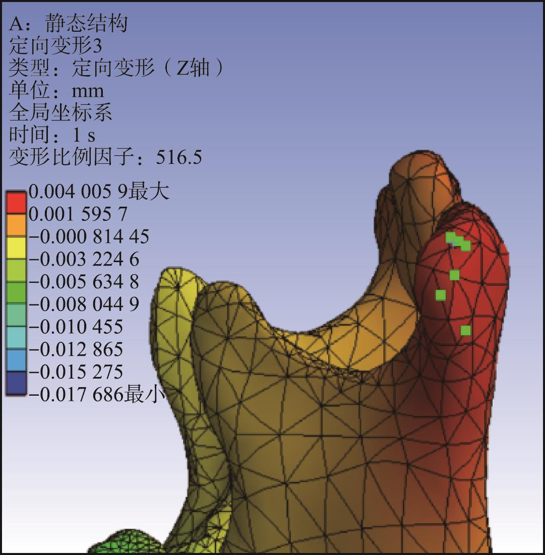
Tab 9
Directional deformation values of the model under different occlusal conditions
| 咬合方式 | 定向变形 | X轴 | Y轴 | Z轴 |
|---|---|---|---|---|
| 前牙咬合 | 最大定向变形值 | 0.002 7 | 0.007 7 | 0.002 7 |
| 最小定向变形值 | |-0.005 9| | |-0.011| | |-0.021| | |
| 正中咬合 | 最大定向变形值 | 0.002 7 | 0.005 5 | 0.004 |
| 最小定向变形值 | |-0.002 9| | |-0.007 3| | 0.017 | |
锁 侧磨牙咬合 侧磨牙咬合 | 最大定向变形值 | 0.001 8 | 0.003 4 | 0.001 6 |
| 最小定向变形值 | |-0.003 2| | |-0.004 4| | |-0.011| | |
| 正常侧磨牙咬合 | 最大定向变形值 | 0.008 4 | 0.002 2 | 0.002 6 |
| 最小定向变形值 | |-0.007 7| | |-0.003| | |-0.007 2| | |
锁 侧前磨牙咬合 侧前磨牙咬合 | 最大定向变形值 | 0.002 1 | 0.005 8 | 0.001 9 |
| 最小定向变形值 | |-0.004 1| | |-0.006 1| | |-0.015| | |
| 正常侧前磨牙咬合 | 最大定向变形值 | 0.007 7 | 0.005 1 | 0.002 |
| 最小定向变形值 | |-0.004 2| | |-0.01| | |-0.016| |
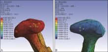
Fig 8
Create local maximum and minimum probes (X-axis)
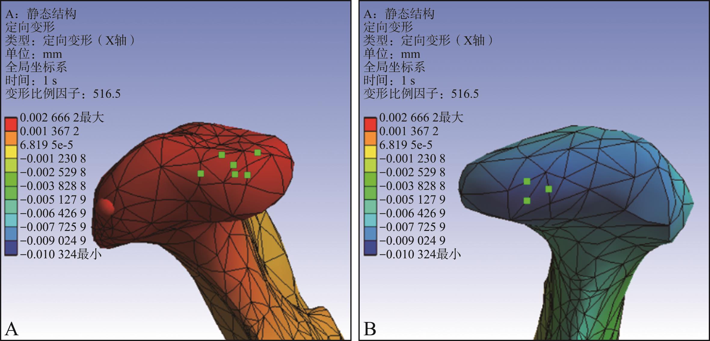
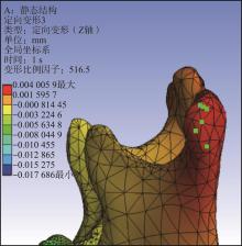
Fig 9
Creating local maximum and minimum probes (Z-axis)
| 1 | 叶莉娜, 何家才. 单侧后牙正锁𬌗、反𬌗对下颌升支及髁突对称性影响的临床观察[J]. 口腔颌面外科杂志, 2020, 30(6): 382-386. |
| Ye LN, He JC. Clinical observation of the effect of unila-teral posterior scissors-bite and crossbite on the symmetry of mandibular ramus and condyle[J]. J Oral Maxillofac Surg, 2020, 30(6): 382-386. | |
| 2 | Baik UB, Kim Y, Sugawara J, et al. Correcting severe scissor bite in an adult[J]. Am J Orthod Dentofac Orthop, 2019, 156(1): 113-124. |
| 3 | Lambourne C, Lampasso J, Buchanan WC Jr, et al. Ma-locclusion as a risk factor in the etiology of headaches in children and adolescents[J]. Am J Orthod Dentofacial Or-thop, 2007, 132(6): 754-761. |
| 4 | Thilander B, Rubio G, Pena L, et al. Prevalence of temporomandibular dysfunction and its association with ma-locclusion in children and adolescents: an epidemiologic study related to specified stages of dental development[J]. Angle Orthod, 2002, 72(2): 146-154. |
| 5 | Egermark I, Magnusson T, Carlsson GE. A 20-year follow-up of signs and symptoms of temporomandibular disorders and malocclusions in subjects with and without orthodontic treatment in childhood[J]. Angle Orthod, 2003, 73(2): 109-115. |
| 6 | Tomonari H, Kubota T, Yagi T, et al. Posterior scissors-bite: masticatory jaw movement and muscle activity[J]. J Oral Rehabil, 2014, 41(4): 257-265. |
| 7 | Manfredini D, Lombardo L, Siciliani G. Temporomandibular disorders and dental occlusion. A systematic review of association studies: end of an era[J]. J Oral Rehabil, 2017, 44(11): 908-923. |
| 8 | 郭维鹏, 李亚兰, 唐志雄, 等. 包含颞下颌关节的下颌骨有限元建模[J]. 生物医学工程研究, 2013, 32(3): 162-166. |
| Guo WP, Li YL, Tang ZX, et al. Finite element modeling of the mandible with temporomandibular joint[J]. J Biomed Eng Res, 2013, 32(3): 162-166. | |
| 9 | 杨文华, 赵宝莲, 孙庚林, 等. 下颌骨颏部正中骨折二维与三维坚强内固定的三维有限元研究[J]. 实用口腔医学杂志, 2011, 27(4): 495-500. |
| Yang WH, Zhao BL, Sun GL, et al. Three-dimensional finite element study of two-and three-dimensional internal fixation for mandibular symphysis fracture[J]. J Pract Stomatol, 2011, 27(4): 495-500. | |
| 10 | 张渊, 王美青, 凌伟. 用于分析𬌗面形态与颞下颌关节生物力学关系的三维有限元模型的建立[J]. 医用生物力学, 2004, 19(4): 249-252. |
| Zhang Y, Wang MQ, Ling W. Establishment of three-dimensional FEM model for evaluation of biomechanical relationship between temporomadibular joint morphology and bilateral condyles[J]. J Med Biomech, 2004, 19(4): 249-252. | |
| 11 | Tanne K, Tanaka E, Sakuda M. Stress distribution in the temporomandibular joint produced by orthopedic chincup forces applied in varying directions: a three-dimensional analytic approach with the finite element method[J]. Am J Orthod Dentofacial Orthop, 1996, 110(5): 502-507. |
| 12 | 王美青, 姚秀芳, 颜朝云, 等. 咬合与髁状突形态的对称性间的相关关系解剖学[J]. 实用口腔医学杂志, 2001, 17(2): 147-150. |
| Wang MQ, Yao XF, Yan CY, et al. An anatomic study on the relationship between occlusion symmetry and condyle symmetry[J]. J Pract Stomatol, 2001, 17(2): 147-150. | |
| 13 | 李爽, 马啸宙, 谢冰鑫, 等. 单、双侧磨牙正锁𬌗患者颞下颌关节形态和位置的锥形束CT研究[J]. 中华口腔正畸学杂志, 2023, 30(2): 81-85. |
| Li S, Ma XZ, Xie BX, et al. The cone beam CT study of temporomandibular joint morphology and position in patients with unilateral and bilateral molar scissors bite[J]. Chin J Orthod, 2023, 30(2): 81-85. | |
| 14 | 仲晓飞, 杜原宏. 磨牙正锁𬌗对髁突形态的影响[J]. 中国临床实用医学, 2014, 5(5): 8-10. |
| Zhong XF, Du YH. Effect of posterior buccal crossbite on condylar morphology[J]. Chin Clin Pract Med, 2014, 5(5): 8-10. | |
| 15 | 陈志兴, 郑怡, 王瑶, 等. 双侧第二磨牙正锁𬌗对下颌骨发育和位置的影响[J]. 中华口腔正畸学杂志, 2016, 23(2): 89-93. |
| Chen ZX, Zheng Y, Wang Y, et al. The effect of bilateral scissor bite of second molars on the growth and position of mandible[J]. Chin J Orthod, 2016, 23(2): 89-93. | |
| 16 | 李爽, 张洪宇, 易周, 等. 单、双侧第二磨牙正锁𬌗与颞下颌关节退行性关节病的CBCT研究[J]. 实用口腔医学杂志, 2023, 39(6): 774-778. |
| Li S, Zhang HY, Yi Z, et al. A CBCT study on the relationship between unilateral and bilateral second molar scissors bite and temporomandibular joint degenerative joint disease[J]. J Pract Stomatol, 2023, 39(6): 774-778. | |
| 17 | 魏子明, 林丽佳, 李旼劼, 等. 青少年单侧后牙正锁𬌗畸形患者双侧髁突在关节窝内位置及其形态变化研究[J]. 中国实用口腔科杂志, 2019, 12(7): 426-429. |
| Wei ZM, Lin LJ, Li MJ, et al. Analysis of the position and morphology of the bilateral condyles in the articular fossa in adolescent patients with unilateral scissors-bite posterior molar[J]. Chin J Pract Stomatol, 2019, 12(7): 426-429. | |
| 18 | Guercio Monaco E, De Stefano AA, Hernandez-Andara A, et al. Correlation between condylar size on CT and position of the articular disc on MRI of the temporomandibular joint[J]. Cranio, 2022, 40(1): 64-71. |
| 19 | 柴明珠, 李新. 夜磨牙、偏侧咀嚼患者颞下颌关节的锥形束CT研究[C]// 第20次全国颞下颌关节病学及𬌗学研讨会暨第七届亚洲颞下颌关节学术大会. 北京: 中华口腔医学会颞下颌关节病学及𬌗学专业委员会, 2023: 347-348. |
| Chai MZ, Li X. Conical beam CT study of temporomandibular joints in patients with night molar and lateral chewing[C]//Proceedings of the 20th annual meeting of society of temporomandibular disorders & occlusion and the 7th Asian academic congress for temporomandibular joint. Beijing: Professional Committee of Temporomandibular Arthropathy and Occlusion of Chinese Stomatological Association, 2023: 347-348. | |
| 20 | Li CX, Xie X, Li MJ, et al. A pilot investigation of condylar position and asymmetry in patients with unilateral posterior scissors-bite malocclusion based on three-dimensional reconstructive imaging technique[J]. BMC Musculoskelet Disord, 2023, 24(1): 253. |
| 21 | 吕云松, 李朝晖. 颞下颌关节紊乱综合征伴偏侧咀嚼患者锥形束CT特征分析[J]. 上海口腔医学, 2022, 31(6): 653-656. |
| Lü YS, Li ZH. Cone beam CT imaging findings in patients with temporomandibular joint disorder syndrome and unilateral chewing[J]. Shanghai J Stomatol, 2022, 31(6): 653-656. | |
| 22 | de Stefano AA, Guercio-Monaco E, Hernández-Andara A, et al. Association between temporomandibular joint disc position evaluated by magnetic resonance imaging and mandibular condyle inclination evaluated by computed tomography[J]. J Oral Rehabil, 2020, 47(6): 743-749. |
| 23 | Amorim MY, Alves MGO, Almeida JD, et al. Inclination of the condylar long axis is not related to temporomandibular disc displacement[J]. J Invest Clin Dent, 2019, 10(1): e12375. |
| 24 | 韩婧文, 王蕾, 任诗琦, 等. 青少年颞下颌关节形态特征与下颌骨三维方向生长的相关性研究[J]. 国际口腔医学杂志, 2024, 51(4): 456-466. |
| Han JW, Wang L, Ren SQ, et al. Correlation between morphological characteristics of the temporomandibular joint and three-dimensional mandibular growth in adolescents[J]. Int J Stomatol, 2024, 51(4): 456-466. | |
| 25 | Tomonari H, Kubota T, Yagi T, et al. Posterior scissors-bite: masticatory jaw movement and muscle activity[J]. J Oral Rehabil, 2014, 41(4): 257-265. |
| 26 | Sritara S, Matsumoto Y, Lou YX, et al. Association between the temporomandibular joint morphology and chewing pattern[J]. Diagnostics (Basel), 2023, 13(13): 2177. |
| 27 | Sezgin OS, Celenk P, Arici S. Mandibular asymmetry in different occlusion patterns[J]. Angle Orthod, 2007, 77(5): 803-807. |
| 28 | 孙舒寒, 马若晗, 衷尔静, 等. 单侧后牙正锁𬌗治疗前后髁突位置和形态CBCT研究[J]. 中华口腔正畸学杂志, 2020, 27(4): 205-211. |
| Sun SH, Ma RH, Zhong EJ, et al. The CBCT study of condylar positions and morphology changes in unilateral posterior scissors bite before and after orthodontic treatment[J]. Chin J Orthod, 2020, 27(4): 205-211. | |
| 29 | 岳强, 车霄楠, 海云, 等. 基于CBCT分析正畸前后单侧后牙正锁𬌗的髁突变化[J]. 口腔医学, 2017, 37(10): 910-913. |
| Yue Q, Che XN, Hai Y, et al. Study on the condylar morphology and location of unilateral scissors bite posterior molar patients with orthodontic treatment based on CBCT[J]. Stomatology, 2017, 37(10): 910-913. | |
| 30 | Paknahad M, Shahidi S. Association between mandibular condylar position and clinical dysfunction index[J]. J Craniomaxillofac Surg, 2015, 43(4): 432-436. |
| 31 | Chang MS, Choi JH, Yang IH, et al. Association between condylar bone density and disk displacement in the temporomandibular joint[J]. J Clin Densitom, 2022, 25(2): 215-222. |
| 32 | 牟婷琛, 冯剑颖, 阎帆, 等. 不同咀嚼压力对幼兔髁突软骨成骨的影响研究[J]. 口腔医学, 2018, 38(10): 868-871. |
| Mou TC, Feng JY, Yan F, et al. Effect of altered mastication on the osteogenesis of condylar cartilage[J]. Stomatology, 2018, 38(10): 868-871. | |
| 33 | Tanaka E, Tanaka M, Watanabe M, et al. Influences of occlusal and skeletal discrepancies on biomechanical environment in the TMJ during maximum clenching: an analytic approach with the finite element method[J]. J Oral Rehabil, 2001, 28(9): 888-894. |
| 34 | Feng Y, Shu J, Liu Y, et al. Biomechanical analysis of temporomandibular joints during mandibular protrusion and retraction motions: a 3D finite element simulation[J]. Comput Methods Programs Biomed, 2021, 208: 106299. |
| 35 | 武付花, 黄迪炎, 郭振国, 等. 三维有限元分析下颌骨不同部位受力髁突的力学应变[J]. 中国组织工程研究, 2015, 19(29): 4667-4671. |
| Wu FH, Huang DY, Guo ZG, et al. Three-dimensional finite element analysis of stress distribution of mandibular condylar under indirect force[J]. Chin J Tissue Eng Res, 2015, 19(29): 4667-4671. | |
| 36 | 孙健, 张富强, 王冬梅, 等. 3种加载方式下正常人下颌骨三维有限元应力分布分析[J]. 上海口腔医学, 2004, 13(1): 41-43. |
| Sun J, Zhang FQ, Wang DM, et al. Stress analysis of the mandible by 3D FEA in normal human being under th-ree loading conditions[J]. Shanghai J Stomatol, 2004, 13(1): 41-43. | |
| 37 | Manfredini D, Perinetti G, Stellini E, et al. Prevalence of static and dynamic dental malocclusion features in subgroups of temporomandibular disorder patients: implications for the epidemiology of the TMD-occlusion association[J]. Quintessence Int, 2015, 46(4): 341-349. |
| [1] | Ma Bowen, Huang Dongzong, Xu Xinyu, Wang Yihan, Li Xiaoxing, Hu Yifan, Yang Shuzhi, Li Hongbo, Hu Min, Liu Hongchen, Jiang Hua. Preliminary study on the correlation between the clinical symptoms of temporomandibular disorder with tinnitus and chewing-side preference habits [J]. West China Journal of Stomatology, 2025, 43(3): 416-421. |
| [2] | Li Shensui, Tian Xudong, Wu Yadong, Wang Weili, Tang Zhenglong. Clinical analysis of changes in the position of the condyle and temporomandibular joint after repair of mandibular defects [J]. West China Journal of Stomatology, 2025, 43(3): 422-430. |
| [3] | Huang Chao, Wu Xingsheng, Zhan Zhen, Zhang Lin, Shi Lianshui. Preliminary evaluation of modified anterior splint combined with anterior repositioning splint after successful mandibular manipulation in treatment of acute anterior disc displacement without reduction of temporomandibular joint [J]. West China Journal of Stomatology, 2025, 43(2): 262-268. |
| [4] | Hu Yifan, Ma Bowen, Zhai Xiaoting, Xu Xinyu, Wang Yihan, Li Hongbo, Hu Min, Liu Hongchen, Jiang Hua. A preliminary analysis of the clinical characteristics of patients with temperature-sensitive temporomandibular joint disorder syndrome [J]. West China Journal of Stomatology, 2025, 43(2): 269-274. |
| [5] | Sun Junhui, Lan Duoduo, Wang Dong, Xu Yao, Wang Zeyu, Zhang Chenchen, Zhang Kai, Xu Tao. Biomechanical analysis of three kinds of rigid internal fixation methods for condylar head fractures [J]. West China Journal of Stomatology, 2025, 43(1): 126-132. |
| [6] | Bi Ruiye, Zhu Songsong. Application of temporomandibular joint prosthesis in oral and maxillofacial surgery: strategic thinking and prospects [J]. West China Journal of Stomatology, 2024, 42(5): 551-557. |
| [7] | Sun Yanping, Liao Li. Effects of surface nanomorphology on the senescence of periodontal ligament stem cells [J]. West China Journal of Stomatology, 2024, 42(2): 172-180. |
| [8] | Yan Sen, Qiao Yongming, Duan Liangwei. Analysis of clinical changes and magnetic resonance imaging features of 37 patients with temporomandibular joint disc condylar complex with anterior disc displacement without reduction [J]. West China Journal of Stomatology, 2024, 42(1): 82-88. |
| [9] | Wang Liangtao, Li Shan, Lu Doudou, Chen Zheng.. Structural design of gradient porous dental implant based on orthogonal test [J]. West China Journal of Stomatology, 2023, 41(6): 647-652. |
| [10] | Li Chenxi, PAREKEJIANG·Pataer , Gong Zhongcheng. Digitalized diagnosis and treatment of pigmented villonodular synovitis of temporomandibular joint: a case report [J]. West China Journal of Stomatology, 2023, 41(6): 725-730. |
| [11] | Luo En.. Treatment of dentofacial deformities secondary to condylar hyperplasia [J]. West China Journal of Stomatology, 2023, 41(4): 369-376. |
| [12] | Kang Fujia, Yu Lei, Zhang Qi, Li Xinpeng, Hu Zhiqiang, Zhu Xianchun.. Three-dimensional finite element study of mandibular first molar distalization with clear aligner [J]. West China Journal of Stomatology, 2023, 41(4): 405-413. |
| [13] | Wang Tiebiao, Zhou Wuchao, Xiao Yin, Cheng Jialong, Ouyang Zhoucheng, Cheng Chen, Xi Weihong.. Application of modified articular disc anchorage in treating the perforation and rupture of temporomandibular joint disc [J]. West China Journal of Stomatology, 2023, 41(4): 434-442. |
| [14] | Fu Yu, Li Ziwei, Zhao Menghan, Shi Ruixin.. Study on the effect of chin morphology on orthodontic treatment [J]. West China Journal of Stomatology, 2023, 41(4): 443-449. |
| [15] | Zhong Jiawei, Fan Peidi, Hu Shoushan, Gao Xinlin, Li Yijun, Wang Jun, Xiong Xin.. Imaging study on the relationship between anterior and posterior occlusal planes and temporomandibular osteoarthrosis [J]. West China Journal of Stomatology, 2023, 41(3): 297-304. |
| Viewed | ||||||
|
Full text |
|
|||||
|
Abstract |
|
|||||
 This work is licensed under a Creative Commons Attribution 3.0 License.
