West China Journal of Stomatology ›› 2025, Vol. 43 ›› Issue (3): 422-430.doi: 10.7518/hxkq.2025.2024337
Previous Articles Next Articles
Li Shensui1( ), Tian Xudong1, Wu Yadong1, Wang Weili2(), Tang Zhenglong1()
Received:2024-09-10
Revised:2025-02-27
Online:2025-06-01
Published:2025-06-10
Contact:
Wang Weili,Tang Zhenglong
E-mail:lishensui@gmc.edu.cn;weili533@163.com;tangzhenglong@hotmail.com
Supported by:CLC Number:
Li Shensui, Tian Xudong, Wu Yadong, Wang Weili, Tang Zhenglong. Clinical analysis of changes in the position of the condyle and temporomandibular joint after repair of mandibular defects[J]. West China Journal of Stomatology, 2025, 43(3): 422-430.
Add to citation manager EndNote|Ris|BibTeX
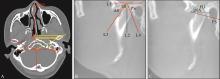
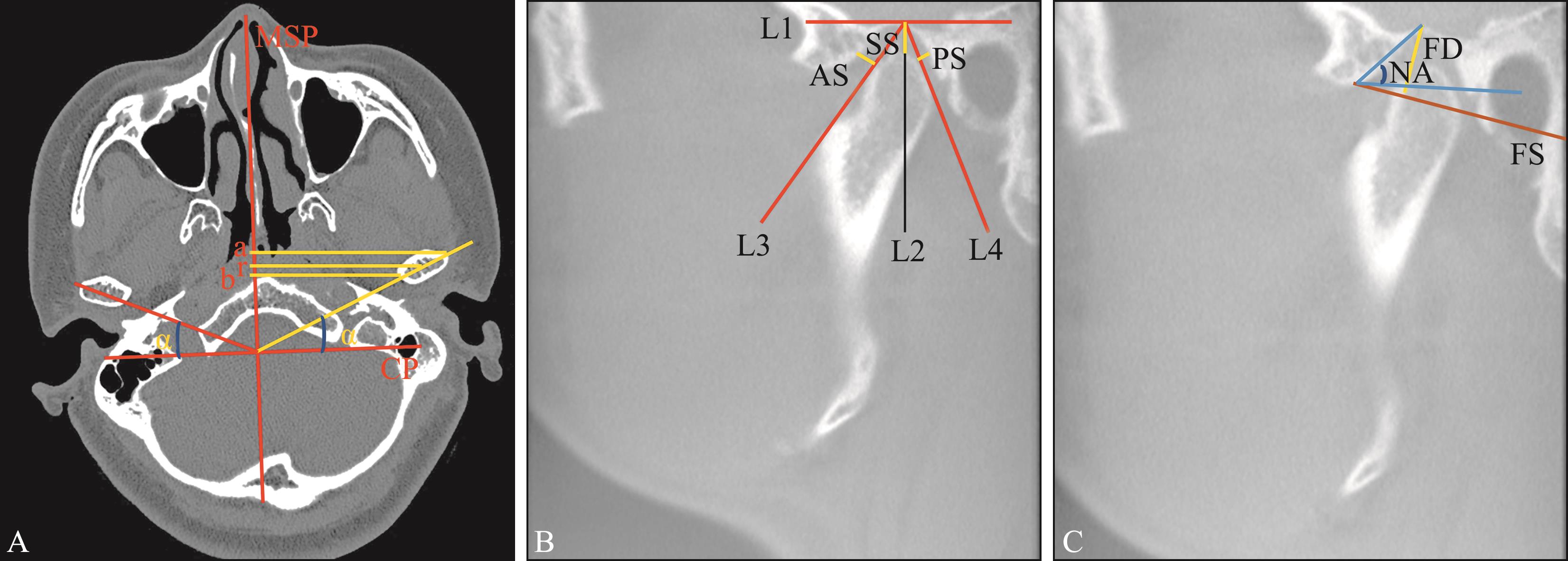
Fig 1
Schematic diagram of temporomandibular joint and condyle measurement method
Tab 1
Measurement parameters and definition of mandible and TMJ
| 指标 | 定义 |
|---|---|
| 下颌升支长度(Co-Go) | 髁顶点到下颌角点的距离 |
| 关节上间隙(super articular space,SS) | 关节窝顶点至髁突顶点的距离 |
| 关节前间隙(anterior articular space,AS) | 过关节窝顶点作髁突前缘切线,切点与关节窝前缘的最短距离 |
| 关节后间隙(retroarticular space,PS) | 过关节窝顶点作髁突后缘切线,切点与关节窝后缘的最短距离 |
| 关节窝宽度(articular fossa width,FS) | 外耳道与关节结节最低点间距离 |
| 关节窝深度(articular fossa depth,FD) | 关节窝顶点至外耳道与关节结节最低点间连线的最短距离 |
| 关节结节角(articular nodular angle,NA) | 关节窝顶点与关节结节最低点间连线与眶耳平面的夹角 |
| a | 髁突外极点到正中矢状面的垂直距离 |
| b | 髁突内极点到正中矢状面的垂直距离 |
| r | 髁突中心点到正中矢状面的垂直距离 |
| α | 髁突长轴与冠状轴的夹角 |
| 正中矢状面(median sagittal plane,MSP) | 鼻尖-鼻中隔-枕骨大孔中点连线,作为矢状面基线 |
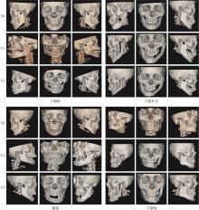
Fig 2
Three-dimensional reconstruction of mandibular defect location and after prosthetic reconstruction

Tab 2
Assessment of patients’ postoperative clinical outcomes
| 变量 | 术后随访时间 | ||
|---|---|---|---|
| T1 | T2 | T3 | |
| 钛板外露 | 0 | 2(4%) | 2(4%) |
| TMJ疼痛 | 0 | 2(4%) | 2(4%) |
| TMJ弹响 | 0 | 0 | 0 |
| 咬合紊乱 | 0 | 1(2%) | 1(2%) |
| 张口受限 | 0 | 0 | 0 |
Tab 3
Postoperative patient satisfaction evaluation
| 满意度 | 术后随访时间 | ||
|---|---|---|---|
| T1 | T2 | T3 | |
| 满意率 | 100% | 100% | 100% |
| 满意 | 44 | 43 | 40 |
| 比较满意 | 6 | 7 | 10 |
| 不满意 | 0 | 0 | 0 |
Tab 4
Effect of patients’ mandibular defects and restorative modalities on postoperative condylar position
| 变量 | T3/n | P值 | ||
|---|---|---|---|---|
| 髁突前移 | 中间 | 髁突后移 | ||
| 骨缺损/cm | 0.030 | |||
| <8.44 | 13 | 8 | 3 | |
| ≥8.44 | 13 | 10 | 3 | |
| 下颌骨缺损修复方式 | 0.700 | |||
| 腓骨瓣 | 25 | 15 | 6 | |
| 髂骨瓣 | 1 | 3 | 0 | |
| 肿瘤类型 | 0.400 | |||
| 良性肿瘤 | 18 | 15 | 3 | |
| 恶性肿瘤 | 8 | 3 | 1 | |
| 是否行数字化技术 | 0.400 | |||
| 是 | 18 | 15 | 3 | |
| 否 | 8 | 3 | 3 | |
| 下颌骨Brown分类 | 0.020 | |||
| BrownⅠ类缺损 | 7 | 4 | 0 | |
| BrownⅠc类缺损 | 4 | 2 | 1 | |
| BrownⅡ类缺损 | 6 | 4 | 0 | |
| BrownⅡc类缺损 | 3 | 2 | 3 | |
| Brown Ⅲ类缺损 | 3 | 3 | 1 | |
| Brown Ⅳ类缺损 | 3 | 3 | 0 | |
| Brown Ⅳc类缺损 | 0 | 0 | 1 | |
Tab 5
Morphometric results of mandibular positions
| 指标 | T0 | T1 | T2 | T3 | 4组间比较P值 |
|---|---|---|---|---|---|
| Co-Go/cm | 6.96±0.99 | 6.12±1.00** | 6.36±0.73 | 6.86±0.73 | 0.003 |
| SS/cm | 0.20±0.10 | 0.44±0.29**** | 0.25±0.14 | 0.32±0.26 | <0.000 1 |
| AS/cm | 0.08±0.04 | 0.21±0.20**** | 0.15±0.13 | 0.09±0.06 | 0.000 1 |
| PS/cm | 0.15±0.10 | 0.30±0.22*** | 0.25±0.16 | 0.22±0.16 | 0.001 |
| FS/cm | 2.79±0.25 | 2.81±0.32 | 2.72±0.22 | 2.81±0.18 | 0.130 |
| FD/cm | 0.88±0.22 | 0.86±0.21 | 0.84±0.12 | 0.99±0.34 | 0.060 |
| NA/° | 38.21±7.27 | 37.34±6.89 | 32.81±5.95* | 41.19±6.64 | 0.006 |
| a/cm | 6.08±0.38 | 6.12±0.58 | 5.96±0.56 | 5.93±0.51 | 0.300 |
| b/cm | 4.54±0.35 | 4.85±0.63* | 4.75±0.59 | 4.62±0.58 | 0.030 |
| r/cm | 5.31±0.36 | 5.50±0.56 | 5.37±0.52 | 5.27±0.49 | 0.160 |
| α/° | 25.85±8.07 | 31.64±9.09** | 30.33±8.31 | 28.99±10.64 | 0.013 |
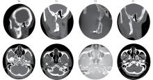
Fig 3
Changes in condyle and joint space after mandibular angle defect reconstruction

Tab 6
Measurements of condylar position after mandibular reconstruction with involved condyles
| 指标 | T0 | T1 | T2 | T3 | 4组间比较P值 |
|---|---|---|---|---|---|
| SS/cm | 0.20±0.09 | 0.80±0.25**** | 0.29±0.09 | 0.35±0.26 | <0.000 1 |
| AS/cm | 0.08±0.08 | 0.46±0.25**** | 0.25±0.17 | 0.24±0.16 | 0.003 |
| PS/cm | 0.13±0.04 | 0.51±0.16**** | 0.44±0.06**** | 0.47±0.10**** | <0.000 1 |
| NA/° | 35.39±5.28 | 34.28±5.20 | 28.79±3.91* | 30.5±5.82 | 0.040 |
| a/cm | 6.20±0.35 | 6.18±0.74 | 5.88±0.68 | 5.95±0.68 | 0.590 |
| b/cm | 4.57±0.58 | 5.40±0.67** | 5.15±0.72 | 5.29±0.88 | 0.010 |
| r/cm | 5.35±0.28 | 5.77±0.68 | 5.46±0.69 | 5.55±0.74 | 0.400 |
| α/° | 28.77±4.82 | 37.55±11.22 | 35.99±8.01 | 38.10±13.36 | 0.090 |
Tab 7
Condylar position measurements after mandibular reconstruction without condylar involvement
| 指标 | T0 | T1 | T2 | T3 | 4组间比较P值 |
|---|---|---|---|---|---|
| SS/cm | 0.20±0.10 | 0.30±0.16**** | 0.22±0.15 | 0.28±0.15 | <0.000 1 |
| AS/cm | 0.09±0.04 | 0.13±0.08** | 0.10±0.05 | 0.09±0.06 | 0.002 |
| PS/cm | 0.16±0.10 | 0.22±0.19 | 0.16±0.11 | 0.18±0.07 | 0.370 |
| NA/° | 39.36±7.57 | 38.92±6.46 | 34.83±5.77 | 40.97±7.32 | 0.140 |
| a/cm | 6.05±0.38 | 6.12±0.51 | 6.00±0.52 | 5.96±0.31 | 0.610 |
| b/cm | 4.52±0.36 | 4.60±0.45 | 4.55±0.40 | 4.52±0.24 | 0.410 |
| r/cm | 5.29±0.39 | 5.39±0.48 | 5.32±0.43 | 5.21±0.29 | 0.480 |
| α/° | 24.91±8.81 | 28.67±5.43 | 28.52±6.94 | 28.86±5.60 | 0.110 |
Tab 8
Measurement results of postoperative changes in mandibular defect repair surgery
| 指标 | T0-T1 | T1-T2 | T2-T3 |
|---|---|---|---|
| Co-Go变化量/cm | -0.88±1.81 | -1.68±2.32 | -1.27±1.70 |
| SS变化量/cm | 0.22±0.27 | 0.02±0.16 | 0.04±0.22 |
| AS变化量/cm | 0.13±0.19 | 0.05±0.15 | -0.005±0.07 |
| PS变化量/cm | 0.14±0.24 | 0.09±0.19 | 0.03±0.16 |
| FS变化量/cm | -0.03±0.50 | -2.66±0.22 | 0.59±1.20 |
| FD变化量/cm | -0.04±0.19 | -0.15±0.27 | -0.10±0.67 |
| NA变化量/° | -1.67±9.38 | -7.77±12.86 | -3.77±19.61 |
| a变化量/cm | -0.07±1.06 | -0.31±1.35 | -1.32±2.35 |
| b变化量/cm | 0.22±1.03 | -0.003±1.16 | -1.02±1.82 |
| r变化量/cm | 0.09±1.02 | -0.16±1.28 | -1.24±2.1 |
| α变化量/° | 5.40±10.83 | 3.36±12.10 | -3.20±16.34 |
Tab 9
Correlation analysis of the amount of preoperative and postoperative changes in mandibular defect repair
| 组别 | SS变化量 | AS变化量 | PS变化量 | NA变化量 | b变化量 | α变化量 | ||||||
|---|---|---|---|---|---|---|---|---|---|---|---|---|
| r值 | P值 | r值 | P值 | r值 | P值 | r值 | P值 | r值 | P值 | r值 | P值 | |
| T0-T1 | -0.06 | 0.700 | -0.07 | 0.640 | -0.34 | 0.020 | -0.49 | 0.001 | -0.42 | 0.003 | -0.47 | 0.001 |
| T1-T2 | -0.41 | 0.040 | -0.54 | 0.007 | -0.39 | 0.060 | -0.15 | 0.300 | -0.52 | 0.010 | -0.69 | 0.001 |
| T2-T3 | 0.14 | 0.680 | -0.38 | 0.270 | -0.14 | 0.690 | -0.48 | 0.150 | -0.28 | 0.430 | -0.64 | 0.040 |
| 1 | Song IS, Ryu JJ, Choi YJ, et al. Pre-contoured reconstruction plate fabricated via three-dimensional printed bending support[J]. J Korean Assoc Oral Maxillofac Surg, 2021, 47(3): 233-236. |
| 2 | Kurlander DE, Garvey PB, Largo RD, et al. The cost utility of virtual surgical planning and computer-assisted design/computer-assisted manufacturing in mandible reconstruction using the free fibula osteocutaneous flap[J]. J Reconstr Microsurg, 2023, 39(3): 221-230. |
| 3 | van Baar GJC, Forouzanfar T, Liberton NPTJ, et al. Accuracy of computer-assisted surgery in mandibular reconstruction: a systematic review[J]. Oral Oncol, 2018, 84: 52-60. |
| 4 | Nham TT, Koudougou C, Piot B, et al. Prosthetic rehabilitation in patients with jaw reconstruction by fibula free flap: a systematic review[J]. J Stomatol Oral Maxillofac Surg, 2024, 125(3): 101735. |
| 5 | Tang QC, Li YX, Yu T, et al. Association between condylar position changes and functional outcomes after condylar reconstruction by free fibular flap[J]. Clin Oral Investig, 2021, 25(1): 95-103. |
| 6 | Swendseid B, Philips R, Rimmer R, et al. Postoperative anatomic position of mandibular free flap neocondyles affects patient symptoms[J]. Facial Plast Surg Aesthet Med, 2021, 23(1): 36-41. |
| 7 | Liu S, Zhang WB, Yu Y, et al. Three-dimensional accuracy of bone contouring surgery for zygomaticomaxillary fibrous dysplasia using virtual planning and surgical na-vigation[J]. J Oral Maxillofac Surg, 2020, 78(12): 2328-2338. |
| 8 | Kamelchuk LS, Grace MG, Major PW. Post-imaging temporomandibular joint space analysis[J]. Cranio, 1996, 14(1): 23-29. |
| 9 | Vitral RW, Telles Cde S. Computed tomography evaluation of temporomandibular joint alterations in classⅡDivision 1 subdivision patients: condylar symmetry[J]. Am J Orthod Dentofacial Orthop, 2002, 121(4): 369-375. |
| 10 | Krisjane Z, Urtane I, Krumina G, et al. Three-dimensio-nal evaluation of TMJ parameters in ClassⅡand Class Ⅲ patients[J]. Stomatologija, 2009, 11(1): 32-36. |
| 11 | Pullinger A, Hollender L. Variation in condyle-fossa relationships according to different methods of evaluation in tomograms[J]. Oral Surg Oral Med Oral Pathol, 1986, 62(6): 719-727. |
| 12 | Brown JS, Barry C, Ho M, et al. A new classification for mandibular defects after oncological resection[J]. Lancet Oncol, 2016, 17(1): e23-e30. |
| 13 | Kang SH, Lee S, Nam W. Condyle dislocation following mandibular reconstruction using a fibula free flap: complication cases[J]. Maxillofac Plast Reconstr Surg, 2019, 41(1): 14. |
| 14 | Al-Wesabi SN, Abotaleb B, Al-Shujaa EA, et al. Three dimensional condylar positional and morphological chan-ges following mandibular reconstruction based on CB-CT analysis: a prospective study[J]. Head Face Med, 2023, 19(1): 3. |
| 15 | 肖晨亮, 孙丽君, 徐路, 等. 下颌骨节段性缺损腓骨修复重建后分段垂直牵张成骨及种植修复1例报告[J]. 口腔颌面外科杂志, 2024, 34(4): 319-323. |
| Xiao CL, Sun LJ, Xu L, et al. Segmental mandibular defect fibula graft repair and reconstruction with vertical segmented distraction osteogenesis and implant rehabilitation: a case report[J]. J Oral Maxillofac Surg, 2024, 34(4): 319-323. | |
| 16 | Yu KH, Lim HJ, Kim SM, et al. Comparison of condylar displacement after sagittal split ramus osteotomy depen-ding on the glenoid fossa depth[J]. J Craniomaxillofac Surg, 2021, 49(1): 9-16. |
| 17 | Wang Y, Li B, Liao J, et al. Comparison of condylar position after free fibular flap mandibular reconstruction using computer-assisted and traditional techniques[J]. BMC Oral Health, 2024, 24(1): 452. |
| 18 | 李云龙, 刘晓琳, 张晓燕, 等. 游离腓骨瓣重建下颌骨术后对称性的CBCT评价[J]. 现代口腔医学杂志, 2022, 36(6): 383-391. |
| Li YL, Liu XL, Zhang XY, et al. CBCT evaluation of symmetry after mandibular reconstruction with free fibula flap[J]. J Modern Stomatol, 2022, 36(6): 383-391. | |
| 19 | Wang W, Shan XF, Liang J, et al. Changes in condylar position after mandibular reconstruction with condylar head preservation by computed tomography[J]. J Oral Maxillofac Surg, 2019, 77(6): 1286-1292. |
| 20 | Swendseid B, Philips R, Rimmer R, et al. Postoperative anatomic position of mandibular free flap neocondyles affects patient symptoms[J]. Facial Plast Surg Aesthet Med, 2021, 23(1): 36-41. |
| 21 | Goormans F, Sun Y, Bila M, et al. Accuracy of computer-assisted mandibular reconstructions with free fibula flap: results of a single-center series[J]. Oral Oncol, 2019, 97: 69-75. |
| 22 | Tabrizi R, Shahidi S, Bahramnejad E, et al. Evaluation of condylar position after orthognathic surgery for treatment of class Ⅱ vertical maxillary excess and mandibular deficiency by using cone-beam computed tomography[J]. J Dent (Shiraz), 2016, 17(4): 318-325. |
| 23 | Schulz KL, Kesting MR, Nobis CP, et al. Three-dimensional evaluation of condylar position after mandibular reconstruction with a fibula free flap-comparison of different surgical techniques[J]. Int J Oral Maxillofac Surg, 2023, 52(6): 648-655. |
| [1] | Chen Jie, Jiang Canhua, Min Anjie, Ren Hui, Gao Zhengyang, Jian Xinchun.. Chimeric deep circumflex iliac artery perforator flap for the simultaneous reconstruction of the composite oromandibular defect [J]. West China Journal of Stomatology, 2015, 33(3): 276-280. |
| [2] | Xie Fuqiang, Sun Jian.. Analysis of reconstruction using non-vascularized iliac bone graft for patients with mandibular defects [J]. West China Journal of Stomatology, 2012, 30(4): 411-413. |
| [3] | Ma Gaoqi, Li Yan, Zhang Gang, Tan Yinghui. The effects of transection of inferior alveolar nerve on inflammatory factors during haematoma phases in mandibular defect healing of denervated rabbits [J]. West China Journal of Stomatology, 2011, 29(06): 640-642. |
| [4] | DENG Xiao1, WAN Zhe2, HE Shu-shu3, Peter Wamalwa3,4, CHEN Song3, ZHANG Zhi-yi5. The centric relation-maximum intercuspation discrepancy in adult Angle’s Class Ⅱ pretreatment patients [J]. West China Journal of Stomatology, 2011, 29(01): 48-52. |
| [5] | WANG Rui-yong1,2, MA Xu-chen2,3, ZHANG Wan-lin3, LIU Deng-gao3. Radiographic study on joint space changes of patients with anterior disc displacement of temporomandibular disorders [J]. West China Journal of Stomatology, 2010, 28(03): 303-305. |
| [6] | LI Yi1, RAN Wei2, WANG Gai-ling1, JING Xiang-dong1. Biocompatibility of new bone tissue engineering scaffolds in vivo [J]. West China Journal of Stomatology, 2009, 27(04): 447-450. |
| [7] | WANG Dong1,2, YANG Zhuang- qun1, HU Xiao- yi1. Thr ee- dimensional finite element analysis of thr ee conjunctive methods of fr ee iliac bone gr aft for established mandibular body defects [J]. West China Journal of Stomatology, 2007, 25(04): 345-348. |
| [8] | KANGHong*,CHAO Yonglie,YIXinzhu.. Reproducibility of Centric Relation of the Patient with Severe Dental Attrition for Oral Rehabilitation [J]. West China Journal of Stomatology, 2003, 21(06): 457-459. |
| Viewed | ||||||
|
Full text |
|
|||||
|
Abstract |
|
|||||
 This work is licensed under a Creative Commons Attribution 3.0 License.
