West China Journal of Stomatology ›› 2022, Vol. 40 ›› Issue (5): 513-521.doi: 10.7518/hxkq.2022.05.003
Previous Articles Next Articles
Li Dexiong1,2( ), Cao Runyuan2, Chen Jiang1,2()
Received:2022-01-26
Revised:2022-07-05
Online:2022-10-01
Published:2022-10-17
Contact:
Chen Jiang
E-mail:li331200@outlook.com;jiangchen@fjmu.edu.cn
Supported by:CLC Number:
Li Dexiong, Cao Runyuan, Chen Jiang. Preliminary study of silk fibroin porous scaffold for oral soft-tissue thickening[J]. West China Journal of Stomatology, 2022, 40(5): 513-521.
Add to citation manager EndNote|Ris|BibTeX
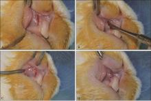
Fig 1
Surgical method of animal experiment

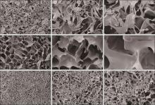
Fig 2
SEM characterization of three groups of scaffold materials
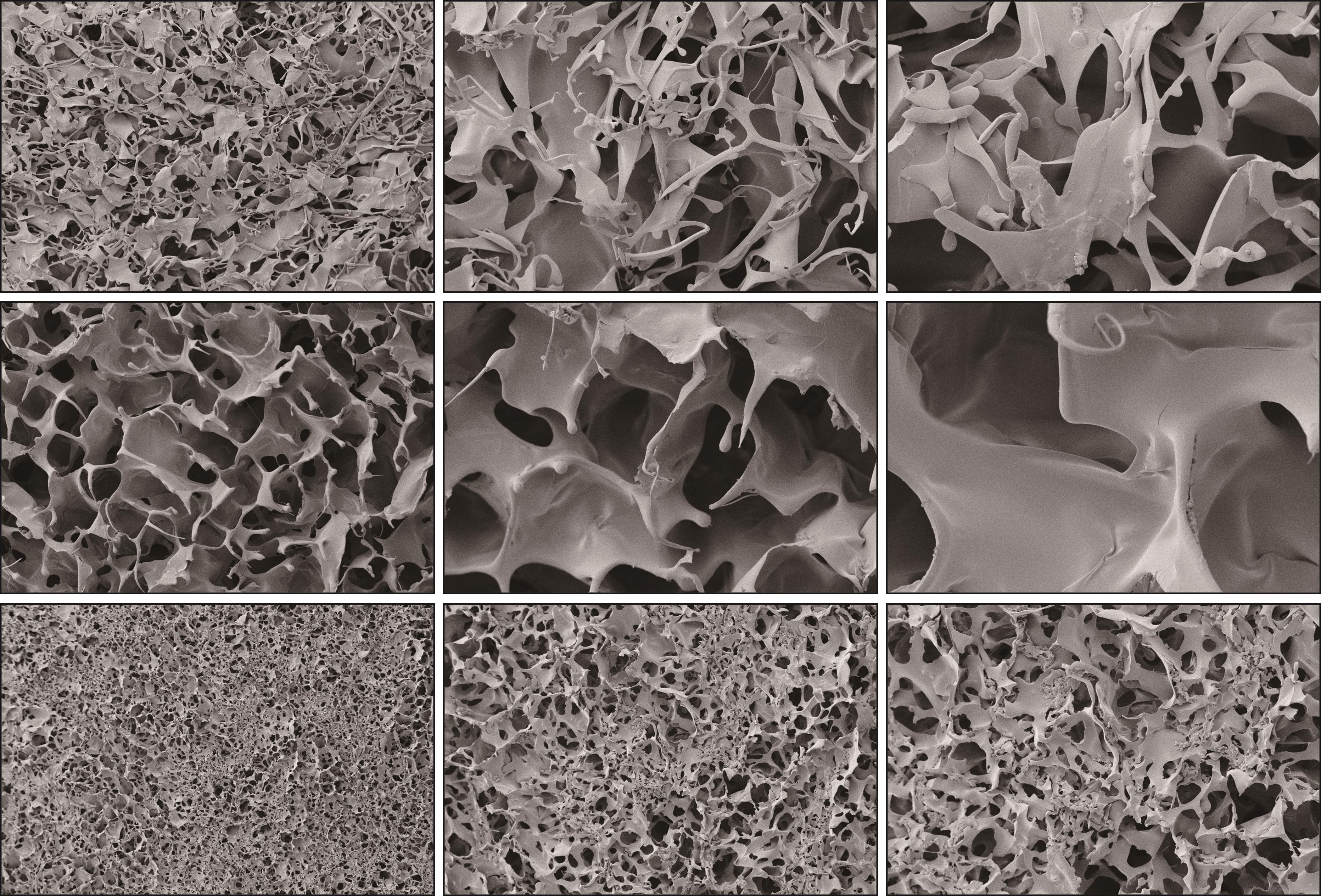
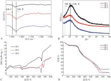
Fig 3
Characterization of scaffolds

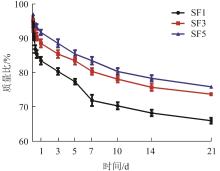
Fig 4
The residual mass ratio of scaffold materials in each group after immersion in pronase E-PBS solution for 3 weeks
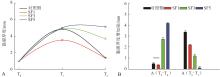
Fig 5
The results of changes in mucosal thickness in the surgical site and the increase in postoperative mucosal thickness before surgery, immediately after surgery, and 3 months after surgery on the experimental side and the control side

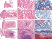
Fig 6
HE staining and Masson staining results of mucosal tissue in each group 3 months after operation

| 1 | Vallecillo C, Toledano-Osorio M, Vallecillo-Rivas M, et al. Collagen matrix vs. autogenous connective tissue graft for soft tissue augmentation: a systematic review and meta-analysis[J]. Polymers (Basel), 2021, 13(11): 1810. |
| 2 | 孟逍逸, 薛毅. 前牙美学区种植的软组织增量研究进展[J]. 口腔颌面修复学杂志, 2018, 19(1): 59-64. |
| Meng XY, Xue Y. Soft tissue augmentation of dental implant in esthetic zone of anterior teeth[J]. Chin J Prosthodont, 2018, 19(1): 59-64. | |
| 3 | Puzio M, Hadzik J, Błaszczyszyn A, et al. Soft tissue augmentation around dental implants with connective tissue graft (CTG) and xenogenic collagen matrix (XC-M). 1-year randomized control trail[J]. Ann Anat, 2020, 230: 151484. |
| 4 | Bakkali S, Rizo-Gorrita M, Romero-Ruiz MM, et al. Efficacy of different surgical techniques for peri-implant tissue preservation in immediate implant placement: a systematic review and meta-analysis[J]. Clin Oral Invest, 2021, 25(4):1655-1675. |
| 5 | Panwar M, Kosala M, Malik D, et al. Comparison of acellular dermal matrix allografts and connective tissue autografts in soft-tissue augmentation around immediate implants: a pilot study[J]. Med J Armed Forces India, 2021(7): 029. |
| 6 | Kroiss S, Rathe F, Sader R, et al. Acellular dermal matrix allograft versus autogenous connective tissue grafts for thickening soft tissue and covering multiple gingival recessions: a 5-year preference clinical study[J]. Quintessence Int, 2019, 50(4): 278-285. |
| 7 | Cairo F, Barbato L, Tonelli P, et al. Xenogeneic collagen matrix versus connective tissue graft for buccal soft tissue augmentation at implant site. A randomized, controlled clinical trial[J]. J Clin Periodontol, 2017, 44(7): 769-776. |
| 8 | Puzio M, Błaszczyszyn A, Hadzik J, et al. Ultrasound assessment of soft tissue augmentation around implants in the aesthetic zone using a connective tissue graft and xenogeneic collagen matrix-1-year randomised follow-up[J]. Ann Anat, 2018, 217: 129-141. |
| 9 | Rothamel D, Benner M, Fienitz T, et al. Biodegradation pattern and tissue integration of native and cross-linked porcine collagen soft tissue augmentation matrices—an experimental study in the rat[J]. Head Face Med, 2014, 10: 10. |
| 10 | Rothamel D, Schwarz F, Sager M, et al. Biodegradation of differently cross-linked collagen membranes: an experimental study in the rat[J]. Clin Oral Implants Res, 2005, 16(3): 369-378. |
| 11 | Melke J, Midha S, Ghosh S, et al. Silk fibroin as biomaterial for bone tissue engineering[J]. Acta Biomater, 2016, 31: 1-16. |
| 12 | Holland C, Numata K, Rnjak-Kovacina J, et al. The biomedical use of silk: past, present, future[J]. Adv Healthc Mater, 2019, 8(1): e1800465. |
| 13 | Zhang Q, Zhao Y, Yan S, et al. Preparation of uniaxial multichannel silk fibroin scaffolds for guiding primary neurons[J]. Acta Biomater, 2012, 8(7): 2628-2638. |
| 14 | Huang W, Ling S, Li C, et al. Silkworm silk-based materials and devices generated using bio-nanotechnology[J]. Chem Soc Rev, 2018, 47(17): 6486-6504. |
| 15 | Li X, Yan S, Qu J, et al. Soft freezing-induced self-assembly of silk fibroin for tunable gelation[J]. Int J Biol Macromol, 2018, 117: 691-695. |
| 16 | Cheng G, Davoudi Z, Xing X, et al. Advanced silk fibroin biomaterials for cartilage regeneration[J]. ACS Biomater Sci Eng, 2018, 4(8): 2704-2715. |
| 17 | Wang XL, Guo CC, Guo LN, et al. Radially aligned porous silk fibroin scaffolds as functional templates for engineering human biomimetic hepatic lobules[J]. ACS Appl Mater Interfaces, 2022, 14(1): 201-213. |
| 18 | Kim UJ, Park J, Kim HJ, et al. Three-dimensional aqueous-derived biomaterial scaffolds from silk fibroin[J]. Biomaterials, 2005, 26(15): 2775-2785. |
| 19 | Nazarov R, Jin HJ, Kaplan DL. Porous 3-D scaffolds from regenerated silk fibroin[J]. Biomacromolecules, 2004, 5(3): 718-726. |
| 20 | Lu Q, Wang X, Lu S, et al. Nanofibrous architecture of silk fibroin scaffolds prepared with a mild self-assembly process[J]. Biomaterials, 2011, 32(4): 1059-1067. |
| 21 | Yao D, Qian Z, Zhou J, et al. Facile incorporation of REDV into porous silk fibroin scaffolds for enhancing vascularization of thick tissues[J]. Mater Sci Eng C Mater Biol Appl, 2018, 93: 96-105. |
| 22 | Han H, Ning H, Liu S, et al. Silk biomaterials with vascularization capacity[J]. Adv Funct Mater, 2016, 26(3): 421-436. |
| 23 | Sang Y, Li M, Liu J, et al. Biomimetic silk scaffolds with an amorphous structure for soft tissue engineering[J]. ACS Appl Mater Interfaces, 2018, 10(11): 9290-9300. |
| 24 | Berry CC, Campbell G, Spadiccino A, et al. The influence of microscale topography on fibroblast attachment and motility[J]. Biomaterials, 2004, 25(26): 5781-5788. |
| 25 | Salem AK, Stevens R, Pearson RG, et al. Interactions of 3T3 fibroblasts and endothelial cells with defined pore features[J]. J Biomed Mater Res, 2002, 61(2): 212-217. |
| 26 | Ho MH, Kuo PY, Hsieh HJ, et al. Preparation of porous scaffolds by using freeze-extraction and freeze-gelation methods[J]. Biomaterials, 2004, 25(1): 129-138. |
| 27 | Zeltinger J, Sherwood JK, Graham DA, et al. Effect of pore size and void fraction on cellular adhesion, proliferation, and matrix deposition[J]. Tissue Eng, 2001, 7(5): 557-572. |
| 28 | Cui L, Li J, Long Y, et al. Vascularization of LBL structured nanofibrous matrices with endothelial cells for tissue regeneration[J]. RSC Adv, 2017, 7(19): 11462-11477. |
| 29 | Han H, Ning H, Liu S, et al. Silk biomaterials with vascularization capacity[J]. Adv Funct Mater, 2016, 26(3): 421-436. |
| 30 | Bhardwaj N, Chakraborty S, Kundu SC. Freeze-gelled silk fibroin protein scaffolds for potential applications in soft tissue engineering[J]. Int J Biol Macromol, 2011, 49(3): 260-267. |
| 31 | Etienne O, Schneider A, Kluge JA, et al. Soft tissue augmentation using silk gels: an in vitro and in vivo study[J]. J Periodontol, 2009, 80(11): 1852-1858. |
| 32 | Jansen RG, van Kuppevelt TH, Daamen WF, et al. Tissue reactions to collagen scaffolds in the oral mucosa and skin of rats: environmental and mechanical factors[J]. Arch Oral Biol, 2008, 53(4): 376-387. |
| 33 | Schenk RK, Buser D, Hardwick WR, et al. Healing pattern of bone regeneration in membrane-protected defects: a histologic study in the canine mandible[J]. Int J Oral Maxillofac Implants, 1994, 9(1): 13-29. |
| 34 | Lundgren AK, Sennerby L, Lundgren D. Guided jaw-bone regeneration using an experimental rabbit model[J]. Int J Oral Maxillofac Surg, 1998, 27(2): 135-140. |
| 35 | Mathes SH, Wohlwend L, Uebersax L, et al. A bioreactor test system to mimic the biological and mechanical environment of oral soft tissues and to evaluate substitutes for connective tissue grafts[J]. Biotechnol Bioeng, 2010, 107(6): 1029-1039. |
| 36 | Marzadori M, Stefanini M, Mazzotti C, et al. Soft-tissue augmentation procedures in edentulous esthetic areas[J]. Periodontol 2000, 2018, 77(1): 111-122. |
| [1] | Liu Yiping, Wang Jue, Tian Zilu, Zhai Peisong, Wang Zhanqi, Zhou Yanmin, Ni Shilei. Effects of scaffold microstructure and mechanical properties on regeneration of tubular dentin [J]. West China Journal of Stomatology, 2020, 38(3): 314-318. |
| [2] | Xinxin Ding, Yanmin Zhou, Xing-chen Xiang, Lin Meng, Qin Qin, Shan Ye. Research progress on chitosan composite scaffolds in bone tissue engineering [J]. West China Journal of Stomatology, 2018, 36(4): 441-446. |
| [3] | Binhong Teng, Yanhong Zhao, Lianyong Wang, Qiang Yang, Hongfa Li, Yunjie Li. Preparation and characterization of oriented scaffolds derived from cartilage extracellular matrix and silk fibroin [J]. West China Journal of Stomatology, 2018, 36(1): 17-22. |
| [4] | Kun Li, Yanhong Zhao, Chen Xu, Lianyong Wang, Qiang Yang, Hongfa Li, Binhong Teng. Development and characterization of oriented scaffolds derived from cartilage extracellular matrix [J]. West China Journal of Stomatology, 2017, 35(1): 51-56. |
| [5] | Xi Weihong, Wang Zhen, Zhu Hongshui, Li Xiaofeng, Xiong Yuanfei. Synthesis and characteristics of integrated bionic mandibular condylar scaffold [J]. West China Journal of Stomatology, 2016, 34(1): 68-72. |
| [6] | Zhang Yan, Jiang Xinquan, Zhang Xiuli, Wang Deping, Zhen Lei.. Cytocompatibility of two porous bioactive glass-ceramic in vitro [J]. West China Journal of Stomatology, 2013, 31(3): 294-299. |
| [7] | Zhang Kefu1, Zhang Shu1, Luo Zhiqiang1, Wang Jing1,2, Wang Tao3, Ou Guomin1,4, Wang Hu1,5. Biocompatibility of porous calcium phosphate ceramic nanocomposite [J]. West China Journal of Stomatology, 2012, 30(2): 209-213. |
| [8] | LAI Ren-fa, ZHAO Qing-tong, LIU Xiang-ning, SHEN Shan. Study on osteogenic ability of chitosan/β-tricalcium phosphate scaffold combined with human bone morphogenetic protein [J]. West China Journal of Stomatology, 2010, 28(05): 464-467. |
| [9] | WANG Lei1, LI Yao -jun2, ZHANG Yan3, PAN Ke -feng3, HUANG Yuan -liang2, LIU Chang -sheng4, JIANG Xin-quan5. The experimental study on porous calcium phosphate cement with bone marrow stromal cells for bone tissue engineering [J]. West China Journal of Stomatology, 2010, 28(03): 315-318. |
| [10] | LI Yi1, RAN Wei2, WANG Gai-ling1, JING Xiang-dong1. Biocompatibility of new bone tissue engineering scaffolds in vivo [J]. West China Journal of Stomatology, 2009, 27(04): 447-450. |
| [11] | WANG Lixia1,2, ZHAO Huan1, JIANG Bo3, DING Yi1. Adhesion and growth of human periodontal ligament cells on hyaluronic acid/collagen scaffold [J]. West China Journal of Stomatology, 2009, 27(02): 220-223. |
| [12] | LIU Shao-hua1, WEI Feng-cai1, ZHANG Dong1, SUN Shan-zhen2, ZHAO Hua-qiang2, LI Guo-ju2. Experimental Study of Mandibular Periosteal Distraction in Rabbits [J]. West China Journal of Stomatology, 2006, 24(03): 273-275. |
| [13] | NONG Xiao-lin1, WANG Da-zhang2,MENGMin1,ZHOUNuo1,MENGNing1,LIJia-quan3,ZHANGHong4. Combining of TNP-470 and 5-Fu in Inhibition of Adenoid Cystic Carcinoma in Nude Mice Model [J]. West China Journal of Stomatology, 2004, 22(04): 267-270. |
| Viewed | ||||||
|
Full text |
|
|||||
|
Abstract |
|
|||||
 This work is licensed under a Creative Commons Attribution 3.0 License.
