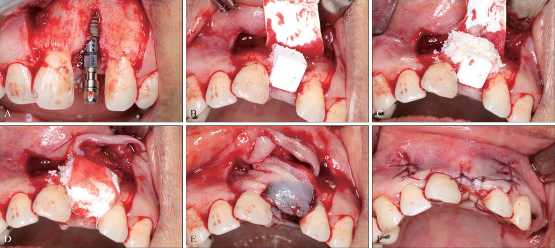
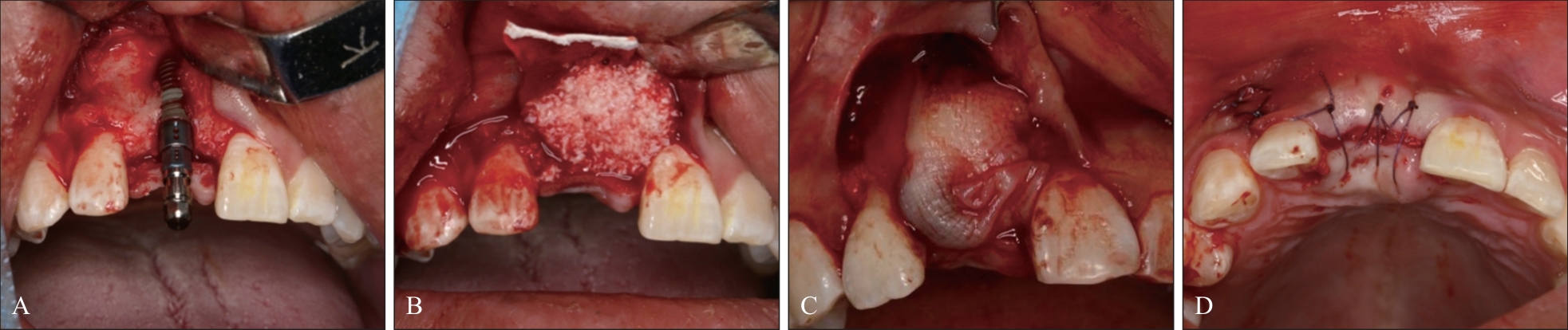
| 1 |
Jung RE, Brügger LV, Bienz SP, et al. Clinical and radiographical performance of implants placed with simultaneous guided bone regeneration using resorbable and nonresorbable membranes after 22-24 years, a prospective, controlled clinical trial[J]. Clin Oral Implants Res, 2021, 32(12): 1455-1465.
|
| 2 |
Mir-Mari J, Benic GI, Valmaseda-Castellón E, et al. Influence of wound closure on the volume stability of particulate and non-particulate GBR materials: an in vitro cone-beam computed tomographic examination. PartⅡ[J]. Clin Oral Implants Res, 2017, 28(6): 631-639.
|
| 3 |
McAllister BS, Haghighat K. Bone augmentation techniques[J]. J Periodontol, 2007, 78(3): 377-396.
|
| 4 |
Islam MT, Felfel RM, Abou Neel EA, et al. Bioactive calcium phosphate-based glasses and ceramics and their biomedical applications: a review[J]. J Tissue Eng, 2017, 8: 2041731417719170.
|
| 5 |
Benic GI, Eisner BM, Jung RE, et al. Hard tissue changes after guided bone regeneration of peri-implant defects comparing block versus particulate bone substitutes: 6-month results of a randomized controlled clinical trial[J]. Clin Oral Implants Res, 2019, 30(10): 1016-1026.
|
| 6 |
Troeltzsch M, Troeltzsch M, Kauffmann P, et al. Clinical efficacy of grafting materials in alveolar ridge augmentation: a systematic review[J]. J Craniomaxillofac Surg, 2016, 44(10): 1618-1629.
|
| 7 |
Wang D, Jin J, Qi W, et al. The two-dimensional size of peri-implant soft tissue in the anterior maxilla and some relevance: a 1-to 7-year cross-sectional study[J]. J Clin Periodontol, 2020, 47(4): 509-517.
|
| 8 |
Oh S, Chung SH, Han JY. Periodontal regenerative the-rapy in endo-periodontal lesions: a retrospective study over 5 years[J]. J Periodontal Implant Sci, 2019, 49(2): 90-104.
|
| 9 |
Silva CGB, Sapata VM, Llanos AH, et al. Peri-implant tissue changes at sites treated with alveolar ridge preservation in the aesthetic zone: twenty-two months follow-up of a randomized clinical trial[J]. J Clin Periodontol, 2022, 49(1): 39-47.
|
| 10 |
Thoma DS, Bienz SP, Lim HC, et al. Explorative randomized controlled study comparing soft tissue thickness, contour changes, and soft tissue handling of two ridge preservation techniques and spontaneous healing two months after tooth extraction[J]. Clin Oral Implants Res, 2020, 31(6): 565-574.
|
| 11 |
Gabay E, Katorza A, Zigdon-Giladi H, et al. Histological and dimensional changes of the alveolar ridge following tooth extraction when using collagen matrix and collagen-embedded xenogenic bone substitute: a randomized clinical trial[J]. Clin Implant Dent Relat Res, 2022, 24(3): 382-390.
|
| 12 |
Rohner D, Hailemariam S, Hammer B. Le Fort I osteotomies using Bio-Oss® collagen to promote bony union: a prospective clinical split-mouth study[J]. Int J Oral Maxillofac Surg, 2013, 42(5): 585-591.
|
| 13 |
Jung RE, Philipp A, Annen BM, et al. Radiographic evaluation of different techniques for ridge preservation after tooth extraction: a randomized controlled clinical trial[J]. J Clin Periodontol, 2013, 40(1): 90-98.
|
| 14 |
Arahira T, Todo M. Effects of proliferation and differentiation of mesenchymal stem cells on compressive mechanical behavior of collagen/β-TCP composite scaffold[J]. J Mech Behav Biomed Mater, 2014, 39: 218-230.
|
| 15 |
Zuercher AN, Mancini L, Naenni N, et al. The L-shape technique in guided bone regeneration with simulta-neous implant placement in the esthetic zone: a step-by-step protocol and a 2-14 year retrospective study[J]. J Esthet Restor Dent, 2023, 35(1): 197-205.
|
| 16 |
Ohnhaus EE, Adler R. Methodological problems in the measurement of pain: a comparison between the verbal rating scale and the visual analogue scale[J]. Pain, 1975, 1(4): 379-384.
|
| 17 |
Wachtel H, Schenk G, Böhm S, et al. Microsurgical access flap and enamel matrix derivative for the treatment of periodontal intrabony defects: a controlled clinical study[J]. J Clin Periodontol, 2003, 30(6): 496-504.
|
| 18 |
Jung EH, Jeong SN, Lee JH. Augmentation stability and early wound healing outcomes of guided bone regeneration in peri-implant dehiscence defects with L-and I-shaped soft block bone substitutes: a clinical and radiographic study[J]. Clin Oral Implants Res, 2021, 32(11): 1308-1317.
|
| 19 |
Wang HL, Boyapati L. “PASS” principles for predictable bone regeneration[J]. Implant Dent, 2006, 15(1): 8-17.
|
| 20 |
朱悦萌, 贾克文, 焦俊杰, 等. 自体血小板浓缩物在角化黏膜增量中的临床应用[J]. 中国口腔种植学杂志, 2023, 28(4): 250-259.
|
|
Zhu YM, Jia KW, Jiao JJ, et al. Clinical applications of autologous platelet-rich concentrate in keratinized mucosal augmentation[J]. Chin J Oral Implant, 2023, 28(4): 250-259.
|
| 21 |
Kao CH. Use of concentrate growth factors gel or membrane in chronic wound healing: description of 18 cases[J]. Int Wound J, 2020, 17(1): 158-166.
|
| 22 |
Chen Y, Cai Z, Zheng D, et al. Inlay osteotome sinus floor elevation with concentrated growth factor application and simultaneous short implant placement in severely atrophic maxilla[J]. Sci Rep, 2016, 6: 27348.
|
| 23 |
Elayah SA, Younis H, Cui H, et al. Alveolar ridge pre-servation in post-extraction sockets using concentrated growth factors: a split-mouth, randomized, controlled clinical trial[J]. Front Endocrinol (Lausanne), 2023, 14: 1163696.
|
| 24 |
刘雨蒙, 袁长永. 浓缩生长因子软组织封闭功能在口腔种植中的应用[J]. 中国口腔种植学杂志, 2021, 26(6): 396-399.
|
|
Liu YM, Yuan CY. Application of concentrated growth factor in soft tissue sealing of oral implantation[J]. Chin J Oral Implant, 2021, 26(6): 396-399.
|
| 25 |
Yu B, Wang Z. Effect of concentrated growth factors on beagle periodontal ligament stem cells in vitro [J]. Mol Med Rep, 2014, 9(1): 235-242.
|

 ), 杨虎1, 刘月1, 史一林1, 张圣锛1, 刘煜1, 宋丰1, 兰晶1(