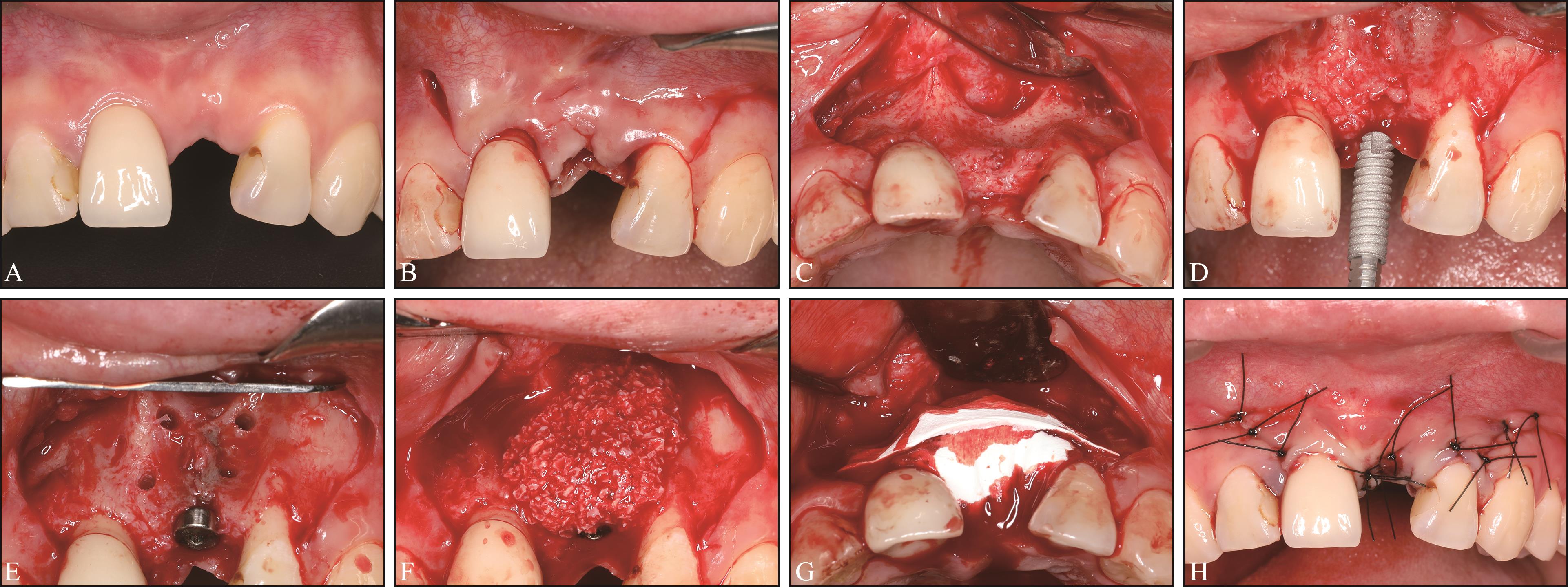
| [1] |
Ramasamy SK, Kusumbe AP, Schiller M, et al. Blood flow controls bone vascular function and osteogenesis[J]. Nat Commun, 2016, 7: 13601.
|
| [2] |
Huang L, Zou R, He J, et al. Comparing osteogenic effects between concentrated growth factors and the acellular dermal matrix[J]. Braz Oral Res, 2018, 32: e29.
|
| [3] |
Feng SW, Su YH, Lin YK, et al. Small blood stem cells for enhancing early osseointegration formation on dental implants: a human phaseⅠsafety study[J]. Stem Cell Res Ther, 2021, 12(1): 380.
|
| [4] |
Braut V, Bornstein MM, Belser U, et al. Thickness of the anterior maxillary facial bone wall—a retrospective radiographic study using cone beam computed tomography[J]. Int J Periodontics Restorative Dent, 2011, 31(2): 125-131.
|
| [5] |
Scarano A, Lorusso F, Arcangelo M, et al. Lateral sinus floor elevation performed with trapezoidal and modified triangular flap designs: a randomized pilot study of post-operative pain using thermal infrared imaging[J]. Int J Environ Res Public Health, 2018, 15(6): 1277.
|
| [6] |
Koymen R, Karacayli U, Gocmen-Mas N, et al. Flap and incision design in implant surgery: clinical and anatomical study[J]. Surg Radiol Anat, 2009, 31(4): 301-306.
|
| [7] |
Bhide A, Acharya G, Baschat A, et al. ISUOG Practice Guidelines (updated): use of Doppler velocimetry in obstetrics[J]. Ultrasound Obstet Gynecol, 2021, 58(2): 331-339.
|
| [8] |
Jiang L, Zhang D, Chen YN, et al. The value of conventional ultrasound combined with superb microvascular imaging and color Doppler flow imaging in the diagnosis of thyroid malignant nodules: a systematic review and meta-analysis[J]. Front Endocrinol (Lausanne), 2023, 14: 1182259.
|
| [9] |
Yu TF, He W, Gan CG, et al. Deep learning applied to two-dimensional color Doppler flow imaging ultrasound images significantly improves diagnostic performance in the classification of breast masses: a multicenter study[J]. Chin Med J (Engl), 2021, 134(4): 415-424.
|
| [10] |
Zarzecki M, Obuchowska I, Ustymowicz A, et al. Glaucoma surgery and ocular blood flow in colour Doppler imaging: is there a link[J]. Clin Ophthalmol, 2024, 18: 49-60.
|
| [11] |
Xue F, Wu BZ, Zhang R, et al. Analyses of gingival papilla blood flow via color doppler flow imaging and micro-flow imaging in patients with advanced periodontitis: a clinical pilot study[J]. Eur J Med Res, 2024, 29(1): 527.
|
| [12] |
Wu C, Liu X, Zhang H, et al. Response of human perio-dontal ligament to orthodontic force using superb microvascular imaging[J]. Am J Orthod Dentofacial Orthop, 2022, 162(5): e257-e266.
|
| [13] |
Abu Alhaija ES, Taha NA. A comparative study of initial changes in pulpal blood flow between conventional and self-ligating fixed orthodontic brackets during leveling and alignment stage[J]. Clin Oral Investig, 2021, 25(3): 971-981.
|
| [14] |
Urban IA, Lozada JL, Wessing B, et al. Vertical bone grafting and periosteal vertical mattress suture for the fi-xation of resorbable membranes and stabilization of particulate grafts in horizontal guided bone regeneration to achieve more predictable results: a technical report[J]. Int J Periodontics Restorative Dent, 2016, 36(2): 153-159.
|
| [15] |
Guo R, Yu Q, Lin Y, et al. Pulp blood flow changes in maxillary and mandibular anterior teeth after orthodontic retraction: a prospective study[J]. BMC Oral Health, 2022, 22(1): 508.
|
| [16] |
Retzepi M, Tonetti M, Donos N. Gingival blood flow changes following periodontal access flap surgery using laser Doppler flowmetry[J]. J Clin Periodontol, 2007, 34(5): 437-443.
|
| [17] |
Kijsamanmith K, Vongsavan N, Matthews B. Pulpal blood flow recorded from exposed dentine with a laser Doppler flow meter using red or infrared light[J]. Arch Oral Biol, 2018, 87: 163-167.
|
| [18] |
Rendell MS, Johnson ML, Smith D, et al. Skin blood flow response in the rat model of wound healing: expression of vasoactive factors[J]. J Surg Res, 2002, 107(1): 18-26.
|
| [19] |
Ahmed MV, Rastogi S, Baad RK, et al. Comparative study between two flaps-trapezoidal flap (TZF) and ocshenbein-leubke flap (OLF) in periapical surgeries[J]. J Maxillofac Oral Surg, 2013, 12(4): 440-446.
|
| [20] |
Schenk RK, Buser D, Hardwick WR, et al. Healing pattern of bone regeneration in membrane-protected defects: a histologic study in the canine mandible[J]. Int J Oral Maxillofac Implants, 1994, 9(1): 13-29.
|
| [21] |
Nobuto T, Suwa F, Kono T, et al. Microvascular response in the periosteum following mucoperiosteal flap surgery in dogs: 3-dimensional observation of an angiogenic process[J]. J Periodontol, 2005, 76(8): 1339-1345.
|
| [22] |
Pazzaglia UE. Periosteal and endosteal reaction to rea-ming and nailing: the possible role of revascularization on the endosteal anchorage of cementless stems[J]. Biomaterials, 1996, 17(10): 1009-1014.
|
| [23] |
Donos N, Akcali A, Padhye N, et al. Bone regeneration in implant dentistry: Which are the factors affecting the clinical outcome[J]. Periodontol 2000, 2023, 93(1): 26-55.
|
| [24] |
Marenzi G, Riccitiello F, Tia M, et al. Influence of leukocyte- and platelet-rich fibrin (L-PRF) in the healing of simple postextraction sockets: a split-mouth study[J]. Biomed Res Int, 2015, 2015: 369273.
|
| [25] |
Botticelli D, Berglundh T, Lindhe J. Hard-tissue alterations following immediate implant placement in extraction sites[J]. J Clin Periodontol, 2004, 31(10): 820-828.
|
| [26] |
Sanz M, Cecchinato D, Ferrus J, et al. A prospective, randomized-controlled clinical trial to evaluate bone pre-servation using implants with different geometry placed into extraction sockets in the maxilla[J]. Clin Oral Implants Res, 2010, 21(1): 13-21.
|
| [27] |
Grassi FR, Grassi R, Rapone B, et al. Dimensional chan-ges of buccal bone plate in immediate implants inserted through open flap, open flap and bone grafting and flapless techniques: a cone-beam computed tomography randomized controlled clinical trial[J]. Clin Oral Implants Res, 2019, 30(12): 1155-1164.
|
| [28] |
Degidi M, Daprile G, Nardi D, et al. Buccal bone plate in immediately placed and restored implant with Bio-Oss® collagen graft: a 1-year follow-up study[J]. Clin Oral Implants Res, 2013, 24(11): 1201-1205.
|
| [29] |
Fu JH, Yeh CY, Chan HL, et al. Tissue biotype and its relation to the underlying bone morphology[J]. J Periodontol, 2010, 81(4): 569-574.
|
| [30] |
Nagaraj KR, Savadi RC, Savadi AR, et al. Gingival biotype—Prosthodontic perspective[J]. J Indian Prosthodont Soc, 2010, 10(1): 27-30.
|
| [31] |
Barone A, Alfonsi F, Derchi G, et al. The effect of insertion torque on the clinical outcome of single implants: a randomized clinical trial[J]. Clin Implant Dent Relat Res, 2016, 18(3): 588-600.
|
| [32] |
Monje A, Roccuzzo A, Buser D, et al. Influence of buccal bone wall thickness on the peri-implant hard and soft tissue dimensional changes: a systematic review[J]. Clin Oral Implants Res, 2023, 34(3): 157-176.
|

 ), 张贤月2, 贾晓凤1, 夏荣1(