| 1 |
Ting HN. The 5th Kuala Lumpur International Conferen-ce on Biomedical Engineering[C]. Kuala Lumpur: Sprin-ger Science & Business Media, 2011.
|
| 2 |
杨文东, 李倩, 王威, 等. 不同参数的Er: YAG激光离体牙窝洞预备的形态学观察[J]. 牙体牙髓牙周病学杂志, 2014, 24(10): 585, 596-598.
|
|
Yang WD, Li Q, Wang W, et al. Morphological changes of dental hard tissues irradiated by Er: YAG laser with different parameters: an in vitro study[J]. Chin J Conserv Dent, 2014, 24(10): 585, 596-598.
|
| 3 |
侯玉一, 侯玉泽, 张涛, 等. Er: YAG激光处理釉质表面对树脂黏结牙釉质剪切强度的影响[J].黑龙江医药科学, 2020, 43(4): 76-77, 80.
|
|
Hou YY, Hou YZ, Zhang T, et al. Effect of Er: YAG laser treatment on the shear strength of resin-bonded tooth enamel[J]. Heilongjiang Med Pharm, 2020, 43(4): 76-77, 80.
|
| 4 |
卢青青, 李淑娟, 许丛琳, 等. Er: YAG激光对Constic自粘接流动树脂预防性充填的边缘封闭效果[J]. 北京口腔医学, 2021, 29(4): 223-227.
|
|
Lu QQ, Li SJ, Xu CL, et al. Microleakage of Constic self-adhensive flowable composites in preventive restoration: Er: YAG laser versus conventional preparation[J]. Beijing J Stomatol, 2021, 29(4): 223-227.
|
| 5 |
王婕, 陈亚明. Er: YAG激光在口腔医学领域的应用及研究进展[J]. 激光杂志, 2015, 36(9): 5-9.
|
|
Wang J, Chen YM. The application and research progress of Er: YAG laser on stomatology[J]. Laser J, 2015, 36(9): 5-9.
|
| 6 |
李乾, 赵彬, 武峰. Er: YAG激光和酸蚀作用于牙釉质表面后剪切强度的比较研究[J]. 中国实用口腔科杂志, 2011, 4(1): 36-38.
|
|
Li Q, Zhao B, Wu F. Comparative study on shear streng-th after Er: YAG laser and acid etching effect on enamel surface[J]. Chin J Pract Stomatol, 2011, 4(1): 36-38.
|
| 7 |
Van As GA. Using the erbium laser to remove porcelain veneers in 60 seconds[J]. J Cosmet Dent, 2013, 28(4): 22-34.
|
| 8 |
Pich O, Franzen R, Gutknecht N, et al. Laser treatment of dental ceramic/cement layers: transmitted energy, temperature effects and surface characterisation[J]. Lasers Med Sci, 2015, 30(2): 591-597.
|
| 9 |
Hayakawa K. Nd: YAG laser for debonding ceramic or-thodontic brackets[J]. Am J Orthod Dentofacial Orthop, 2005, 128(5): 638-647.
|
| 10 |
Zhang Y, Rocca JP, Fornaini C, et al. Erbium-doped, yttrium-aluminum-garnet laser debonding of porcelain la-minate veneers: an ex vivo study[J]. Contemp Clin Dent, 2018, 9(4): 570-573.
|
| 11 |
Bishara SE, Fonseca JM, Boyer DB. The use of debon-ding pliers in the removal of ceramic brackets: force le-vels and enamel cracks[J]. Am J Orthod Dentofacial Orthop, 1995, 108(3): 242-248.
|
| 12 |
Buu N, Morford C, Finzen F, et al. Er:YAG laser de-bonding of porcelain veneers[C]//Rechmann P, Fried D. Lasers in dentistry ⅩⅥ. San Francisco: International Society for Optics and Photonics, 2010: 1-8.
|
| 13 |
许庆, 杨烁, 张宁. Er: YAG激光去除Zenostar T全瓷冠的能量评估方法研究[J]. 口腔材料器械杂志, 2021, 30(1): 7-12.
|
|
Xu Q, Yang S, Zhang N. Energy assessment of Zenostar T all-ceramic crown removal using Er: YAG laser[J]. Chin J Dent Mater Dev, 2021, 30(1): 7-12.
|
| 14 |
Shahmiri RA, Standard OC, Hart JN, et al. A review of the characteristics and optimization of optical properties of zirconia ceramics for aesthetic dental restorations[J]. Int J Med Health Sci, 2017, 11(8): 1-9.
|
| 15 |
Ghoveizi R, Parsirad R, Tavakolizadeh S, et al. Effect of different Nd: YAG laser power outputs on bond strength of resin cement to zirconia in comparison to sandblasting[J]. J Lasers Med Sci, 2021, 12: e6.
|
| 16 |
Fried WA, Chan KH, Darling CL,et al. Use of a DPSS Er: YAG laser for the selective removal of composite from tooth surfaces[J]. Biomed Opt Express, 2018, 9(10): 5026-5036.
|
| 17 |
郑向明, 刘筱琳, 胡德渝, 等. Er: YAG激光在预防性树脂充填中的应用——扫描电镜研究[J]. 中国激光医学杂志, 2003, 12(4): 24-28.
|
|
Zheng XM, Liu XL, Hu DY, et al. Application of Er: YAG Laser to preventive resin restoration-scanning electron microscopy study[J]. Chin J Laser Med Surg, 2003, 12(4): 24-28.
|

 贴面的实验研究
 ), 洪菲菲1, 何良航2, 温玮2, 雷贤林2, 张志升1, 尹路1,3(
 贴面进行体外照射去粘接,通过分析比较Er:Yag激光对于
 贴面、牙体组织微观结构的影响,为激光无创拆除
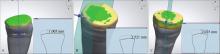
 贴面提供理论依据。 方法 选取新鲜拔除正畸下颌前磨牙,标准化牙体预备后制作3种不同厚度(1.0、1.5、2.0 mm)和不同材质(Vita琥珀瓷、Vita MarkⅡ、润瓷)
 贴面进行粘接,1周后使用Er: Yag激光(2.5、3.5 W)照射
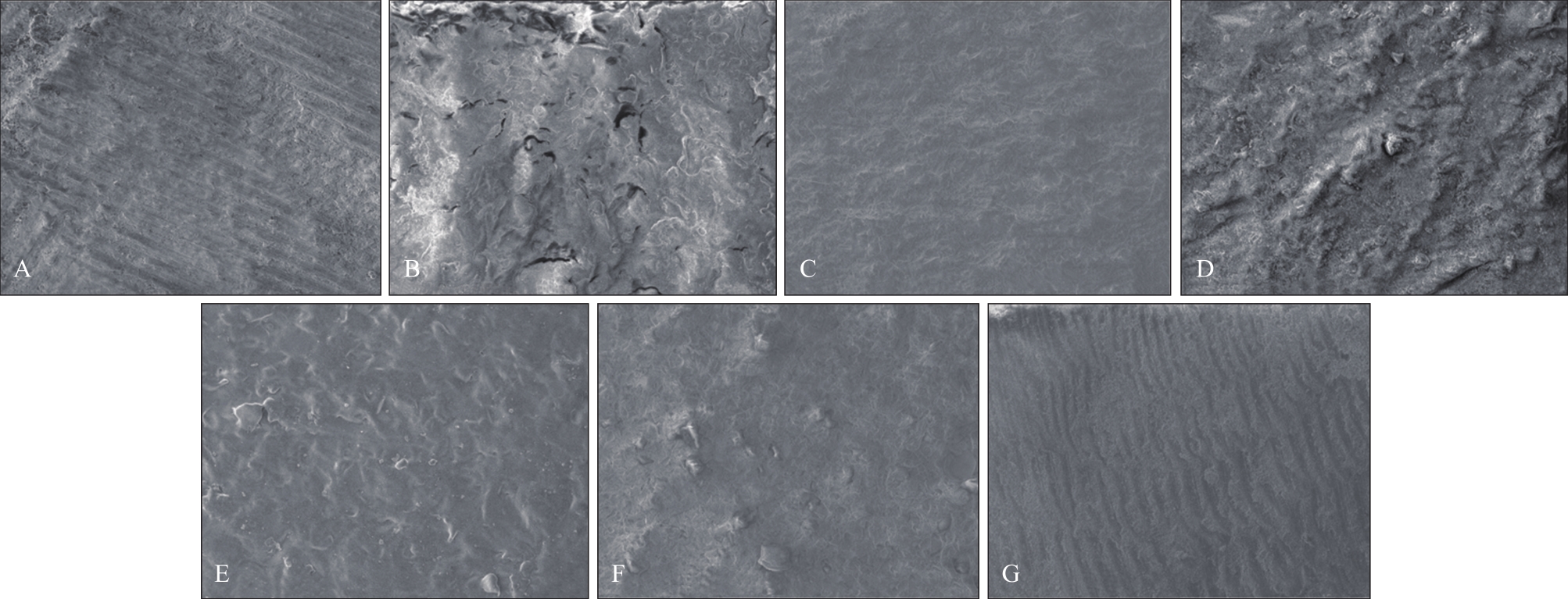
 贴面并记录时间。扫描电镜(SEM)观察去除后的微观形态。 结果 润瓷
 贴面经2.5或3.5 W Er: Yag激光长时间(>20 min)照射后仍无法取下;2.5 W Er: Yag激光去除粘接时间:1.0 mm Vita琥珀瓷组(96.0 s±16.0 s)大于1.0 mm Vita MarkⅡ组(84.5 s±19.5 s)(P<0.05);1.5 mm Vita琥珀瓷组(246.5 s±13.5 s)大于1.5 mm Vita MarkⅡ组(170.0 s±14.0 s)(P<0.05);3.5 W Er: Yag激光去除粘接时间:2.0 mm Vita琥珀瓷组(381.0 s±24.0 s)大于2.0 mm Vita MarkⅡ组(341.5 s±26.5 s)。 结论 同种材质、相同厚度情况下:激光功率越大,拆除时间越短,当功率较小时,可能导致
 贴面无法拆除。相同厚度、相同功率情况下:激光穿透瓷块到达粘接层可能对瓷块结构产生影响。同种材料、相同功率情况下:瓷块厚度越厚,拆除所需时间越长,所需功率越高。激光无法直接拆除树脂类
 贴面。
贴面。