华西口腔医学杂志 ›› 2017, Vol. 35 ›› Issue (6): 583-587.doi: 10.7518/hxkq.2017.06.004

• • 上一篇    下一篇

增殖细胞核抗原和P53在萎缩性腮腺再生中的表达及意义

魏月端1(), 左金华1(), 丁长玲2, 朱玉红3, 王丽芳1, 宋冰1, 王晶4   

  1. 1.滨州医学院附属医院口腔颌面外科
    2.药学部
    3.病理科
    4.口腔内科,滨州 256600
  • 收稿日期:2017-06-15 修回日期:2017-09-12 出版日期:2017-12-20 发布日期:2017-12-01
  • 作者简介:

    魏月端,硕士,E-mail:1363686792@qq.com

  • 基金资助:
    山东省医药卫生科技发展计划(2011HW004,2015WS0-487)

Significance of the expression of proliferating cell nuclear antigen and P53 in the regeneration process of an atrophic parotid gland

Yueduan Wei1(), Jinhua Zuo1(), Changling Ding2, Yuhong Zhu3, Lifang Wang1, Bing Song1, Jing Wang4   

  1. 1. Dept. of Oral and Maxillofacial Surgery, The Affiliated Hospital of Binzhou Medical College, Binzhou 256600, China
    2. Dept. of Pharmacy, The Affiliated Hospital of Binzhou Medical College, Binzhou 256600, China
    3. Dept. of Pathology, The Affiliated Hospital of Binzhou Medical College, Binzhou 256600, China
    4. Dept. of Oral Medicine, The Affiliated Hospital of Binzhou Medical College, Binzhou 256600, China
  • Received:2017-06-15 Revised:2017-09-12 Online:2017-12-20 Published:2017-12-01
  • Supported by:
    Supported by: Shandong Medicine and Health Science and Technology Development Plan (2011HW004, 2015WS0487).

摘要:

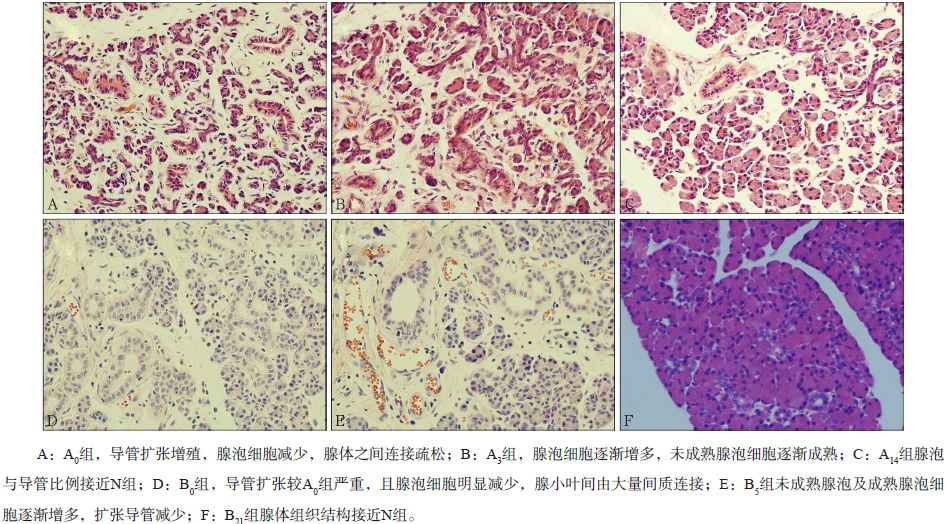
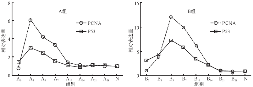
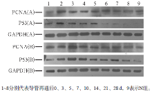
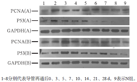
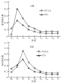
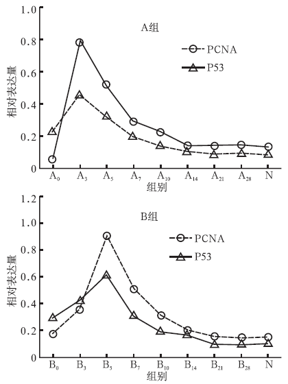
目的 从基因和蛋白水平研究增殖细胞核抗原(PCNA)和P53在萎缩性腮腺再生过程中的表达及意义。方法 将102只Wistar大鼠分为实验组和对照组,结扎实验组大鼠腮腺主导管,分别于结扎7 d(A组)、14 d(B组)后实现腮腺导管再通,并于再通后第0、3、5、7、10、14、21、28天处死大鼠获取新鲜腮腺组织标本。采用苏木精-伊红(HE)染色法观察腮腺组织形态学变化,实时荧光定量聚合酶链式反应和Western blot法检测两组腮腺组织中PCNA和P53的表达。结果 两实验组在腮腺导管结扎第7、14天,腺泡凋亡,导管细胞增殖,同组P53相对表达量高于PCNA;随着再通时间延长,腺泡细胞逐渐增多,A组在导管再通后第3天、B组在导管再通后第5天,PCNA和P53相对表达量达到峰值,与对照组比较,差异有统计学意义(P<0.01);其后PCNA和P53相对表达量逐渐减少,A组于再通后第21天、B组于再通后第28天接近对照组。结论 腮腺导管结扎后,P53相对表达量增多,诱导腮腺腺泡凋亡;导管再通后,PCNA相对表达量逐渐增多,达峰值后减少,可促进腺泡增殖。

关键词: 腮腺, 萎缩, 再生, 增殖细胞核抗原, P53

Abstract:

Objective This research aims to further explore the expression and significance of proliferating cell nuclear antigen (PCNA) and P53 in regenerating rat atrophy parotid gland from the gene and protein levels. Methods One hundred and two Wistar rats were randomly divided into experimental and control groups; the former group’s duct was ligated and then released respectively in 7 (Group A) and 14 days (Group B). Fresh parotid specimens were obtained at 0, 3, 5, 7, 10, 14, 21, and 28 days after being released. Hematoxylin-eosin staining method was used to observe the morphological changes of the parotid gland. The significance of P53 and PCNA in two groups was resolved by real-time fluorescence quantitative polymerase china reaction and Western blot. Results Acinar cells aoptosis and duct cells proliferation occurred when the occlusion of the parotid duct was reversed on days 7 and 14. The expression of P53 was higher than that of PCNA, and they reached the peak at the third and fifth days after groups A and B regenerated, respectively. This finding was significantly different compared with the control (P<0.01). P53 and PCNA contents decreased gradually; acinar and duct gradually returned to normal morphology; PCNA and P53 contents gradually close to the normal control group. Conclusion After ligating the parotid duct, P53 was highly expressed, and induced parotid gland atrophy. Mean-while, PCNA was highly expressed, which then decreased inducing gland recovery.

Key words: parotid gland, atrophy, regeneration, proliferating cell nuclear antigen, P53

中图分类号: