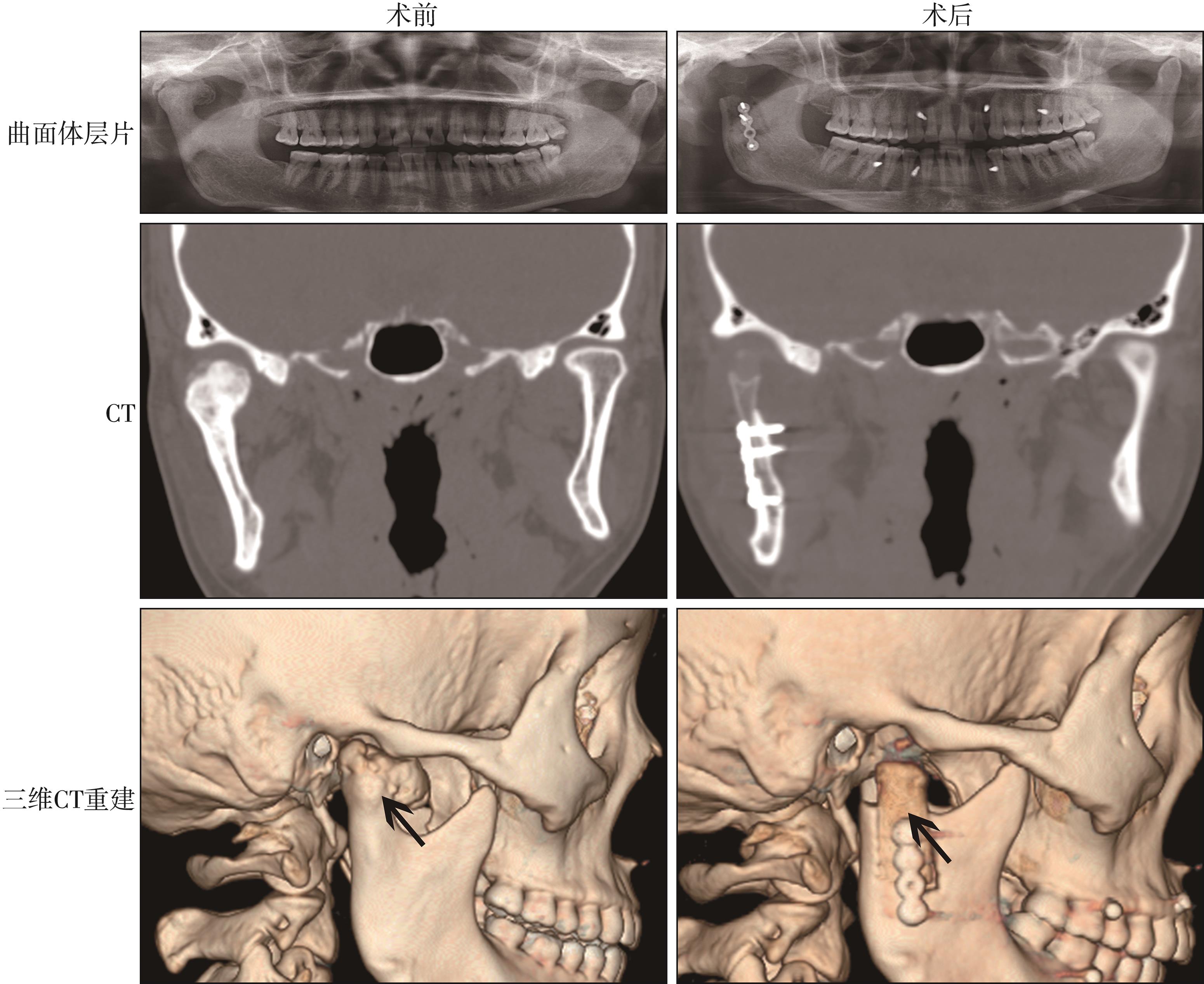
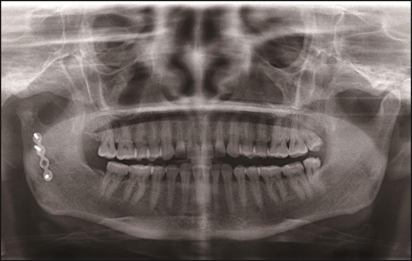
| 1 |
Dowgierd K, Pokrowiecki R, Kulesa Mrowiecka M, et al. Protocol for multi-stage treatment of temporomandibular joint ankylosis in children and adolescents[J]. J Clin Med, 2022, 11(2): 428.
|
| 2 |
陈浩哲, 祝颂松. 利用自体下颌骨重建下颌髁突的研究进展[J]. 临床口腔医学杂志, 2018, 34(3): 183-186.
|
|
Chen HZ, Zhu SS. Progress in reconstruction of mandibular condyle with autogenous mandible[J]. J Clin Stomatol, 2018, 34(3): 183-186.
|
| 3 |
Güzel MZ, Arslan H, Saraç M. Mandibular condyle reconstruction with inlay application of autogenous costochondral graft after condylectomy: Cerrahpaşa’s techni-que[J]. J Oral Maxillofac Surg, 2007, 65(4): 615-620.
|
| 4 |
Koneru G, Bhargava D, Somuri AV, et al. Temporomandibular joint alloplastic reconstruction of post-traumatic joint degeneration with Sawhney Type Ⅰ ankylosis using 3D-custom GD-condylar cap prosthesis to restore condylar form and function[J]. J Stomatol Oral Maxillofac Surg, 2021, 122(3): 315-318.
|
| 5 |
Qiu YT, Yang C, Chen MJ. Endoscopically assisted reconstruction of the mandibular condyle with a costochondral graft through a modified preauricular approach[J]. Br J Oral Maxillofac Surg, 2010, 48(6): 443-447.
|
| 6 |
MacIntosh RB, Henny FA. A spectrum of application of autogenous costochondral grafts[J]. J Maxillofac Surg, 1977, 5(4): 257-267.
|
| 7 |
Al-Moraissi EA, Louvrier A, Colletti G, et al. Does the surgical approach for treating mandibular condylar fractures affect the rate of seventh cranial nerve injuries? A systematic review and meta-analysis based on a new classification for surgical approaches[J]. J Craniomaxillofac Surg, 2018, 46(3): 398-412.
|
| 8 |
Koirala U, Subedi S. Retromandibular transparotid approach for subcondylar mandibular fracture: a retrospective study[J]. Dent Traumatol, 2021, 37(2): 314-320.
|
| 9 |
Kumar P, Jeyaraman B, Rajiah D, et al. Evaluation of mini-preauricular incision in the surgical management of condylar fracture[J]. Cureus, 2022, 14(11): e31725.
|
| 10 |
Mandal J, Bhutia O, Roychoudhury A, et al. Does the retromandibular transparotid approach provide quicker access to fracture of mandibular subcondyle compared with the retromandibular transmasseteric anterior paro-tid approach[J]. J Oral Maxillofac Surg, 2021, 79(3): 644-651.
|
| 11 |
Parihar VS, Bandyopadhyay TK, Chattopadhyay PK, et al. Retromandibular transparotid approach compared wi-th transmasseteric anterior parotid approach for the ma-nagement of fractures of the mandibular condylar process: a prospective randomised study[J]. Br J Oral Maxillofac Surg, 2019, 57(9): 880-885.
|
| 12 |
Li H, Zhang G, Cui J, et al. A modified preauricular approach for treating intracapsular condylar fractures to prevent facial nerve injury: the supratemporalis approa-ch[J]. J Oral Maxillofac Surg, 2016, 74(5): 1013-1022.
|
| 13 |
Singh PK, Singh G, Vignesh U, et al. Comparative eva-luation of modified tragus edge approach and retromandibular approach to mid- or low-level mandibular condylar fractures[J]. J Maxillofac Oral Surg, 2022, 21(1): 184-190.
|
| 14 |
Girhe V, Patil V, Bhujbal R, et al. Pre-auricular transparotid approach for the management of mandibular condylar fracture: an experience of 82 cases[J]. J Maxillofac Oral Surg, 2022, 21(3): 916-922.
|
| 15 |
Robiony M, Sembronio S. Comment on “A modified preauricular approach for treating intracapsular condylar fractures to prevent facial nerve injury: the supratemporalis approach”[J]. J Oral Maxillofac Surg, 2016, 74(11): 2114-2115.
|
| 16 |
Tang M, Wang L, Zhang M, et al. Modified tragus edge and transmasseteric anteroparotid approach for intracapsular and condylar neck fractures[J]. J Craniofac Surg, 2020, 31(6): 1822-1826.
|
| 17 |
Kochhar A, Larian B, Azizzadeh B. Facial nerve and parotid gland anatomy[J]. Otolaryngol Clin North Am, 2016, 49(2): 273-284.
|
| 18 |
Anderson SR, Pak KY, Vincent AG, et al. Reconstruction of the mandibular condyle[J]. Facial Plast Surg, 2021, 37(6): 728-734.
|
| 19 |
Goerke D, Sampson DE, Tibesar RJ, et al. Rib reconstruction of the absent mandibular condyle in children[J]. Otolaryngol Head Neck Surg, 2013, 149(3): 372-376.
|
| 20 |
Prasad C, Uma Maheswari G, Karthikeyan D. Fate of costochondral graft in temporomandibular joint reconstruction: a histological study[J]. J Maxillofac Oral Surg, 2016, 15(2): 179-183.
|
| 21 |
Awal DH, Jaffer M, Charan G, et al. Costochondral graf-ting for paediatric temporomandibular joint reconstruction: 10-year outcomes in 55 cases[J]. Int J Oral Maxillofac Surg, 2018, 47(11): 1433-1438.
|

 ), 王浪1,2, 王雷1, 饶鹏程1,3, 罗道文1, 付光新1,4, 肖金刚1,3,4(