| [1] |
Kaczor-Urbanowicz K, Zadurska M, Czochrowska E . Impacted teeth: an interdisciplinary perspective[J]. Adv Clin Exp Med, 2016,25(3):575-585.
doi: 10.17219/acem/37451
URL
|
| [2] |
Juodzbalys G, Daugela P . Mandibular third molar impaction: review of literature and a proposal of a classification[J]. J Oral Maxillofac Res, 2013,4(2):e1.
|
| [3] |
胡圣望, 熊炎斌, 肖菊芳 , 等. 成人下颌骨第三磨牙阻生关系的研究[J]. 咸宁医学院学报, 2001,15(3):153-156.
|
|
Hu SW, Xiong YB, Xiao JF , et al. A study of the relationship between the mandible and the impacted mandibular third molar[J]. J Xianning Med Coll, 2001,15(3):153-156.
|
| [4] |
Cho SY, Ki Y, Chu V , et al. Impaction of permanent mandibular second molars in ethnic Chinese schoolchildren[J]. J Can Dent Assoc, 2008,74(6):521.
|
| [5] |
Evans R . Incidence of lower second permanent molar impaction[J]. Br J Orthod, 1988,15(3):199-203.
|
| [6] |
Ferro F, Funiciello G, Perillo L , et al. Mandibular lip bumper treatment and second molar eruption disturbances[J]. Am J Orthod Dentofacial Orthop, 2011,139(5):622-627.
doi: 10.1016/j.ajodo.2009.07.024
URL
|
| [7] |
Kritzler K . CBCT imaging vs conventional radiography[J]. Am J Orthod Dentofac Orthop, 2017,152(2):146-148.
doi: 10.1016/j.ajodo.2017.04.018
URL
|
| [8] |
Tsolakis AI, Kalavritinos M, Bitsanis E , et al. Reliability of different radiographic methods for the localization of displaced maxillary canines[J]. Am J Orthod Dentofacial Orthop, 2018,153(2):308-314.
doi: 10.1016/j.ajodo.2017.06.026
URL
|
| [9] |
Thiesen G, Freitas MPM, Araújo EA , et al. Three-dimensional evaluation of craniofacial characteristics related to mandibular asymmetries in skeletal Class Ⅰ patients[J]. Am J Orthod Dentofacial Orthop, 2018,154(1):91-98.
|
| [10] |
沈晓玲, 刘敏, 黄海霞 , 等. 三维数字化解剖测量颞下颌关节的骨性结构[J]. 中国组织工程研究, 2016,20(20):2893-2898.
|
|
Shen XL, Liu M, Huang HX , et al. Three-dimensional digital model measurements of the temporomandibular joint[J]. Chin J Tis Eng Res, 2016,20(20):2893-2898.
|
| [11] |
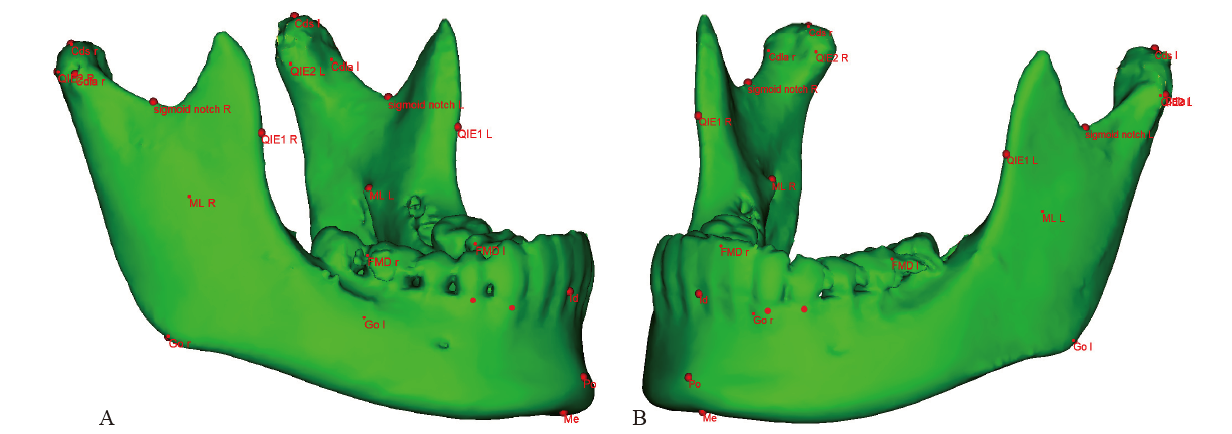
Kelly MP, Vorperian HK, Wang Y , et al. Characterizing mandibular growth using three-dimensional imaging techniques and anatomic landmarks[J]. Arch Oral Biol, 2017,77:27-38.
|
| [12] |
Cassetta M, Altieri F, Di Mambro A , et al. Impaction of permanent mandibular second molar: a retrospective study[J]. Med Oral Patol Oral Cir Bucal, 2013,18(4):e564-e568.
|
| [13] |
Capelli J Jr . Mandibular growth and third molar impaction in extraction cases[J]. Angle Orthod, 1991,61(3):223-229.
|
| [14] |
Balraj L, Nagaraj T, Irugu K , et al. Radiographic assessment of distribution of mandibular third molar impaction: a retrospective study[J]. J Indian Acad Oral Med Radiol, 2016,28(2):145.
doi: 10.4103/0972-1363.195125
URL
|
| [15] |
Fu PS, Wang JC, Wu YM , et al. Impacted mandibular second molars[J]. Angle Orthod, 2012,82(4):670-675.
doi: 10.2319/102111-656.1
URL
|
| [16] |
Je E . Gender variation in pattern of mandibular third molar impaction[J]. J Dent Oral Disord Ther, 2017,5(2):1-4.
|
| [17] |
Neychev D, Chenchev I, Atanasov D . Mandibular second molar impaction—literature review and case reports[J]. Scr Sci Med Dent, 2017,3(1):70.
|
| [18] |
Sawchuk D, Currie K, Vich ML , et al. Diagnostic methods for assessing maxillary skeletal and dental transverse deficiencies: a systematic review[J]. Korean J Orthod, 2016,46(5):331-342.
|
| [19] |
Shapira Y, Finkelstein T, Shpack N , et al. Mandibular second molar impaction. PartⅠ: genetic traits and characteristics[J]. Am J Orthod Dentofacial Orthop, 2011,140(1):32-37.
|

 )
)


 平面与颞下颌骨关节病关系的影像研究