West China Journal of Stomatology ›› 2024, Vol. 42 ›› Issue (3): 286-295.doi: 10.7518/hxkq.2024.2023336
• Basic Research • Previous Articles Next Articles
Li Yue( ), Xu Chunmei, Xie Xudong, Shi Peilei, Wang Jun(), Ding Yi()
Received:2023-10-10
Revised:2023-11-09
Online:2024-06-01
Published:2024-05-24
Contact:
Wang Jun,Ding Yi
E-mail:lant59liyue@163.com;junwang@scu.edu.cn;yiding2000@126.com
Supported by:CLC Number:
Li Yue, Xu Chunmei, Xie Xudong, Shi Peilei, Wang Jun, Ding Yi. Temporal and spatial expression analysis of periostin in mice periodontitis model[J]. West China Journal of Stomatology, 2024, 42(3): 286-295.
Add to citation manager EndNote|Ris|BibTeX
Tab 1
Primer sequences
| 基因 | 引物序列 |
|---|---|
| GAPDH | Forward:5’-GCACCGTCAAGGCTGAGAAC-3’ |
| Reverse:5’-TGGTGAAGACGCCAGTGGA-3’ | |
| TGF-β1 | Forward:5’-GCCTTTCCTGCTTCTCATGG-3’ |
| Reverse:5’-TCCTTGCGGAAGTCAATGTAC-3’ | |
| MMP2 | Forward:5’-ACCCATTTACACCTACACCAAG-3’ |
| Reverse:5’-TGTTTGCAGATCTCAGGAGTG-3’ | |
| 骨膜蛋白 | Forward:5’-CAACGGGCAAATACTGGAAAC-3’ |
| Reverse:5’-TCTCGCGGAATATGTGAATCG-3’ |
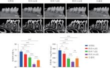
Fig 1
Micro-CT images and statistical analysis results of bone parameters of mandibular alveolar bone in each group

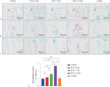
Fig 2
TRAP staining and statistical analyses results in the first mandibular molar of mice in each group

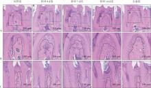
Fig 3
Images of HE staining in the first mandibular molar of mice in each group

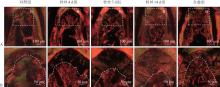
Fig 4
Polarized light scanning images of Sirius red staining in the first mandibular molar of mice in each group


Fig 5
Changes of RNA expression of periostin in the periodontal tissue of the first mandibular molar of mice in each group

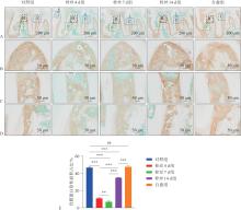
Fig 6
Changes in periostin expression in the periodontal tissue of the first mandibular molar of mice in each group

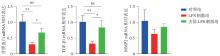
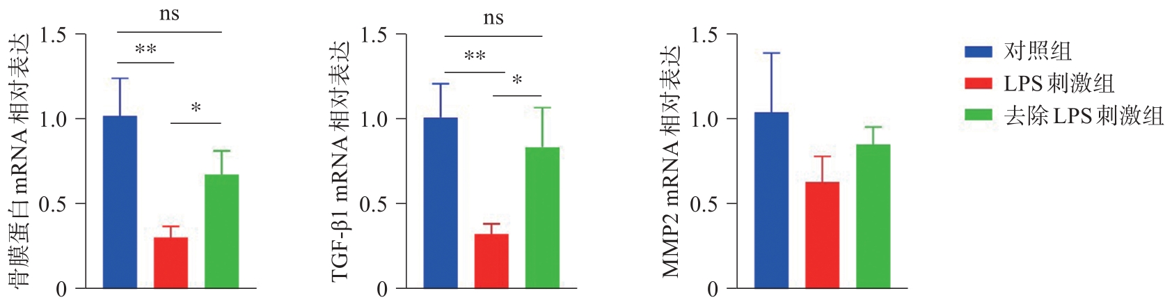
Fig 7
Changes of mRNA expression of periostin and related factors in periodontal ligament cells in each group
| 1 | Slots J. Periodontitis: facts, fallacies and the future[J]. Periodontol 2000, 2017, 75(1): 7-23. |
| 2 | Chien WC, Fu E, Chung CH, et al. Type 2 diabetes mellitus and periodontitis: bidirectional association in population-based 15-year retrospective cohorts[J]. J Clin Endocrinol Metab, 2023, 108(11): e1289-e1297. |
| 3 | Sun J, Guo G. Association between atherogenic index of plasma and periodontitis among US adults[J]. BMC Oral Health, 2023, 23(1): 166. |
| 4 | Lazar L, Dako T, Bortoc R, et al. Periostin as a marker of periodontal status. A narrative review[J]. Romanian J Oral Rehabil, 2022, 14(4): 228-238. |
| 5 | Sophia K, Suresh S, Sudhakar U, et al. Comparative eva-luation of serum and gingival crevicular fluid periostin levels in periodontal health and disease: a biochemical study[J]. Cureus, 2020, 12(3): e7218. |
| 6 | Arslan R, Karsiyaka Hendek M, Kisa U, et al. The effect of non-surgical periodontal treatment on gingival crevicular fluid periostin levels in patients with gingivitis and periodontitis[J]. Oral Dis, 2021, 27(6): 1478-1486. |
| 7 | Kii I, Ito H. Periostin and its interacting proteins in the construction of extracellular architectures[J]. Cell Mol Life Sci, 2017, 74(23): 4269-4277. |
| 8 | Du J, Li M. Functions of Periostin in dental tissues and its role in periodontal tissues’ regeneration[J]. Cell Mol Life Sci, 2017, 74(23): 4279-4286. |
| 9 | Padial-Molina M, Volk SL, Rios HF. Periostin increases migration and proliferation of human periodontal ligament fibroblasts challenged by tumor necrosis factor-α and Porphyromonas gingivalis lipopolysaccharides[J]. J Periodontal Res, 2014, 49(3): 405-414. |
| 10 | Wu Z, Dai W, Wang P, et al. Periostin promotes migration, proliferation, and differentiation of human perio-dontal ligament mesenchymal stem cells[J]. Connect Tissue Res, 2018, 59(2): 108-119. |
| 11 | Yan Y, Zhang H, Liu L, et al. Periostin reverses high glucose-inhibited osteogenesis of periodontal ligament stem cells via AKT pathway[J]. Life Sci, 2020, 242: 117184. |
| 12 | Ma D, Zhang R, Sun Y, et al. A novel role of periostin in postnatal tooth formation and mineralization[J]. J Biol Chem, 2011, 286(6): 4302-4309. |
| 13 | Rios H, Koushik SV, Wang H, et al. Periostin null mice exhibit dwarfism, incisor enamel defects, and an early-onset periodontal disease-like phenotype[J]. Mol Cell Biol, 2005, 25(24): 11131-11144. |
| 14 | Rios HF, Ma D, Xie Y, et al. Periostin is essential for the integrity and function of the periodontal ligament during occlusal loading in mice[J]. J Periodontol, 2008, 79(8): 1480-1490. |
| 15 | Rangiani A, Jing Y, Ren YS, et al. Critical roles of periostin in the process of orthodontic tooth movement[J]. Eur J Orthod, 2015, 38(4): 373-378. |
| 16 | Balli U, Keles ZP, Avci B, et al. Assessment of periostin levels in serum and gingival crevicular fluid of patients with periodontal disease[J]. J Periodontal Res, 2015, 50(6): 707-713. |
| 17 | Padial-Molina M, Volk SL, Taut AD, et al. Periostin is down-regulated during periodontal inflammation[J]. J Dent Res, 2012, 91(11): 1078-1084. |
| 18 | Tang HN, Xia Y, Yu Y, et al. Stem cells derived from “inflamed” and healthy periodontal ligament tissues and their sheet functionalities: a patient-matched comparison[J]. J Clin Periodontol, 2016, 43(1): 72-84. |
| 19 | Jolly S, Lang V, Koelzer VH, et al. Single-cell quantification of mRNA expression in the human brain[J]. Sci Rep, 2019, 9(1): 12353. |
| 20 | Socransky SS, Haffajee AD, Goodson JM, et al. New concepts of destructive periodontal disease[J]. J Clin Pe-riodontol, 1984, 11(1): 21-32. |
| 21 | Daines SM, Wang Y, Orlandi RR. Periostin and osteopontin are overexpressed in chronically inflamed sinuses[J]. Int Forum Allergy Rhinol, 2011, 1(2): 101-105. |
| 22 | Uchida M, Shiraishi H, Ohta S, et al. Periostin, a matricellular protein, plays a role in the induction of chemokines in pulmonary fibrosis[J]. Am J Respir Cell Mol Biol, 2012, 46(5): 677-686. |
| 23 | Kii I, Amizuka N, Minqi L, et al. Periostin is an extracellular matrix protein required for eruption of incisors in mice[J]. Biochem Biophys Res Commun, 2006, 342(3): 766-772. |
| 24 | Tabata C, Hongo H, Sasaki M, et al. Altered distribution of extracellular matrix proteins in the periodontal ligament of periostin-deficient mice[J]. Histol Histopathol, 2014, 29(6): 731-742. |
| 25 | Suzuki H, Amizuka N, Kii I, et al. Immunohistochemical localization of periostin in tooth and its surrounding tissues in mouse mandibles during development[J]. Anat Rec A Discov Mol Cell Evol Biol, 2004, 281(2): 1264-1275. |
| 26 | Afanador E, Yokozeki M, Oba Y, et al. Messenger RNA expression of periostin and Twist transiently decrease by occlusal hypofunction in mouse periodontal ligament[J]. Arch Oral Biol, 2005, 50(12): 1023-1031. |
| 27 | Park CH, Rios HF, Jin Q, et al. Tissue engineering bone-ligament complexes using fiber-guiding scaffolds[J]. Bio-materials, 2012, 33(1): 137-145. |
| 28 | Jiang Y, Yang P, Li C, et al. Periostin regulates LPS-induced apoptosis via Nrf2/HO-1 pathway in periodontal li-gament fibroblasts[J]. Oral Dis, 2023, 29(5): 2188-2204. |
| 29 | Lodyga M, Hinz B. TGF‑β1—A truly transforming grow-th factor in fibrosis and immunity[J]. Semin Cell Dev Biol, 2020, 101: 123-139. |
| 30 | Moreau JM, Velegraki M, Bolyard C, et al. Transforming growth factor-β1 in regulatory T cell biology[J]. Sci Immunol, 2022, 7(69): eabi4613. |
| 31 | Wei Q, Liu D, Chu G, et al. TGF-β1-supplemented decellularized annulus fibrosus matrix hydrogels promote annulus fibrosus repair[J]. Bioact Mater, 2022, 19: 581-593. |
| 32 | Khurshid Z, Mali M, Adanir N, et al. Periostin: immunomodulatory effects on oral diseases[J]. Eur J Dent, 2020, 14(3): 462-466. |
| 33 | Yin SL, Qin ZL, Yang X. Role of periostin in skin wound healing and pathologic scar formation[J]. Chin Med J (Engl), 2020, 133(18): 2236-2238. |
| [1] | Xin Yu, Fu Ruobing, Xin Xirui, Shang Yaqi, Liu Xinchan, Yu Weixian. Role of connexin 43 in a rat model of periodontitis-induced renal injury [J]. West China Journal of Stomatology, 2024, 42(3): 296-303. |
| [2] | Liu Hua, Yue Wanyuan, Shao Shuai, Sun Jiaping, Yang Ying, Dai Xiaoming. Global analysis of DNA methylation changes during experimented lingual carcinogenesis [J]. West China Journal of Stomatology, 2024, 42(3): 319-328. |
| [3] | Chen Hong, Zhang Ronghua, Zhao Yuan. Non-surgical treatment of maxillary lateral incisor double dens invaginatus type Ⅲ with apical periodontitis [J]. West China Journal of Stomatology, 2024, 42(3): 409-414. |
| [4] | Ma Haonan, Li Qiong, Shang Yaqi, Xin Xirui, Liu Xinchan, Wu Zhou, Yu Weixian. Impact of circadian clock protein Bmal1 on experimentally-induced periodontitis-associated renal injury [J]. West China Journal of Stomatology, 2024, 42(2): 163-171. |
| [5] | Sun Jinmeng, Zhang Ying, Zheng Zejun, Ding Xiaoling, Sun Minmin, Ding Gang. Potential mechanism of ginseng in the treatment of periodontitis based on network pharmacology and molecular docking [J]. West China Journal of Stomatology, 2024, 42(2): 181-191. |
| [6] | Ye Changchang, Yang He, Huang Ping. Application of intentional replantation in advanced periodontitis involving teeth preservation [J]. West China Journal of Stomatology, 2024, 42(1): 12-18. |
| [7] | Wang Jun.. Vital pulp therapy of permanent teeth with irreversible pulpitis [J]. West China Journal of Stomatology, 2023, 41(6): 622-627. |
| [8] | Wang Qintao, Ma Zhiwei, Wang Jinjin.. Personal understanding of the extraction or rescue on severe periodontitis teeth [J]. West China Journal of Stomatology, 2023, 41(6): 635-640. |
| [9] | Zhang Yanbiao, Wei Meirong, Xia Tianyong, Yin Wenting, Mao Shumei. Association between serum Galectin-3 and periodontitis in patients with type 2 diabetes mellitus [J]. West China Journal of Stomatology, 2023, 41(6): 653-661. |
| [10] | Cai Hongxuan, Wang Zheng’an, Zhang Zan, Dai Jingyi, Si Weixing, Fu Qiya, Yang Jingwen, Tian Yaguang. Morinda officinalis polysaccharides inhibit the expression and activity of NOD-like receptor thermal protein domain associated protein 3 in inflammatory periodontal ligament cells by upregulating silent information regulator sirtuin 1 [J]. West China Journal of Stomatology, 2023, 41(6): 662-670. |
| [11] | Jiang Jianhong, Shi Xinglian, He Quanmin, Gao Li, Yang Kun, Wang Taiping, Li Zhezhen, Liu Mei. Correlation between health literacy and life quality in elderly patients with chronic periodontitis [J]. West China Journal of Stomatology, 2023, 41(6): 694-700. |
| [12] | Lin Li, Li Zhaorong, Jin Yining, Yin Shou-cheng.. Treatment strategies for periodontitis patients with systemic disease [J]. West China Journal of Stomatology, 2023, 41(5): 502-511. |
| [13] | Liu Yuting, Yuan Quan.. Clinical decision and related factors influencing implant direction in the esthetic area [J]. West China Journal of Stomatology, 2023, 41(5): 512-520. |
| [14] | Zhang Chen, Hou Zhenzhen, Zong Yingrui.. Exploratory research on the probable shared molecular mechanism and transcription factors between chronic periodontitis and chronic obstructive pulmonary disease [J]. West China Journal of Stomatology, 2023, 41(5): 533-540. |
| [15] | Luo Xiao, Chen Yu, Shi Bing, Zheng Qian, Li Chenghao.. Three-dimensional reconstruction reveals the correlation between the extent of alveolar clefts and secondary nasal deformity in adults [J]. West China Journal of Stomatology, 2023, 41(4): 421-425. |
| Viewed | ||||||
|
Full text |
|
|||||
|
Abstract |
|
|||||
 This work is licensed under a Creative Commons Attribution 3.0 License.
