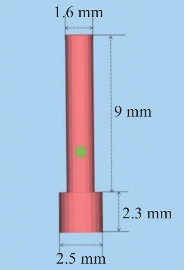
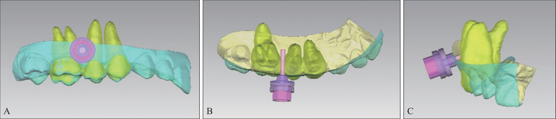
| [1] |
Imburgia M, Logozzo S, Hauschild U, et al. Accuracy of four intraoral scanners in oral implantology: a comparative in vitro study[J]. BMC Oral Health, 2017, 17(1): 92.
|
| [2] |
Tepedino M, Masedu F, Chimenti C. Comparative evaluation of insertion torque and mechanical stability for self-tapping and self-drilling orthodontic miniscrews—an in vitro study[J]. Head Face Med, 2017, 13(1): 10.
|
| [3] |
Mohammed H, Wafaie K, Rizk MZ, et al. Role of anatomical sites and correlated risk factors on the survival of orthodontic miniscrew implants: a systematic review and meta-analysis[J]. Prog Orthod, 2018, 19(1): 36.
|
| [4] |
Murugesan A, Sivakumar A. Comparison of bone thickness in infrazygomatic crest area at various miniscrew insertion angles in Dravidian population—A cone beam computed tomography study[J]. Int Orthod, 2020, 18(1): 105-114.
|
| [5] |
仇玲玲, 厉松, 白玉兴. 基于锥形束CT的正畸种植体导板设计及其引导下种植体植入安全性和稳定性的初步评价[J]. 中华口腔医学杂志, 2016, 51(6): 336-340.
|
|
Qiu LL, Li S, Bai YX. Preliminary safety and stability assessment of orthodontic miniscrew implantation gui-ded by surgical template based on cone-beam CT images[J]. Chin J Stomatol, 2016, 51(6): 336-340.
|
| [6] |
Estelita S, Janson G, Chiqueto K, et al. Predictable drill-free screw positioning with a graduated 3-dimensional radiographic-surgical guide: a preliminary report[J]. Am J Orthod Dentofacial Orthop, 2009, 136(5): 722-735.
|
| [7] |
张永清, 陈昕, 闫寒松, 等. 正畸支抗种植体定位尺的研制和应用[J]. 口腔医学研究, 2006, 22(4): 459.
|
|
Zhang YQ, Chen X, Yan HS, et al. Development and application of orthodontic anchorage implant positioning ruler[J]. J Oral Sci Res, 2006, 22(4): 459.
|
| [8] |
王晓波, 张梦洁, 孙应明, 等. 正畸微型种植体支抗的三维导向植入研究[J]. 现代口腔医学杂志, 2011, 25(4): 261-264.
|
|
Wang XB, Zhang MJ, Sun YM, et al. Study on three-dimensional guided orthodontic micro-implant anchorage[J]. J Modern Stomatol, 2011, 25(4): 261-264.
|
| [9] |
陈妍曲, 唐敏, 黄旋平, 等. 高精度三维整合牙颌模型个体化微种植体手术导板的计算机辅助设计与制作[J]. 中国组织工程研究, 2018, 22(10): 1529-1533.
|
|
Chen YQ, Tang M, Huang XP, et al. The computer-aided design and manufacturing of individualized miniscrew surgical guides based on a high-precision three-dimensional integrated digital maxillodental model[J]. Chin J Tis Eng Res, 2018, 22(10): 1529-1533.
|
| [10] |
Liu H, Liu DX, Wang G, et al. Accuracy of surgical positioning of orthodontic miniscrews with a computer-ai-ded design and manufacturing template[J]. Am J Orthod Dentofacial Orthop, 2010, 137(6): 728.e1-728.e10.
|
| [11] |
Endo T, Uchikura K, Ishida K, et al. Thresholds for cli-nically significant tooth-size discrepancy[J]. Angle Orthod, 2009, 79(4): 740-746.
|
| [12] |
刘静. 口内扫描仪全牙列扫描精度及不同操作者间扫描精度差异的研究[D]. 济南: 山东大学, 2017.
|
|
Liu J. Accuracy of full arch scans with intraoral scanner and accuracy of scan results between examiners[D]. Jinan: Shandong University, 2017.
|
| [13] |
Sivamurthy G, Sundari S. Stress distribution patterns at mini-implant site during retraction and intrusion-a three-dimensional finite element study[J]. Prog Orthod, 2016, 17: 4.
|
| [14] |
武明彤, 唐素霞, 彭玲燕, 等. 仿真头颅模型口内和手持牙颌模型条件下四种口内扫描仪全牙弓扫描时间和精度的对比研究[J]. 中华口腔医学杂志, 2021, 56(6): 570-575.
|
|
Wu MT, Tang SX, Peng LY, et al. Scan time and accuracy of full-arch scans with intraoral scanners: a comparative study on conditions of the intraoral head-simulator and the hand-held model[J]. Chin J Stomatol, 2021, 56(6): 570-575.
|
| [15] |
Favero R, Volpato A, Francesco M, et al. Accuracy of 3D digital modeling of dental arches[J]. Dental Press J Orthod, 2019, 24(1): 38e1-38e7.
|
| [16] |
汤槟晖, 徐婷, 孙志丹, 等. 金属激光三维打印技术研究现状及其发展趋势[J]. 材料保护, 2020, 53(6): 132-138.
|
|
Tang BH, Xu T, Sun ZD, et al. Research status and deve-lopment trend of metal laser three-dimensional printing technology[J]. Mater Prot, 2020, 53(6): 132-138.
|
| [17] |
周庆军, 严振宇, 韩旭, 等. 激光熔化沉积TC11钛合金的组织与力学性能[J]. 中国激光, 2018, 45(11): 1102005.
|
|
Zhou QJ, Yan ZY, Han X, et al. Microstructure and mechanical properties of laser melting deposition TC11 titanium alloy[J]. Chin J Laser, 2018, 45(11): 1102005.
|

 ), 温奥楠1, 高梓翔1, 李志华2, 张晟2, 王勇1(