华西口腔医学杂志 ›› 2024, Vol. 42 ›› Issue (2): 268-276.doi: 10.7518/hxkq.2024.2023327

• 临床新技术 • 上一篇    

数字孪生辅助调整颌位流程的临床新技术和应用效果研究

龚衍吉1(), 刘洋1(), 尹德强2   

  1. 1.口腔疾病防治全国重点实验室 国家口腔医学中心 国家口腔疾病临床医学研究中心 四川大学华西口腔医院颞下颌关节科,成都 610041
    2.重庆大学航空航天学院工程力学系,重庆 400044
  • 收稿日期:2023-10-02 修回日期:2024-01-17 出版日期:2024-04-01 发布日期:2024-03-26
  • 通讯作者: 刘洋 E-mail:gongyanji_kq@163.com;liu@scu.edu.cn
  • 作者简介:龚衍吉,硕士,E-mail:gongyanji_kq@163.com
  • 基金资助:
    国家重点研发计划(2023YFC3605600);国家自然科学基金(11972097);四川省自然科学基金(23NSFSC1708);四川大学华西口腔医院探索与研发项目(RD-03-202005);四川大学华西口腔医院临床医学研究项目(LCYJ2023-YF-3)

Effect of the application of digital technology-assisted optimization in the process of adjusting jaw position

Gong Yanji1(), Liu Yang1(), Yin Deqiang2   

  1. 1.State Key Laboratory of Oral Diseases & National Center for Stomatology & National Clinical Research Center for Oral Diseases & Dept. of Temporomandibular Joint, West China Hospital of Stomatology, Sichuan University, Chengdu 610041, China
    2.Dept. of Engineering Mechanics, College of Aerospace Engineering, Chongqing University, Chong-qing 400044, China
  • Received:2023-10-02 Revised:2024-01-17 Online:2024-04-01 Published:2024-03-26
  • Contact: Liu Yang E-mail:gongyanji_kq@163.com;liu@scu.edu.cn
  • Supported by:
    National Key Research and Development Program of China(2023YFC3605600);The National Natural Science Foundation of China(11972097);Natural Science Foundation of Sichuan Province(23NSFSC1708);Research and Development Program of West China Hospital of Stomatology, Sichuan University(RD-03-202005);Clinical Research Project of West China Hospital of Stomatology, Sichuan University(LCYJ2023-YF-3)

摘要:

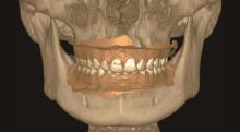
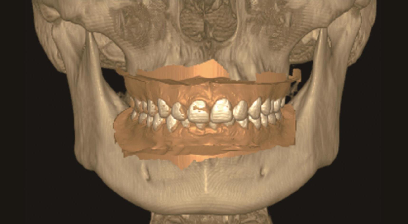
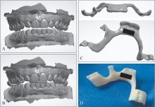
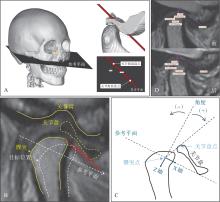
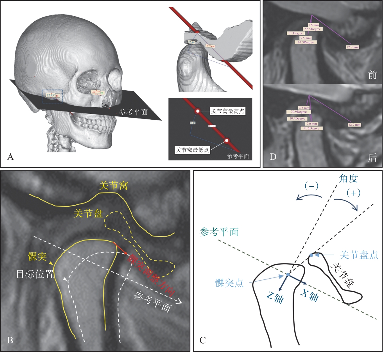
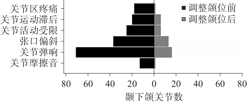
目的 介绍一种衍生自数字孪生的颌位调整新技术,并评价其辅助临床治疗颞下颌关节紊乱病(TMD)的效果。 方法 纳入2022年6月至2023年5月于四川大学华西口腔医院颞下颌关节科就诊的TMD患者74例,收集患者的初诊计算机断层扫描(CT)和双侧颞下颌关节磁共振成像(MRI)数据。根据MRI数据进行评估将148个关节分为正常盘-髁关系组(正常组)、可复性盘移位(DDWR)组以及不可复性盘移位(DDWoR)组,用CT数据重建患者口颌系统三维模型并构建个性化参考系进行颌位调整,将调整后的咬合关系输出打印为咬合导板,患者佩戴后行MRI复查。测量不同组别调整颌位前后患者髁突及关节盘的移位量和方向以及髁突与关节盘的夹角,评估其与髁突移位的相关性。 结果 在正常组中,关节盘沿X轴和Z轴分别向后、下移动(-0.60±0.62)、(0.51±0.71)mm;在DDWR组中,关节盘沿X轴和Z轴分别向后、上移动(-1.33±1.38)、(-0.09±1.31)mm;在DDWoR组中,关节盘沿X轴和Z轴分别向前、下移动(0.49±1.76)、(1.35±1.76)mm。在3组中,患者的髁突与关节盘的夹角在调整颌位后都减小。所有患者在调整颌位后症状均得到了改善。 结论 使用数字化软件辅助调整颌位能够简化流程、降低技术的敏感性,改善患者盘-髁结构及症状,在治疗TMD患者中的应用具有重要的临床意义。

关键词: 颌位调整, 数字化技术, AVIA软件, 颞下颌关节紊乱病

Abstract:

Objective The aim of this study was to demonstrate a novel jaw position adjustment technique derived from digital twins and evaluate the application effect of digital technology-assisted optimization in the process of adjusting jaw position on patients with temporomandibular disorders (TMD). Methods A total of 74 patients with TMD who attended the Department of Temporomandibular Joint, West China Hospital of Stomatology, Si-chuan University, between June 2022 and May 2023 were selected. The patient’s initial computed tomography (CT) and bilateral temporomandibular joint data obtained by magnetic resonance imaging (MRI) were collected. The 148 joints were divided into the normal disc-condyle relationship (N) group, disc displacement with reduction (DDWR) group, and disc displacement without reduction (DDWoR) group. Assisted by digital technology, the patient’s CT data were reconstructed, and a personalized reference plane was established to adjust the jaw position. A three-point bite guiding splint was designed by the adjusted occlusal space and then fabricated by 3D printing technology. It was worn by the patients and then reviewed by MRI. Before and after the adjustment of jaw position, the amount and direction of condyle and disc displacement and the angle between condyle and disc were measured as the evaluation indexes of the effect of the adjustment. The correlation with condylar displacement was evaluated. Results In the N group, the disc moved backward and downward along the X and Z axes by (-0.60±0.62) and (0.51±0.71) mm, respectively. In the DDWR group, the disc moved backward and upward along the X and Z axes by (-1.33±1.38) and (-0.09±1.31) mm, respectively. In the DDWoR group, the disc moved forward and downward along the X and Z axes by (0.49±1.76) and (1.35±1.76) mm, respectively. The angle between the condyle and the disc decreased after adjustment of the jaw position in all three groups. All patients showed improvement in symptoms after adjustment. Conclusion Digital technology-assisted jaw position adjustment can simplify the process, reduce the sensitivity of the technique, and improve patients’ disc-condyle structure and symptoms. Therefore, its application in the treatment of patients with TMD is of great clinical significance.

Key words: jaw position adjustment, digital technology, AVIA software, temporomandibular disorders

中图分类号: