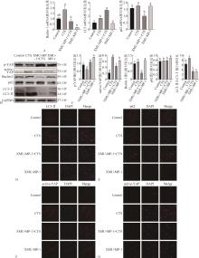
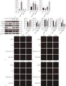
| 1 |
Chang M, Lin H, Luo M, et al. Integrated miRNA and mRNA expression profiling of tension force-induced bone formation in periodontal ligament cells[J]. In Vitro Cell Dev Biol Anim, 2015, 51(8): 797-807.
|
| 2 |
Xu J, Zhao X, Zeng J, et al. Role of autophagy in the periodontal ligament reconstruction during orthodontic tooth movement in rats[J]. J Dent Sci, 2020, 15(3): 351-363.
|
| 3 |
Dohmen M, Krieg S, Agalaridis G, et al. AMPK-dependent activation of the Cyclin Y/CDK16 complex controls autophagy[J]. Nat Commun, 2020, 11(1): 1032.
|
| 4 |
Blawat K, Mayr A, Hardt M, et al. Regulation of auto-phagic signaling by mechanical loading and inflammation in human PDL fibroblasts[J]. Int J Mol Sci, 2020, 21(24): E9446.
|
| 5 |
Deretic V, Levine B. Autophagy balances inflammation in innate immunity[J]. Autophagy, 2018, 14(2): 243-251.
|
| 6 |
Pedro JMB, Sica V, Madeo F, et al. Acyl-CoA-binding protein (ACBP): the elusive ‘hunger factor’ linking autophagy to food intake[J]. Cell Stress, 2019, 3(10): 312-318.
|
| 7 |
Feng X, Zhang H, Meng LB, et al. Hypoxia-induced acetylation of PAK1 enhances autophagy and promotes brain tumorigenesis via phosphorylating ATG5[J]. Autophagy, 2021, 17(3): 723-742.
|
| 8 |
Keith SA, Maddux SK, Zhong Y, et al. Graded proteasome dysfunction in caenorhabditis elegans activates an adaptive response involving the conserved SKN-1 and ELT-2 transcription factors and the autophagy-lysosome pathway[J]. PLoS Genet, 2016, 12(2): e1005823.
|
| 9 |
Vidoni C, Ferraresi A, Secomandi E, et al. Autophagy drives osteogenic differentiation of human gingival mesenchymal stem cells[J]. Cell Commun Signal, 2019, 17(1): 98.
|
| 10 |
Elbediwy A, Vincent-Mistiaen ZI, Thompson BJ. YAP and TAZ in epithelial stem cells: a sensor for cell polarity, mechanical forces and tissue damage[J]. Bioessays, 2016, 38(7): 644-653.
|
| 11 |
Fu HD, Wang BK, Wan ZQ, et al. Wnt5a mediated canonical Wnt signaling pathway activation in orthodontic tooth movement: possible role in the tension force-induced bone formation[J]. J Mol Histol, 2016, 47(5): 455-466.
|
| 12 |
Gong Y, Li SJ, Liu R, et al. Inhibition of YAP with si-RNA prevents cartilage degradation and ameliorates osteoarthritis development[J]. J Mol Med (Berl), 2019, 97(1): 103-114.
|
| 13 |
Wang D, He J, Huang B, et al. Emerging role of the Hippo pathway in autophagy[J]. Cell Death Dis, 2020, 11(10): 880.
|
| 14 |
Elosegui-Artola A, Andreu I, Beedle AEM, et al. Force triggers YAP nuclear entry by regulating transport across nuclear pores[J]. Cell, 2017, 171(6): 1397-1410.e14.
|
| 15 |
Piccolo S, Dupont S, Cordenonsi M. The biology of YAP/TAZ: Hippo signaling and beyond[J]. Physiol Rev, 2014, 94(4): 1287-1312.
|
| 16 |
Pei T, Huang X, Long Y, et al. Increased expression of YAP is associated with decreased cell autophagy in the eutopic endometrial stromal cells of endometriosis[J]. Mol Cell Endocrinol, 2019, 491: 110432.
|
| 17 |
Sun B, Wen Y, Wu X, et al. Expression pattern of YAP and TAZ during orthodontic tooth movement in rats[J]. J Mol Histol, 2018, 49(2): 123-131.
|
| 18 |
Pan X, Wu B, Fan X, et al. YAP accelerates vascular senescence via blocking autophagic flux and activating mTOR[J]. J Cell Mol Med, 2021, 25(1): 170-183.
|
| 19 |
Pei T, Luo B, Huang W, et al. Increased expression of YAP inhibited the autophagy level by upregulating m-TOR signal in the eutopic ESCs of endometriosis[J]. Front Endocrinol (Lausanne), 2022, 13: 813165.
|
| 20 |
Jin L, Chen Y, Cheng D, et al. YAP inhibits autophagy and promotes progression of colorectal cancer via upregulating Bcl-2 expression[J]. Cell Death Dis, 2021, 12(5): 457.
|
| 21 |
Ge MK, Zhou CC, Li H, et al. Lithium chloride atte-nuates suppressed differentiation induced by mechanical strain in cementoblasts[J]. Connect Tissue Res, 2019, 60(5): 444-451.
|
| 22 |
朱庆党, 刘丽, 杨艳丽. 张应力刺激下人牙周膜成纤维细胞纤连蛋白、整合素、细胞骨架的变化[J]. 上海口腔医学, 2010, 19(6): 601-606.
|
|
Zhu QD, Liu L, Yang YL. Effect of tensile stress on fibronectin-integrins-cytoskeleton in cultured human pe-riodontal ligament fibroblasts[J]. Shanghai J Stomatol, 2010, 19(6): 601-606.
|
| 23 |
朱庆党, 巢永烈, 陈新民, 等. 机械应力对人牙周膜成纤维细胞整合素β1 mRNA表达的调节[J]. 华西口腔医学杂志, 2008, 26(2): 194-197.
|
|
Zhu QD, Chao YL, Chen XM, et al. Regulation of integrin beta1 mRNA expression by mechanical stress in human periodontal ligament fibroblasts[J]. West China J Stomatol, 2008, 26(2): 194-197.
|
| 24 |
Memmert S, Damanaki A, Weykopf B, et al. Autophagy in periodontal ligament fibroblasts under biomechanical loading[J]. Cell Tissue Res, 2019, 378(3): 499-511.
|
| 25 |
Chen L, Mo S, Hua Y. Compressive force-induced autophagy in periodontal ligament cells downregulates osteoclastogenesis during tooth movement[J]. J Periodontol, 2019, 90(10): 1170-1181.
|
| 26 |
Zhang J, Zhou Y, Tang PMK, et al. Mechanotransduction and cytoskeleton remodeling shaping YAP1 in gastric tumorigenesis[J]. Int J Mol Sci, 2019, 20(7): 1576.
|
| 27 |
Yang Y, Wang BK, Chang ML, et al. Cyclic stretch enhances osteogenic differentiation of human periodontal ligament cells via YAP activation[J]. Biomed Res Int, 2018, 2018: 2174824.
|
| 28 |
Zhao M, Zhang Y, Jiang Y, et al. YAP promotes autophagy and progression of gliomas via upregulating HMGB1[J]. J Exp Clin Cancer Res, 2021, 40(1): 99.
|
| 29 |
Zhou X, Wang H, Li D, et al. MST1/2 inhibitor XMU-MP-1 alleviates the injury induced by ionizing radiation in haematopoietic and intestinal system[J]. J Cell Mol Med, 2022, 26(5): 1621-1628.
|
| 30 |
van Soldt BJ, Cardoso WV. Hippo-Yap/Taz signaling: complex network interactions and impact in epithelial cell behavior[J]. Wiley Interdiscip Rev Dev Biol, 2020, 9(3): e371.
|
| 31 |
Zheng Y, Pan D. The Hippo signaling pathway in development and disease[J]. Dev Cell, 2019, 50(3): 264-282.
|
| 32 |
Pavel M, Park SJ, Frake RA, et al. α-Catenin levels determine direction of YAP/TAZ response to autophagy perturbation[J]. Nat Commun, 2021, 12(1): 1703.
|
| 33 |
Liang N, Zhang C, Dill P, et al. Regulation of YAP by mTOR and autophagy reveals a therapeutic target of tuberous sclerosis complex[J]. J Exp Med, 2014, 211(11): 2249-2263.
|
| 34 |
Wang P, Gong Y, Guo T, et al. Activation of Aurora A kinase increases YAP stability via blockage of autophagy[J]. Cell Death Dis, 2019, 10(6): 432.
|

 ), 何海燕3, 吕佳岭4, 伍宇婕1,2, 钟冠男1,2, 徐晓梅1,2(