West China Journal of Stomatology ›› 2020, Vol. 38 ›› Issue (5): 525-531.doi: 10.7518/hxkq.2020.05.009
Previous Articles Next Articles
Zhang Ting1,2( ), Chen Du3, Miao Leiying1,2, Xie Sijing1,2, Tang Xuna1,2,4()
Received:2020-01-17
Revised:2020-03-15
Online:2020-10-01
Published:2020-10-14
Contact:
Tang Xuna
E-mail:zhangting7700@163.com;xunatang@126.com
Supported by:CLC Number:
Zhang Ting, Chen Du, Miao Leiying, Xie Sijing, Tang Xuna. Guided endodontic access of calcified root canal by laser melting templates[J]. West China Journal of Stomatology, 2020, 38(5): 525-531.
Add to citation manager EndNote|Ris|BibTeX

Fig 1
Design and clinical application of laser melting templates in the first case
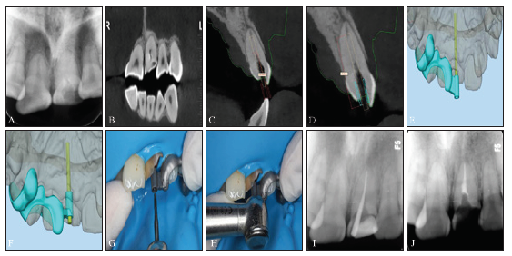

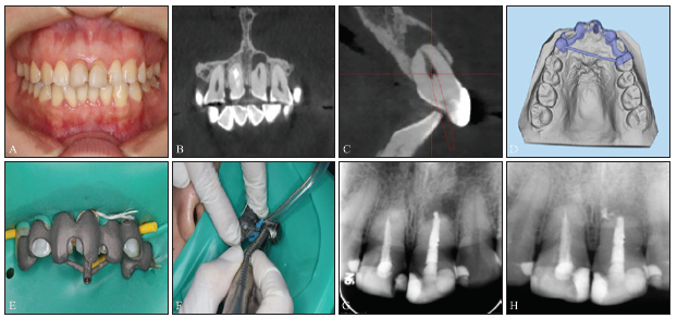
Fig 2
Design and clinical application of laser melting templates in the second case
Tab 1
Deviations of planned and repared access cavity at base and tip of the bur in the first case
| 项目 | 角度偏差/° | 钻基底偏差/mm | 钻尖端偏差/mm | ||||
|---|---|---|---|---|---|---|---|
| 近远中向 | 颊舌向 | 冠根向 | 近远中向 | 颊舌向 | 冠根向 | ||
| 平均值 | 1.770 | 0.403 | 0.463 | 0.497 | 0.433 | 0.493 | 0.537 |
| 最小值 | 1.550 | 0.390 | 0.450 | 0.490 | 0.420 | 0.480 | 0.520 |
| 最大值 | 1.980 | 0.410 | 0.480 | 0.500 | 0.420 | 0.500 | 0.550 |
| 标准差 | 0.215 | 0.012 | 0.015 | 0.006 | 0.015 | 0.012 | 0.015 |
Tab 2
Deviations of planned and repared access cavity at base and tip of the bur in the second case
| 项目 | 角度偏差/° | 钻基底偏差/mm | 钻尖端偏差/mm | ||||
|---|---|---|---|---|---|---|---|
| 近远中向 | 颊舌向 | 冠根向 | 近远中向 | 颊舌向 | 冠根向 | ||
| 平均值 | 3.260 | 0.247 | 0.347 | 0.183 | 0.347 | 0.463 | 0.310 |
| 最小值 | 3.210 | 0.230 | 0.330 | 0.180 | 0.330 | 0.450 | 0.300 |
| 最大值 | 3.320 | 0.260 | 0.360 | 0.190 | 0.360 | 0.480 | 0.320 |
| 标准差 | 0.056 | 0.015 | 0.015 | 0.006 | 0.015 | 0.015 | 0.010 |
| [1] |
Mello-Moura AC, Santos AM, Bonini GA , et al. Pulp calcification in traumatized primary teeth—classification, clinical and radiographic aspects[J]. J Clin Pediatr Dent, 2017,41(6):467-471.
doi: 10.17796/1053-4628-41.6.9 URL pmid: 28937901 |
| [2] |
Delivanis HP, Sauer GJ . Incidence of canal calcification in the orthodontic patient[J]. Am J Orthod, 1982,82(1):58-61.
doi: 10.1016/0002-9416(82)90547-4 URL pmid: 6961778 |
| [3] |
Karteva E, Manchorova N, Petrova N , et al. Effect of ageing and endodontic treatment on the thermal stability of human dentin[J]. Biomed Mater Eng, 2019,30(2):145-156.
doi: 10.3233/BME-191040 URL pmid: 30741663 |
| [4] |
Langeland K, Dowden WE, Tronstad L , et al. Human pulp changes of iatrogenic origin[J]. Oral Surg Oral Med Oral Pathol, 1971,32(6):943-980.
doi: 10.1016/0030-4220(71)90183-6 URL pmid: 4942993 |
| [5] |
Oginni AO, Adekoya-Sofowora CA, Kolawole KA . Evaluation of radiographs, clinical signs and symptoms associated with pulp canal obliteration: an aid to treatment decision[J]. Dent Traumatol, 2009,25(6):620-625.
doi: 10.1111/j.1600-9657.2009.00819.x URL pmid: 19917027 |
| [6] |
van der Meer WJ, Vissink A, Ng YL , et al. 3D Computer aided treatment planning in endodontics[J]. J Dent, 2016,45:67-72.
doi: 10.1016/j.jdent.2015.11.007 URL pmid: 26627596 |
| [7] |
Matherne RP, Angelopoulos C, Kulild JC , et al. Use of cone-beam computed tomography to identify root canal systems in vitro[J]. J Endod, 2008,34(1):87-89.
doi: 10.1016/j.joen.2007.10.016 URL pmid: 18155501 |
| [8] |
Krastl G, Zehnder MS, Connert T , et al. Guided endodontics: a novel treatment approach for teeth with pulp canal calcification and apical pathology[J]. Dent Traumatol, 2016,32(3):240-246.
doi: 10.1111/edt.12235 URL pmid: 26449290 |
| [9] |
Zehnder MS, Connert T, Weiger R , et al. Guided endodontics: accuracy of a novel method for guided access cavity preparation and root canal location[J]. Int Endod J, 2016,49(10):966-972.
doi: 10.1111/iej.12544 URL pmid: 26353942 |
| [10] |
Connert T, Zehnder MS, Amato M , et al. Microguided endodontics: a method to achieve minimally invasive access cavity preparation and root canal location in mandibular incisors using a novel computer-guided technique[J]. Int Endod J, 2018,51(2):247-255.
doi: 10.1111/iej.12809 URL pmid: 28665514 |
| [11] |
Fonseca Tavares WL , Diniz Viana AC, de Carvalho Machado V, et al. Guided endodontic access of calcified anterior teeth[J]. J Endod, 2018,44(7):1195-1199.
doi: 10.1016/j.joen.2018.04.014 URL pmid: 29941111 |
| [12] | 林捷, 林珍香, 郑志强 , 等. 根管导板技术辅助冠修复后磨牙的根管治疗[J]. 实用口腔医学杂志, 2018,34(5):648-651. |
| Lin J, Lin ZX, Zheng ZQ , et al. Endodontic treatment of the molars following crown restoration by using endodontic guide technique[J]. J Pract Stomatol, 2018,34(5):648-651. | |
| [13] |
Bürklein S, Schäfer E . Minimally invasive endodontics[J]. Quintessence Int, 2015,46(2):119-124.
doi: 10.3290/j.qi.a33047 URL pmid: 25500587 |
| [14] | Opal S, Garg S, Dhindsa A , et al. Minimally invasive clinical approach in indirect pulp therapy and healing of deep carious lesions[J]. J Clin Pediatr Dent, 2014,38(3):185-192. |
| [15] |
Ruddle CJ . Endodontic triad for success: the role of minimally invasive technology[J]. Dent Today, 2015, 34(5): 76, 78-80.
URL pmid: 26470575 |
| [16] | 承清, 夏文薇 . 三维打印导板在牙体牙髓专业领域中的研究和应用[J]. 中华口腔医学杂志, 2019,54(1):67-70. |
| Cheng Q, Xia WW . Research and application of three-dimensional printed template in endodontics[J]. Chin J Stomatol, 2019,54(1):67-70. | |
| [17] |
Zubizarreta-Macho Á, Ferreiroa A, Agustín-Panadero R , et al. Endodontic re-treatment and restorative treatment of a dens invaginatus type Ⅱ through new technologies[J]. J Clin Exp Dent, 2019,11(6):e570-e576.
doi: 10.4317/jced.55840 URL pmid: 31346380 |
| [18] | 封琼, 王一舟, 黄雨婷 , 等. 精准微创根管治疗: 3D导板指引下的钙化根管疏通术[J]. 口腔医学研究, 2017,33(4):427-431. |
| Feng Q, Wang YZ, Huang YT , et al. Negotiation of calcified root canal under guidance of 3D guides: precise minimally invasive root canal treatment[J]. J Oral Sci Res, 2017,33(4):427-431. | |
| [19] |
El Kholy K, Janner SF, Schimmel M , et al. The influence of guided sleeve height, drilling distance, and drilling key length on the accuracy of static computer-assisted implant surgery[J]. Clin Implant Dent Relat Res, 2019,21(1):101-107.
doi: 10.1111/cid.12705 URL pmid: 30589502 |
| [20] | Han T, Kundu S, Nag A , et al. 3D printed sensors for biomedical applications: a review[J]. Sensors (Basel), 2019,19(7):E1706. |
| [21] |
Lu YJ, Wu SQ, Gan YL , et al. Microstructure, mechanical property and metal release of As-SLM CoCrW alloy under different solution treatment conditions[J]. J Mech Behav Biomed Mater, 2015,55:179-190.
doi: 10.1016/j.jmbbm.2015.10.019 URL pmid: 26590910 |
| [22] |
Koutsoukis T, Zinelis S, Eliades G , et al. Selective laser melting technique of Co-Cr dental alloys: a review of structure and properties and comparative analysis with other available techniques[J]. J Prosthodont, 2015,24(4):303-312.
doi: 10.1111/jopr.12268 URL pmid: 26129918 |
| [23] |
Schneider D, Marquardt P, Zwahlen M , et al. A systematic review on the accuracy and the clinical outcome of computer-guided template-based implant dentistry[J]. Clin Oral Implants Res, 2009,20(Suppl 4):73-86.
doi: 10.1111/clr.2009.20.issue-s4 URL |
| [24] | 李晋蒙, 欧国敏 . 计算机辅助设计种植导板精确性及其影响因素[J]. 华西口腔医学杂志, 2017,35(1):93-98. |
| Li JM, Ou GM . Accuracy of computer-guided implant placement and influencing factors[J]. West China J Stomatol, 2017,35(1):93-98. | |
| [25] |
Buchgreitz J, Buchgreitz M, Mortensen D , et al. Guided access cavity preparation using cone-beam computed tomography and optical surface scans—an ex vivo study[J]. Int Endod J, 2016,49(8):790-795.
doi: 10.1111/iej.12516 URL pmid: 26201367 |
| [1] | Li Chengxi, Song Weijian.. Root canal treatment of type Ⅱ and ⅢA double dens invaginatus in maxillary lateral incisor: a case report [J]. West China Journal of Stomatology, 2023, 41(2): 232-236. |
| [2] | Yuan Jing, Yu Sijing, You Meng, Zhang Qiong, Ye Ling, Gao Bo. Regenerative endodontic treatment of dens in dente in maxillary lateral incisor with immature root: a case report [J]. West China Journal of Stomatology, 2022, 40(6): 716-720. |
| [3] | Cai Pingping, Chen Xi, Jiang Yi, Lu Zhaojie, Lin Jie, Zheng Zhiqiang.. Comparing accuracy after guide access and microscope-assisted access for fiber post removal [J]. West China Journal of Stomatology, 2022, 40(3): 297-302. |
| [4] | Gao Yuxuan, Wang Liu, Fu Yujie, Yang Fan, Zhang Lan, Huang Dingming. Minimally invasive treatment of calcified root canals in anterior teeth with digital guide technique [J]. West China Journal of Stomatology, 2022, 40(1): 111-122. |
| [5] | Yuan Zhiyao, Zou Xihong, Dai Linlin, Ao Huizhi, Li Houxuan.. Clinical analysis on the root fracture of the maxillary first molar [J]. West China Journal of Stomatology, 2021, 39(5): 555-559. |
| [6] | Guo Meiling, Huang Zhen, Wang Chong, Wang Yujiang. Effect of bilateral sagittal split ramus osteotomy on temporomandibular joint symptom and condylar position in patients with skeletal class Ⅲ malocclusion by cone beam computed tomography [J]. West China Journal of Stomatology, 2020, 38(5): 519-524. |
| [7] | Hu Shuang, Li Chunmei, Zhang Shuaiyuan, Qin Shuo, Xie Chenlu, Niu Zhixing, Sun Minglei. Clinical value of oral repair membrane and β-tricalcium phosphate in the treatment of the postoperative bone defect of jaw cyst [J]. West China Journal of Stomatology, 2020, 38(5): 541-545. |
| [8] | Shi Xiong, Li Shengjiao, Zhou Jianping, Zhang Li. Experimental study on the feasibility of low-dose cone beam computed tomography scanning [J]. West China Journal of Stomatology, 2020, 38(4): 415-418. |
| [9] | Zhao Haiyan, Wang Nan, Ding Yi, Zheng Haiying, Qian Junrong. Accuracy of cone beam computed tomography in assessing maxillary molar furcation involvement [J]. West China Journal of Stomatology, 2020, 38(3): 270-273. |
| [10] | Yao Ye,Qingzhu Li,Jingqiu Tu,Yonghua Lei. Characteristics of mandible and mandibular dentition according to vertical facial skeletal features of adolescents [J]. West China Journal of Stomatology, 2019, 37(1): 76-80. |
| [11] | Yulan Wang,Tiejun Wang,Zhonghao Liu. Changes in root and alveolar bone before and after treatment by retracting the upper incisors [J]. West China Journal of Stomatology, 2018, 36(6): 638-645. |
| [12] | Shu Li,Jie Lei,Kaiyuan Fu. Radiological characteristics of the cyst-like lesion of condyle in temporomandibular joint by cone beam computed tomography [J]. West China Journal of Stomatology, 2018, 36(5): 498-502. |
| [13] | Huirong Zhang, Lefeng Yin, Yanli Liu, Liyi Yan, Ning Wang, Gang Liu, Xiaoli An, Bin Liu. Fabrication and accuracy research on 3D printing dental model based on cone beam computed tomography digital modeling [J]. West China Journal of Stomatology, 2018, 36(2): 156-161. |
| [14] | Xing Ke, Bohan Li, Linlin Chen, Xindi Jiang. A cone beam computed tomography study on the anatomical position of accessory mandibular foramina in Jiangxi adults [J]. West China Journal of Stomatology, 2017, 35(6): 607-612. |
| [15] | Lili Yang, Yan Zhang, Shijun Zhao, Shuai Zhang, Na Wang, Jie Xu, Shue Hu, Zhiyuan Xu. Clinical application of cone beam computed tomography combined with micro-ultrasound technique in treating three mesial canals in mandibular first molars [J]. West China Journal of Stomatology, 2017, 35(4): 384-388. |
| Viewed | ||||||
|
Full text |
|
|||||
|
Abstract |
|
|||||
 This work is licensed under a Creative Commons Attribution 3.0 License.
