West China Journal of Stomatology ›› 2018, Vol. 36 ›› Issue (6): 638-645.doi: 10.7518/hxkq.2018.06.011
Previous Articles Next Articles
Yulan Wang,Tiejun Wang,Zhonghao Liu( )
)
Received:2018-04-05
Revised:2018-06-12
Online:2018-12-01
Published:2018-12-12
Contact:
Zhonghao Liu
E-mail:dentlzh@163.com
CLC Number:
Yulan Wang,Tiejun Wang,Zhonghao Liu. Changes in root and alveolar bone before and after treatment by retracting the upper incisors[J]. West China Journal of Stomatology, 2018, 36(6): 638-645.
Add to citation manager EndNote|Ris|BibTeX
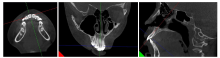
Fig.1
MPR of the teeth
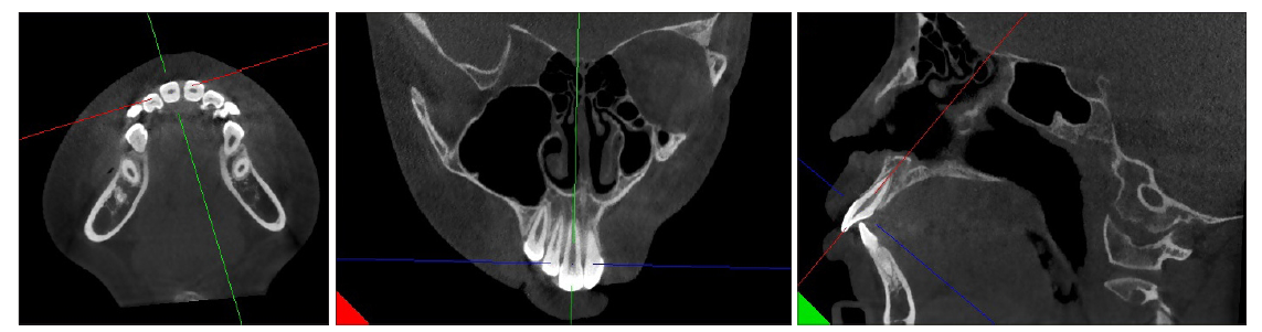
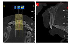
Fig.2
Measurement of root and alveolar bone changes


Fig.3
The trace of the lateral radiograph and the measurement of tooth movement
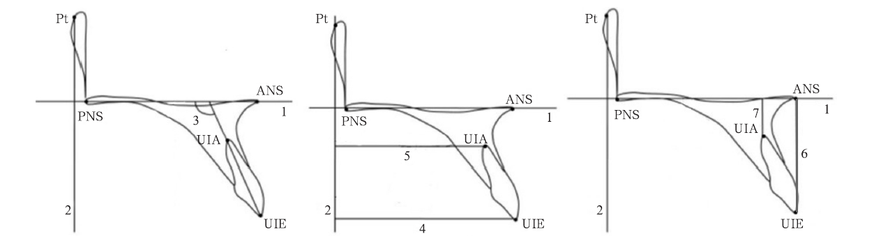

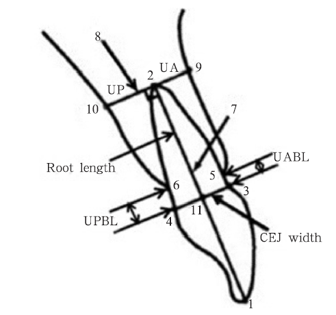
Fig.4
Measurement reference point/line and definition of measured variable.
Tab 1
Statistical description of root absorption and mea-surement before and after treatment for adduc-tion of upper incisors
| 测量项目 | T1 | T2 | ΔT(T1-T2) |
|---|---|---|---|
| U1-SN/° | 116.29±5.92 | 103.38±2.92 | 12.92±6.43 |
| U1E-sag/mm | 53.66±3.08 | 48.13±2.40 | 5.54±2.21 |
| U1E-ver/mm | 28.31±1.83 | 28.90±1.38 | -0.60±0.95 |
| UA/mm | 2.49±0.75 | 2.28±0.75 | 0.21±1.03 |
| UABL/mm | 0.88±0.28 | 1.08±0.35 | -0.20±0.22 |
| UPBL/mm | 0.71±0.26 | 0.73±0.31 | -0.02±0.35 |
| Root length/mm | 12.17±0.86 | 11.36±0.91 | 0.81±0.46 |
| 牙根吸收率/% | 6.80±3.60 |
Tab 2
Paired t test of root and alveolar bone changes before and after treatment of adduction of up-per incisor
| 分析项目 | t值 | P值 |
|---|---|---|
| T2UABL-T1UABL | 5.502 | 0.000* |
| T2UPBL-T1UPBL | 0.329 | 0.744 |
| T1Root length-T2Root length | 10.81 | 0.000* |
Tab 3
Pearson correlation coefficient analysis
| 分析项目 | 统计量 | ΔTU1-SN | ΔTU1E-sag | ΔTU1E-ver | T2UA | ΔTUA |
|---|---|---|---|---|---|---|
| ΔTRoot length | r值 | 0.251 | 0.405* | 0.140 | -0.391* | 0.313 |
| P值 | 0.134 | 0.013 | 0.416 | 0.017 | 0.060 | |
| ΔTRoot absorption | r值 | 0.176 | 0.358* | 0.101 | -0.417* | 0.280 |
| P值 | 0.298 | 0.030 | 0.559 | 0.010 | 0.093 | |
| ΔTUABL | r值 | -0.354* | -0.103 | -0.282 | -0.129 | -0.161 |
| P值 | 0.032 | 0.545 | 0.096 | 0.448 | 0.342 |
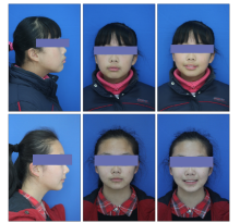
Fig.5
Facial images before and after treatment in typical case
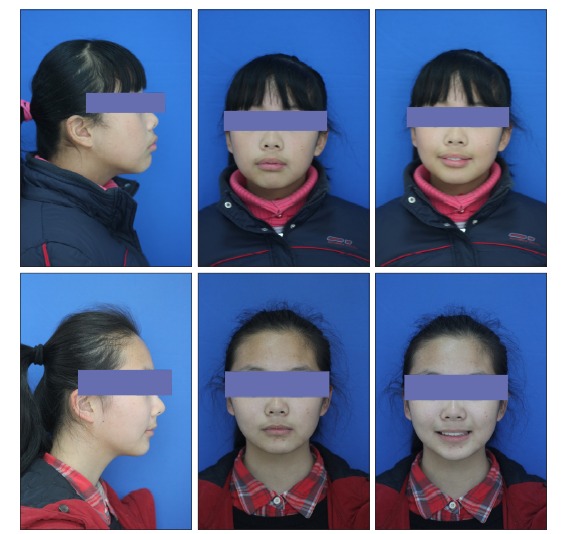
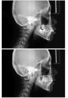
Fig 6
Cranial positioning lateral images before and after treatment in typical case


Fig 7
Intraoral image before and after treatment in typical case
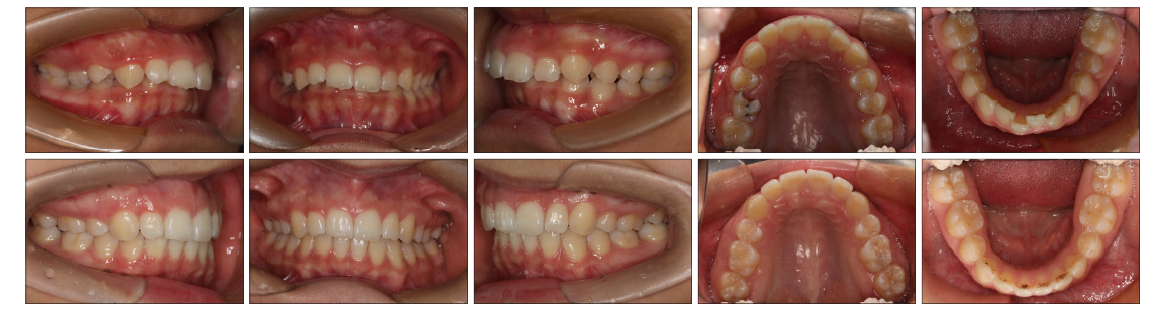
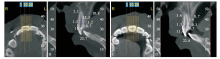
Fig 8
The measurement graphs of the typical patient by CBCT before and after treatment
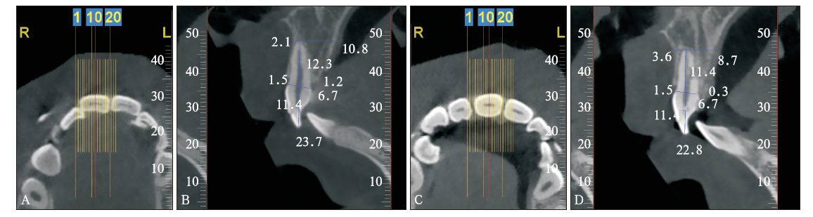
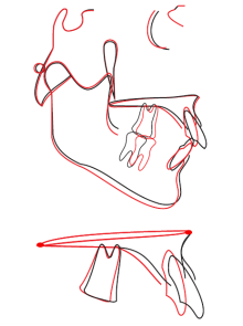
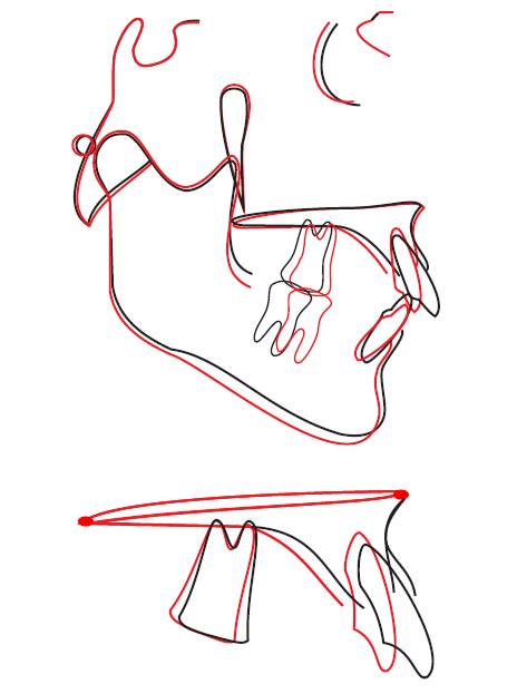
Fig 9
Overlapping graphs of cephalometric measurements before and after treatment
Tab 4
Comparison of cephalometric measurements be-fore and after treatment
| 测量项目 | 正常值 | 治疗前测量值 | 治疗后测量值 |
|---|---|---|---|
| SNA/° | 82.8±4.0 | 87.0 | 87.3 |
| SNB/° | 80.1±3.9 | 81.0 | 80.2 |
| ANB/° | 2.7±2.0 | 6.0 | 7.1 |
| FH-NP/° | 85.4±3.7 | 82.1 | 83.0 |
| NA-PA/° | 6.0±4.4 | 12.6 | 14.9 |
| U1-NA/mm | 5.1±2.4 | 4.8 | -1.2 |
| U1-NA/° | 22.8±5.7 | 22.1 | 8.0 |
| L1-NB/mm | 6.7±2.1 | 6.0 | 6.7 |
| L1-NB/° | 30.3±5.8 | 29.1 | 38.2 |
| U1-L1/° | 125.4±7.9 | 123.9 | 127.4 |
| U1-SN/° | 105.7±6.3 | 109.2 | 95.2 |
| MP-SN/° | 32.5±5.2 | 33.1 | 34.1 |
| MP-FH/° | 31.1±5.6 | 32.0 | 32.2 |
| L1-MP/° | 92.6±7.0 | 96.5 | 104.0 |
| Y轴/° | 66.3±7.1 | 66.4 | 67.1 |
| Pg-NB/mm | 1.0±1.5 | 1.3 | 1.2 |
Tab 5
Statistical description of root absorption and mea-surement before and after treatment for adduc-tion of upper incisors in typical case
| 测量项目 | T1 | T2 | ΔT(T1-T2) |
|---|---|---|---|
| U1-SN/° | 109.2 | 95.2 | 14 |
| U1E-sag/mm | 57.63 | 49.91 | 7.72 |
| U1E-ver/mm | 28.65 | 29.89 | -1.24 |
| UA/mm | 2.1 | 3.6 | -1.5 |
| UABL/mm | 1.5 | 1.5 | 0 |
| UPBL/mm | 1.2 | 0.3 | 0.9 |
| Root length/mm | 23.7 | 22.8 | 0.9 |
| 牙根吸收率/% | 3.80 |
| [1] |
余丽霞, 何姝姝, 陈嵩 . 全景及根尖片对正畸相关牙根吸收诊断准确性的研究[J]. 华西口腔医学杂志, 2012,30(2):169-172.
doi: 10.3969/j.issn.1000-1182.2012.02.014 URL |
|
Yu LX, He SS, Chen S . Diagnostic accuracy of orthopan-tomogram and periapical film in evaluating root resorption associated with orthodontic force[J]. West Chin J Stomatol, 2012,30(2):169-172.
doi: 10.3969/j.issn.1000-1182.2012.02.014 URL |
|
| [2] |
Estrela C, Bueno MR, Leles CR , et al. Accuracy of cone beam computed tomography and panoramic and periapical radiography for detection of apical periodontitis[J]. J Endod, 2008,34(3):273-279.
doi: 10.1016/j.joen.2007.11.023 URL pmid: 18291274 |
| [3] |
Lagravère MO, Carey J, Toogood RW , et al. Three-dimen-sional accuracy of measurements made with software on cone-beam computed tomography images[J]. Am J Orthod Dentofacial Orthop, 2008,134(1):112-116.
doi: 10.1016/j.ajodo.2006.08.024 URL |
| [4] |
Veyre-Goulet S, Fortin T, Thierry A . Accuracy of linear measurement provided by cone beam computed tomography to assess bone quantity in the posterior maxilla: a human cadaver study[J]. Clin Implant Dent Relat Res, 2008,10(4):226-230.
doi: 10.1111/j.1708-8208.2008.00083.x URL pmid: 18384410 |
| [5] |
Lund H, Gröndahl K, Ken HS , et al. Apical root resorption during orthodontic treatment. A prospective study using cone beam CT[J]. Angle Orthod, 2012,82(3):480-487.
doi: 10.2319/061311-390.1 URL pmid: 2767586 |
| [6] |
Castro IO, Alencar AH, Valladares-Neto J , et al. Apical root resorption due to orthodontic treatment detected by cone beam computed tomography[J]. Angle Orthod, 2013,83(2):196-203.
doi: 10.2319/032112-240.1 URL pmid: 22812378 |
| [7] | Lee AY, Kim YH . Comparison of movement of the upper dentition according to anchorage method: orthodontic mini-implant versus conventional anchorage reinforcement in classⅠmalocclusion[J]. ISRN Dent, 2011: 321206. |
| [8] |
Kim Y, Park JU, Kook YA . Alveolar bone loss around inci-sors in surgical skeletal class Ⅲ patients[J]. Angle Orthod, 2009,79(4):676-682.
doi: 10.2319/070308-341.1 URL pmid: 19537864 |
| [9] |
Nelson PA, Artun J . Alveolar bone loss of maxillary anterior teeth in adult orthodontic patients[J]. Am J Orthod Dento-facial Orthop, 1997,111(3):328-334.
doi: 10.1016/S0889-5406(97)70192-6 URL pmid: 9082856 |
| [10] |
Artun J, van’t Hullenaar R, Doppel D , et al. Identification of orthodontic patients at risk of severe apical root resorp-tion[J]. Am J Orthod Dentofacial Orthop, 2009,135(4):448-455.
doi: 10.1016/j.ajodo.2007.06.012 URL pmid: 19361730 |
| [11] |
Simplicio H, da Silva JS, Caldas SG , et al. External apical root resorption in retracted incisors[J]. Orthodontics (Chic), 2012,13(1):86-93.
URL pmid: 22567619 |
| [12] | 马宁, 李巍然, 陈晓红 , 等. 上切牙内收前后的牙根吸收研究[J]. 现代口腔医学杂志, 2015,29(6):330-334. |
| Ma N, Li WR, Chen XH , et al. Root resorption analysis of the maxillary incisors during retraction stage[J]. J Modern Stomatol, 2015,29(6):330-334. | |
| [13] |
许天民 . 青少年期正畸治疗与上中切牙牙根吸收的关系[J]. 中华口腔医学杂志, 2002,37(4):265-268.
doi: 10.3760/j.issn:1002-0098.2002.04.008 URL |
|
Xu TM . The relationship between apical root resorption and orthodontic tooth movement in growing subjects[J]. Chin J Stomatol, 2002,37(4):265-268.
doi: 10.3760/j.issn:1002-0098.2002.04.008 URL |
|
| [14] | de Freitas MR, Soares Beltrão RT, Janson G , et al. Evalua-tion of root resorption after open bite treatment with and without extractions[J]. Am J Orthod Dentofacial Orthop, 2007, 132(2): 143.e15-143.e22. |
| [15] |
Motokawa M, Sasamoto T, Kaku M , et al. Association be-tween root resorption incident to orthodontic treatment and treatment factors[J]. Eur J Orthod, 2012,34(3):350-356.
doi: 10.1093/ejo/cjs064 URL pmid: 23142948 |
| [16] |
Parker RJ, Harris EF . Directions of orthodontic tooth move-ments associated with external apical root resorption of the maxillary central incisor[J]. Am J Orthod Dentofacial Orthop, 1998,114(6):677-683.
doi: 10.1016/S0889-5406(98)70200-8 URL |
| [17] |
Horiuchi A, Hotokezaka H, Kobayashi K . Correlation be-tween cortical plate proximity and apical root resorption[J]. Am J Orthod Dentofacial Orthop, 1998,114(3):311-318.
doi: 10.1016/S0889-5406(98)70214-8 URL |
| [18] |
Ahn HW, Moon SC, Baek SH . Morphometric evaluation of changes in the alveolar bone and roots of the maxillary anterior teeth before and after en masse retraction using cone-beam computed tomography[J]. Angle Orthod, 2013,83(2):212-221.
doi: 10.2319/041812-325.1 URL pmid: 23066654 |
| [19] |
Vardimon AD, Oren E, Ben-Bassat Y . Cortical bone remo-deling/tooth movement ratio during maxillary incisor retrac-tion with tip versus torque movements[J]. Am J Orthod Den-tofacial Orthop, 1998,114(5):520-529.
doi: 10.1016/j.cellsig.2008.05.012 URL pmid: 9810048 |
| [20] |
林薇薇, 陈金武 . 成人前牙内收前后切牙牙槽骨高度变化的研究[J]. 实用口腔医学杂志, 2014,30(6):823-826.
doi: 10.3969/j.issn.1001-3733.2014.06.019 URL |
|
Lin WW, Chen JW . Changes in alveolar bone height due to retraction of anterior teeth in adult patients[J]. J Pract Sto-matol, 2014,30(6):823-826.
doi: 10.3969/j.issn.1001-3733.2014.06.019 URL |
| [1] | Li Chengxi, Song Weijian.. Root canal treatment of type Ⅱ and ⅢA double dens invaginatus in maxillary lateral incisor: a case report [J]. West China Journal of Stomatology, 2023, 41(2): 232-236. |
| [2] | Yuan Jing, Yu Sijing, You Meng, Zhang Qiong, Ye Ling, Gao Bo. Regenerative endodontic treatment of dens in dente in maxillary lateral incisor with immature root: a case report [J]. West China Journal of Stomatology, 2022, 40(6): 716-720. |
| [3] | Cai Pingping, Chen Xi, Jiang Yi, Lu Zhaojie, Lin Jie, Zheng Zhiqiang.. Comparing accuracy after guide access and microscope-assisted access for fiber post removal [J]. West China Journal of Stomatology, 2022, 40(3): 297-302. |
| [4] | Yuan Zhiyao, Zou Xihong, Dai Linlin, Ao Huizhi, Li Houxuan.. Clinical analysis on the root fracture of the maxillary first molar [J]. West China Journal of Stomatology, 2021, 39(5): 555-559. |
| [5] | Guo Meiling, Huang Zhen, Wang Chong, Wang Yujiang. Effect of bilateral sagittal split ramus osteotomy on temporomandibular joint symptom and condylar position in patients with skeletal class Ⅲ malocclusion by cone beam computed tomography [J]. West China Journal of Stomatology, 2020, 38(5): 519-524. |
| [6] | Zhang Ting, Chen Du, Miao Leiying, Xie Sijing, Tang Xuna. Guided endodontic access of calcified root canal by laser melting templates [J]. West China Journal of Stomatology, 2020, 38(5): 525-531. |
| [7] | Hu Shuang, Li Chunmei, Zhang Shuaiyuan, Qin Shuo, Xie Chenlu, Niu Zhixing, Sun Minglei. Clinical value of oral repair membrane and β-tricalcium phosphate in the treatment of the postoperative bone defect of jaw cyst [J]. West China Journal of Stomatology, 2020, 38(5): 541-545. |
| [8] | Shi Xiong, Li Shengjiao, Zhou Jianping, Zhang Li. Experimental study on the feasibility of low-dose cone beam computed tomography scanning [J]. West China Journal of Stomatology, 2020, 38(4): 415-418. |
| [9] | Zhao Haiyan, Wang Nan, Ding Yi, Zheng Haiying, Qian Junrong. Accuracy of cone beam computed tomography in assessing maxillary molar furcation involvement [J]. West China Journal of Stomatology, 2020, 38(3): 270-273. |
| [10] | Yao Ye,Qingzhu Li,Jingqiu Tu,Yonghua Lei. Characteristics of mandible and mandibular dentition according to vertical facial skeletal features of adolescents [J]. West China Journal of Stomatology, 2019, 37(1): 76-80. |
| [11] | Shu Li,Jie Lei,Kaiyuan Fu. Radiological characteristics of the cyst-like lesion of condyle in temporomandibular joint by cone beam computed tomography [J]. West China Journal of Stomatology, 2018, 36(5): 498-502. |
| [12] | Huirong Zhang, Lefeng Yin, Yanli Liu, Liyi Yan, Ning Wang, Gang Liu, Xiaoli An, Bin Liu. Fabrication and accuracy research on 3D printing dental model based on cone beam computed tomography digital modeling [J]. West China Journal of Stomatology, 2018, 36(2): 156-161. |
| [13] | Xing Ke, Bohan Li, Linlin Chen, Xindi Jiang. A cone beam computed tomography study on the anatomical position of accessory mandibular foramina in Jiangxi adults [J]. West China Journal of Stomatology, 2017, 35(6): 607-612. |
| [14] | Lili Yang, Yan Zhang, Shijun Zhao, Shuai Zhang, Na Wang, Jie Xu, Shue Hu, Zhiyuan Xu. Clinical application of cone beam computed tomography combined with micro-ultrasound technique in treating three mesial canals in mandibular first molars [J]. West China Journal of Stomatology, 2017, 35(4): 384-388. |
| [15] | Jingqiu Tu, Jiaqian Fan, Yonghua Lei. Characteristics of mandible and mandibular dentition in patients with near-normal occlusion and different vertical facial skeletal types [J]. West China Journal of Stomatology, 2017, 35(4): 403-407. |
| Viewed | ||||||
|
Full text |
|
|||||
|
Abstract |
|
|||||
 This work is licensed under a Creative Commons Attribution 3.0 License.
