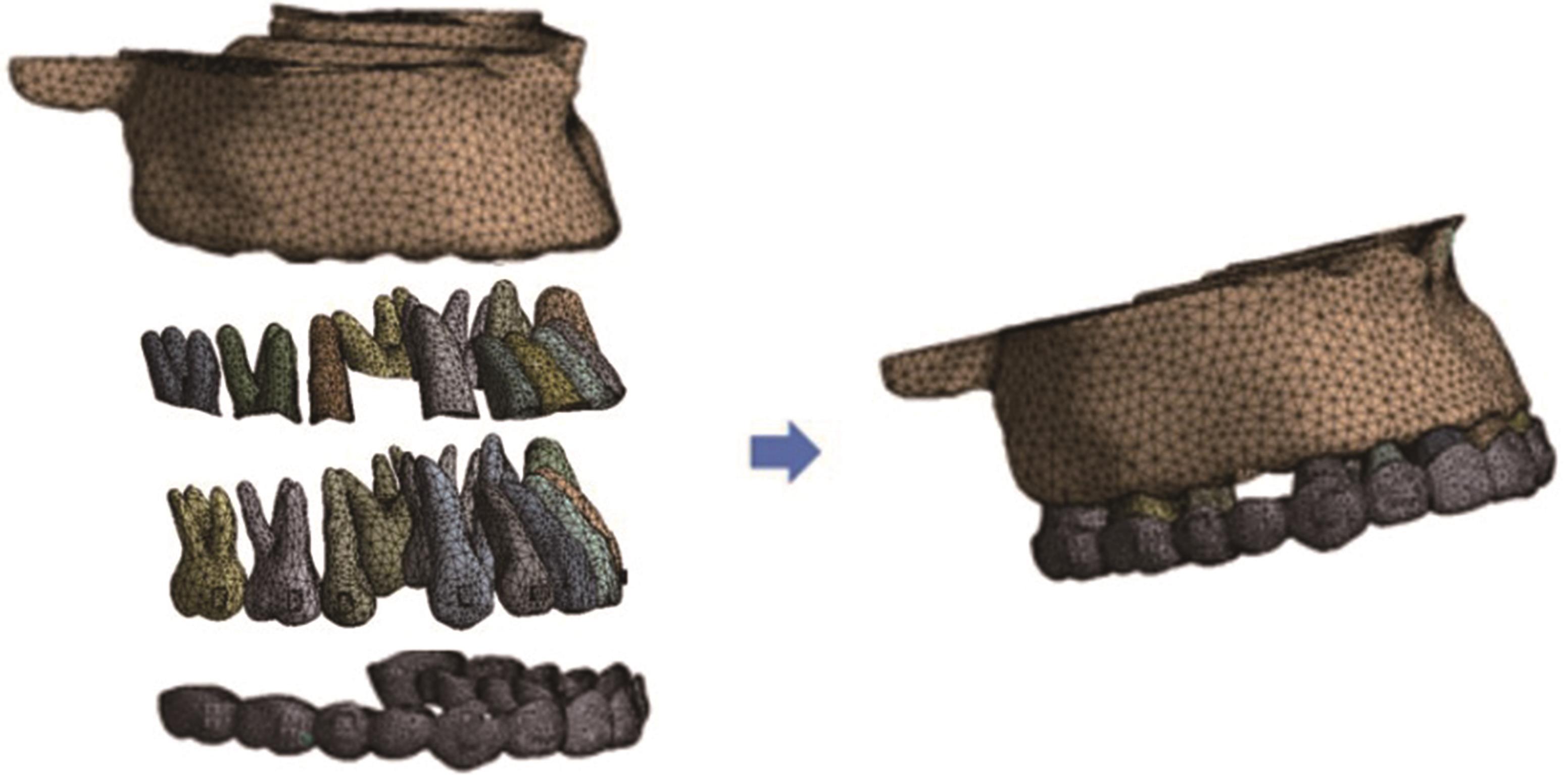
| 1 |
郑钰婷, 陈琳, 吴嘉桦, 等. 无托槽隐形矫治器整体内收上颌前牙的三维有限元分析[J]. 实用口腔医学杂志, 2017, 33(5): 621-624.
|
|
Zheng YT, Chen L, Wu JH, et al. En-masse retraction of maxillary anterior teeth with clear aligner: a finite element analysis[J]. J Pract Stomatol, 2017, 33(5): 621-624.
|
| 2 |
白煜, 冯雪, 刘冬梅, 等. 无托槽隐形矫治器联合微种植钉整体内收上前牙的三维有限元研究[J]. 实用口腔医学杂志, 2019, 35(1): 71-76.
|
|
Bai Y, Feng X, Liu DM, et al. Combined use of miniscrews and clear aligner for en-mass retraction of maxillary anterior teeth: a finite element analysis[J]. J Pract Stomatol, 2019, 35(1): 71-76.
|
| 3 |
Machado RM. Space closure using aligners[J]. Dental Press J Orthod, 2020, 25(4): 85-100.
|
| 4 |
李晓梅, 徐宝华. 无托槽隐形矫治器联合种植支抗内收上前牙的三维有限元研究[J]. 中日友好医院学报, 2021, 35(4): 211-215.
|
|
Li XM, Xu BH. Clear aligner combined with micro-implant anchorage for en-mass retraction of maxillary anterior teeth: a three-dimensional finite element analysis[J]. J China-Japan Friendship Hosp, 2021, 35(4): 211-215.
|
| 5 |
Lin LY, Chang CH, Roberts WE. Bimaxillary protrusion and gummy smile treated with clear aligners: closing premolar extraction spaces with bone screw anchorage[J]. APOS Trend Orthod, 2020, 10: 120-131.
|
| 6 |
张鸿军, 嵇国平, 沈刚. 利用颧牙槽嵴区种植钉矫正边缘性Ⅱ类错𬌗[J]. 上海口腔医学, 2013, 22(3): 310-315.
|
|
Zhang HJ, Ji GP, Shen G. A clinical study on effects of distalization of whole upper arch in borderline Class Ⅱ malocclusion using microscrew anchorages in inferiozygomatic area[J]. Shanghai J Stomatol, 2013, 22(3): 310-315.
|
| 7 |
Yokoi Y, Arai A, Kawamura J, et al. Effects of attachment of plastic aligner in closing of diastema of maxillary dentition by finite element method[J]. J Healthc Eng, 2019, 2019: 1075097.
|
| 8 |
唐娜, 赵志河, 王军, 等. 无托槽隐形矫治技术生物力学效应的有限元法研究[J]. 医用生物力学, 2010, 25(6): 399-405.
|
|
Tang N, Zhao ZH, Wang J, et al. Biomechanical effects of bracketless appliance technology: a finite element me-thod study[J]. J Med Biomech, 2010, 25(6): 399-405.
|
| 9 |
戴宁, 曾照斌, 刘海锋, 等. 两种常用加力方式对种植钉压低上前牙力学行为影响的三维有限元分析[J]. 口腔医学研究, 2011, 27(5): 372-375.
|
|
Dai N, Zeng ZB, Liu HF, et al. Comparison of the intrution effects of two commonly used loading approaches on the maxillary incisors: a 3-dimensional finite element analysis[J]. J Oral Sci Res, 2011, 27(5): 372-375.
|
| 10 |
Ohnishi H, Yagi T, Yasuda Y, et al. A mini-implant for orthodontic anchorage in a deep overbite case[J]. Angle Orthod, 2005, 75(3): 444-452.
|
| 11 |
Upadhyay M, Nagaraj K, Yadav S, et al. Mini-implants for en masse intrusion of maxillary anterior teeth in a severe ClassⅡdivision 2 malocclusion[J]. J Orthod, 2008, 35(2): 79-89.
|
| 12 |
Thresher RW, Saito GE. The stress analysis of human teeth[J]. J Biomech, 1973, 6(5): 443-449.
|
| 13 |
Farah JW, Craig RG, Sikarskie DL. Photoelastic and finite element stress analysis of a restored axisymmetric first molar[J]. J Biomech, 1973, 6(5): 511-520.
|
| 14 |
Chetan S, Keluskar KM, Vasisht VN, et al. En-masse retraction of the maxillary anterior teeth by applying force from four different levels—a finite element study[J]. J Clin Diagn Res, 2014, 8(9): ZC26-ZC30.
|
| 15 |
龙茜, 管晓燕, 刘建国. 正畸牙移动的实验模型研究进展[J]. 遵义医学院学报, 2019, 42(2): 227-237.
|
|
Long Q, Guan XY, Liu JG. Model establishment of or-thodontic tooth movement[J]. J Zunyi Med Univ, 2019, 42(2): 227-237.
|
| 16 |
Jiang T, Wu RY, Wang JK, et al. Clear aligners for maxillary anterior en masse retraction: a 3D finite element study[J]. Sci Rep, 2020, 10(1): 10156.
|
| 17 |
田雪, 王娟, 彭友俭. 三种不同部位微种植体压低上前牙改善露龈微笑的临床疗效对比[J]. 中华口腔正畸学杂志, 2019, 26(1): 2-6.
|
|
Tian X, Wang J, Peng YJ. Comparison of clinical effects of three different positions of micro-implants intruding upper anterior teeth to improve the gummy smile[J]. Chin J Orthod, 2019, 26(1): 2-6.
|
| 18 |
Liu L, Zhan Q, Zhou J, et al. Effectiveness of an anterior mini-screw in achieving incisor intrusion and palatal root torque for anterior retraction with clear aligners[J]. Angle Orthod, 2021, 91(6): 794-803.
|
| 19 |
李晓玮, 任超超, 王喆垚, 等. 舌向整体移位设计量对隐形矫治力影响的研究[J]. 中华口腔正畸学杂志, 2014, 21(4): 200-203.
|
|
Li XW, Ren CC, Wang JY, et al. Effects of amount of activation for lingual bodily movement of maxillary central incisor on orthodontic force generated by thermoplastic aligners[J]. Chin J Orthod, 2014, 21(4): 200-203.
|
| 20 |
Rosvall MD, Fields HW, Ziuchkovski J, et al. Attractiveness, acceptability, and value of orthodontic appliances[J]. Am J Orthod Dentofacial Orthop, 2009, 135(3): 276.e1-e12.
|
| 21 |
Rossini G, Parrini S, Castroflorio T, et al. Efficacy of clear aligners in controlling orthodontic tooth movement: a systematic review[J]. Angle Orthod, 2015, 85(5): 881-889.
|
| 22 |
Clements KM, Bollen AM, Huang G, et al. Activation time and material stiffness of sequential removable or-thodontic appliances. Part 2: dental improvements[J]. Am J Orthod Dentofacial Orthop, 2003, 124(5): 502-508.
|
| 23 |
Kim SH, Hwang YS, Ferreira A, et al. Analysis of temporary skeletal anchorage devices used for en-masse retraction: a preliminary study[J]. Am J Orthod Dentofacial Orthop, 2009, 136(2): 268-276.
|
| 24 |
Jasoria G, Shamim W, Rathore S, et al. Miniscrew implants as temporary anchorage devices in orthodontics: a comprehensive review[J]. J Contemp Dent Pract, 2013, 14(5): 993-999.
|
| 25 |
Suzuki H, Moon W, Previdente LH, et al. Miniscrew-assisted rapid palatal expander (MARPE): the quest for pure orthopedic movement[J]. Dental Press J Orthod, 2016, 21(4): 17-23.
|
| 26 |
Lee J, Miyazawa K, Tabuchi M, et al. Midpalatal miniscrews and high-pull headgear for anteroposterior and vertical anchorage control: cephalometric comparisons of treatment changes[J]. Am J Orthod Dentofacial Orthop, 2013, 144(2): 238-250.
|
| 27 |
Weir T. Clear aligners in orthodontic treatment[J]. Aust Dent J, 2017, 62(): 58-62.
|
| 28 |
安世英, 张继武, 马俐丽, 等. 无托槽隐形矫治技术内收上前牙的三维有限元分析[J]. 临床口腔医学杂志, 2020, 36(10): 583-586.
|
|
An SY, Zhang JW, Ma LL, et al. The biomechanical mechanism of invisible appliance in retracting the maxillary incisors[J]. J Clin Stomatol, 2020, 36(10): 583-586.
|
| 29 |
肖亚萍. 无托槽隐形矫治器加载不同转矩压低上颌前牙的三维有限元分析[D]. 唐山: 华北理工大学, 2021.
|
|
Xiao YP. Three-dimensional finite element analysis of invisible bracketless aligners loaded with different tor-ques depressing maxillary anterior teeth[D]. Tangshan: North China University of Science and Technology, 2021.
|
| 30 |
Shaw AM, Sameshima GT, Vu HV. Mechanical stress generated by orthodontic forces on apical root cementum: a finite element model[J]. Orthod Craniofac Res, 2004, 7(2): 98-107.
|
| 31 |
Han GL, Huang SF, Von den Hoff JW, et al. Root resorption after orthodontic intrusion and extrusion: an intraindividual study[J]. Angle Orthod, 2005, 75(6): 912-918.
|

 ), 夏恺, 罗良语, 赵志河, 刘钧(