| 1 |
Brånemark PI, Hansson BO, Adell R, et al. Osseointegrated implants in the treatment of the edentulous jaw. Experience from a 10-year period[J]. Scand J Plast Reconstr Surg Suppl, 1977, 16: 1-132.
|
| 2 |
Wang Y, Zhang Y, Miron RJ. Health, maintenance, and recovery of soft tissues around implants[J]. Clin Implant Dent Relat Res, 2016, 18(3): 618-634.
|
| 3 |
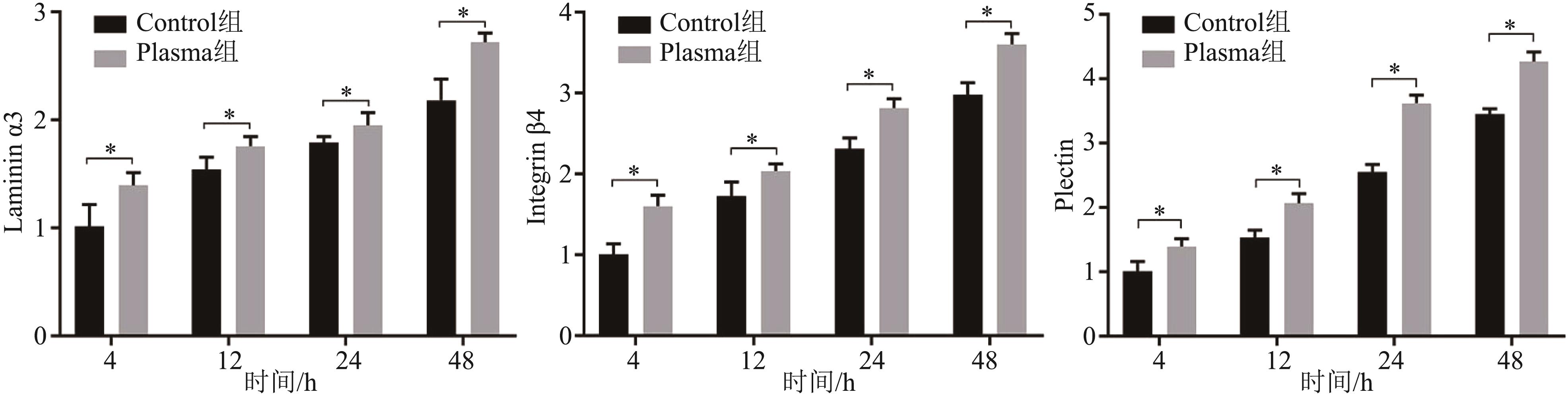
Zhang J, Wang H, Wang Y, et al. Substrate-mediated gene transduction of LAMA3 for promoting biological sealing between titanium surface and gingival epithelium[J]. Colloids Surf B Biointerfaces, 2018, 161: 314-323.
|
| 4 |
Ivanovski S, Lee R. Comparison of peri-implant and periodontal marginal soft tissues in health and disease[J]. Periodontol 2000, 2018, 76(1): 116-130.
|
| 5 |
Atsuta I, Ayukawa Y, Kondo R, et al. Soft tissue sealing around dental implants based on histological interpretation[J]. J Prosthodont Res, 2016, 60(1): 3-11.
|
| 6 |
Kalyoncuoglu UT, Yilmaz B, Koc SG, et al. Investigation of surface structure and biocompatibility of chitosan-coated zirconia and alumina dental abutments[J]. Clin Implant Dent Relat Res, 2018, 20(6): 1022-1029.
|
| 7 |
Xu R, Hu X, Yu X, et al. Micro-/nano-topography of selective laser melting titanium enhances adhesion and proliferation and regulates adhesion-related gene expressions of human gingival fibroblasts and human gingival epithelial cells[J]. Int J Nanomedicine, 2018, 13: 5045-5057.
|
| 8 |
Takebe J, Miyata K, Miura S, et al. Effects of the nanotopographic surface structure of commercially pure titanium following anodization-hydrothermal treatment on ge-ne expression and adhesion in gingival epithelial cells[J]. Mater Sci Eng C Mater Biol Appl, 2014, 42: 273-279.
|
| 9 |
Lee JH, Kim YH, Choi EH, et al. Air atmospheric-pressure plasma-jet treatment enhances the attachment of human gingival fibroblasts for early peri-implant soft tissue seals on titanium dental implant abutments[J]. Acta Odontol Scand, 2015, 73(1): 67-75.
|
| 10 |
Abdallah MN, Badran Z, Ciobanu O, et al. Strategies for optimizing the soft tissue seal around osseointegrated implants[J]. Adv Healthc Mater, 2017, 6(20). .
|
| 11 |
Lou BS, Hsieh JH, Chen CM, et al. Helium/argon-gene-rated cold atmospheric plasma facilitates cutaneous wound healing[J]. Front Bioeng Biotechnol, 2020, 8: 683.
|
| 12 |
Scharf C, Eymann C, Emicke P, et al. Improved wound healing of airway epithelial cells is mediated by cold atmospheric plasma: a time course-related proteome analysis[J]. Oxid Med Cell Longev, 2019, 2019: 7071536.
|
| 13 |
Packer JR, Hirst AM, Droop AP, et al. Notch signalling is a potential resistance mechanism of progenitor cells within patient-derived prostate cultures following ROS-inducing treatments[J]. FEBS Lett, 2020, 594(2): 209-226.
|
| 14 |
Chandrasekhara V, Rhoades D, Kaimakliotis PZ, et al. An endoscopic training and assessment model for argon plasma coagulation[J]. Adv Med Sci, 2019, 64(1): 152-156.
|
| 15 |
Park J, Suh D, Tang T, et al. Non-thermal atmospheric pressure plasma induces epigenetic modifications that activate the expression of various cytokines and growth factors in human mesoderm-derived stem cells[J]. Free Radic Biol Med, 2020, 148: 108-122.
|
| 16 |
Zhao SS, Han R, Li Y, et al. Investigation of the mechanism of enhanced and directed differentiation of neural stem cells by an atmospheric plasma jet: a gene-level study[J]. J Appl Phys, 2019, 125(16): 163301.
|
| 17 |
Wang M, Zhou Y, Shi D, et al. Cold atmospheric plasma (CAP)-modified and bioactive protein-loaded core-shell nanofibers for bone tissue engineering applications[J]. Biomater Sci, 2019, 7(6): 2430-2439.
|
| 18 |
Tominami K, Kanetaka H, Sasaki S, et al. Cold atmospheric plasma enhances osteoblast differentiation[J]. P-LoS One, 2017, 12(7): e0180507.
|
| 19 |
Reitberger HH, Czugala M, Chow C, et al. Argon cold plasma—A novel tool to treat therapy-resistant corneal infections[J]. Am J Ophthalmol, 2018, 190: 150-163.
|
| 20 |
Laroussi M. Low temperature plasma-based sterilization: overview and state-of-the-art[J]. Plasma Process Polym, 2005, 2(5): 391-400.
|
| 21 |
Park JK, Nam SH, Kwon HC, et al. Feasibility of non-thermal atmospheric pressure plasma for intracoronal bleaching[J]. Int Endod J, 2011, 44(2): 170-175.
|
| 22 |
Herbst SR, Hertel M, Ballout H, et al. Bactericidal efficacy of cold plasma at different depths of infected root canals in vitro [J]. Open Dent J, 2015, 9: 486-491.
|
| 23 |
Langmuir I. Oscillations in ionized gases[J]. Proc Natl Acad Sci U S A, 1928, 14(8): 627-637.
|
| 24 |
Haertel B, von Woedtke T, Weltmann KD, et al. Non-thermal atmospheric-pressure plasma possible application in wound healing[J]. Biomol Ther (Seoul), 2014, 22(6): 477-490.
|
| 25 |
Griffin MF, Ibrahim A, Seifalian AM, et al. Argon plasma modification promotes adipose derived stem cells osteogenic and chondrogenic differentiation on nanocomposite polyurethane scaffolds; implications for skeletal tissue engineering[J]. Mater Sci Eng C Mater Biol Appl, 2019, 105: 110085.
|
| 26 |
Zhang Y, Xiong Y, Xie P, et al. Non-thermal plasma reduces periodontitis-induced alveolar bone loss in rats[J]. Biochem Biophys Res Commun, 2018, 503(3): 2040-2046.
|
| 27 |
Borradori L, Sonnenberg A.Structure and function of hemidesmosomes: more than simple adhesion complexes[J]. J Invest Dermatol, 1999, 112(4): 411-418.
|
| 28 |
Atsuta I, Yamaza T, Yoshinari M, et al. Changes in the distribution of laminin-5 during peri-implant epithelium formation after immediate titanium implantation in rats[J]. Biomaterials, 2005, 26(14): 1751-1760.
|
| 29 |
Atsuta I, Yamaza T, Yoshinari M, et al. Ultrastructural localization of laminin-5 (gamma2 chain) in the rat peri-implant oral mucosa around a titanium-dental implant by immuno-electron microscopy[J]. Biomaterials, 2005, 26(32): 6280-6287.
|
| 30 |
TVON Woedtke, Schmidt A, Bekeschus S, et al. Plasma medicine: a field of applied redox biology[J]. In Vivo, 2019, 33(4): 1011-1026.
|
| 31 |
Kalghatgi S, Friedman G, Fridman A, et al. Endothelial cell proliferation is enhanced by low dose non-thermal plasma through fibroblast growth factor-2 release[J]. Ann Biomed Eng, 2010, 38(3): 748-757.
|
| 32 |
Kang SU, Choi JW, Chang JW, et al. N2 non-thermal atmospheric pressure plasma promotes wound healing in vitro and in vivo: potential modulation of adhesion molecules and matrix metalloproteinase-9[J]. Exp Dermatol, 2017, 26(2): 163-170.
|
| 33 |
Kurake N, Tanaka H, Ishikawa K, et al. Cell survival of glioblastoma grown in medium containing hydrogen peroxide and/or nitrite, or in plasma-activated medium[J]. Arch Biochem Biophys, 2016, 605: 102-108.
|

 ), 敖小刚, 郑铮, 陈文川(