| 1 |
Khalaf K, Miskelly J, Voge E, et al. Prevalence of hypodontia and associated factors: a systematic review and Meta-analysis[J]. J Orthod, 2014, 41(4): 299-316.
|
| 2 |
Kjær I. Mechanism of human tooth eruption: review article including a new theory for future studies on the eruption process[J]. Scientifica (Cairo), 2014, 2014: 341905.
|
| 3 |
Becktor JP, Einersen S, Kjaer I. A sella turcica bridge in subjects with severe craniofacial deviations[J]. Eur J Orthod, 2000, 22(1): 69-74.
|
| 4 |
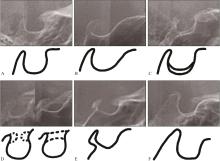
刘军, 杨洁, 李铎, 等. 第二前磨牙先天缺失与蝶鞍的相关性研究[J]. 中国实用口腔科杂志, 2019, 12(2): 92-95.
|
|
Liu J, Yang J, Li D, et al. Study on the relationship between sella and the congenital absence of second premolar[J]. Chin J Pract Stomatol, 2019, 12(2): 92-95.
|
| 5 |
Alqahtani H. Association between sella turcica bridging and congenitally missing maxillary lateral incisors[J]. J Dent Sci, 2020, 15(1): 59-64.
|
| 6 |
Canigur Bavbek N, Arslan Avan B. Morphometric evaluation of cranial base and sella turcica in patients with bilateral agenesis of maxillary lateral incisors[J]. Odonto-logy, 2021, 109(3): 701-709.
|
| 7 |
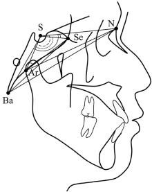
李亚其, 王梓千, 刘家琦, 等. 蝶鞍与颈椎成熟度的相关性研究[J]. 口腔医学研究, 2022, 38(4): 367-371.
|
|
Li YQ, Wang ZQ, Liu JQ, et al. Study on correlation between sella turcica and cervical vertebral maturation[J]. J Oral Sci Res, 2022, 38(4): 367-371.
|
| 8 |
Silverman FN. Roentgen standards fo-size of the pitui-tary fossa from infancy through adolescence[J]. Am J Roentgenol Radium Ther Nucl Med, 1957, 78(3): 451-460.
|
| 9 |
Axelsson S, Storhaug K, Kjaer I. Post-natal size and morphology of the sella turcica. longitudinal cephalometric standards for Norwegians between 6 and 21 years of age[J]. Eur J Orthod, 2004, 26(6): 597-604.
|
| 10 |
Leonardi R, Barbato E, Vichi M, et al. A sella turcica bridge in subjects with dental anomalies[J]. Eur J Orthod, 2006, 28(6): 580-585.
|
| 11 |
Proff P, Will F, Bokan I, et al. Cranial base features in skeletal Class Ⅲ patients[J]. Angle Orthod, 2008, 78(3): 433-439.
|
| 12 |
李蓝, 张博文, 赵志河. 牙缺失基因的研究进展[J]. 华西口腔医学杂志, 2013, 31(4): 436-439.
|
|
Li L, Zhang BW, Zhao ZH. Research progress on gene involved in tooth agenesis[J]. West China J Stomatol, 2013, 31(4): 436-439.
|
| 13 |
Nieminen P, Arte S, Tanner D, et al. Identification of a nonsense mutation in the PAX9 gene in molar oligodontia[J]. Eur J Hum Genet, 2001, 9(10): 743-746.
|
| 14 |
Vieira AR, Modesto A, Meira R, et al. Interferon regulatory factor 6 (IRF6) and fibroblast growth factor receptor 1 (FGFR1) contribute to human tooth agenesis[J]. Am J Med Genet A, 2007, 143A(6): 538-545.
|
| 15 |
Vieira AR, Meira R, Modesto A, et al. MSX1, PAX9, and TGFA contribute to tooth agenesis in humans[J]. J Dent Res, 2004, 83(9): 723-727.
|
| 16 |
Frazier-Bowers SA, Guo DC, Cavender A, et al. A novel mutation in human PAX9 causes molar oligodontia[J]. J Dent Res, 2002, 81(2): 129-133.
|
| 17 |
Fauzi NH, Ardini YD, Zainuddin Z, et al. A review on non-syndromic tooth agenesis associated with PAX9 mutations[J]. Jpn Dent Sci Rev, 2018, 54(1): 30-36.
|
| 18 |
Abdalla EM, Mostowska A, Jagodziński PP, et al. A no-vel WNT10A mutation causes non-syndromic hypodontia in an Egyptian family[J]. Arch Oral Biol, 2014, 59(7): 722-728.
|
| 19 |
Scribante A, Sfondrini MF, Cassani M, et al. Sella turcica bridging and dental anomalies: is there an association[J]. Int J Paediatr Dent, 2017, 27(6): 568-573.
|
| 20 |
Canigur Bavbek N, Dincer M. Dimensions and morphologic variations of sella turcica in type 1 diabetic patients[J]. Am J Orthod Dentofacial Orthop, 2014, 145(2): 179-187.
|
| 21 |
Korayem M, AlKofide E. Size and shape of the sella turcica in subjects with Down syndrome[J]. Orthod Craniofac Res, 2015, 18(1): 43-50.
|
| 22 |
Jankowski T, Jedliński M, Schmeidl K, et al. Sella turcica abnormalities, dental age and dental abnormalities in Polish children[J]. Int J Environ Res Public Health, 2021, 18(19): 10101.
|
| 23 |
Antonarakis GS, Huanca Ghislanzoni L, La Scala GC, et al. Sella turcica morphometrics in children with unilateral cleft lip and palate[J]. Orthod Craniofac Res, 2020, 23(4): 398-403.
|
| 24 |
Sato D, Endo T. Size and bridging of the sella turcica in Japanese orthodontic patients with tooth agenesis[J]. O-dontology, 2020, 108(4): 730-737.
|
| 25 |
Arntsen T, Kjær I, Sonnesen L. Lengths of the maxillary central incisor, the nasal bone, and the anterior cranial base in different skeletal malocclusions[J]. Acta Odontol Scand, 2009, 67(5): 265-270.
|
| 26 |
Endo T, Yoshino S, Ozoe R, et al. Association of advanced hypodontia and craniofacial morphology in Japanese orthodontic patients[J]. Odontology, 2004, 92(1): 48-53.
|
| 27 |
Kjaer I, Becktor KB, Lisson J, et al. Face, palate, and craniofacial morphology in patients with a solitary median maxillary central incisor[J]. Eur J Orthod, 2001, 23(1): 63-73.
|
| 28 |
Miletich I, Sharpe PT. Neural crest contribution to mammalian tooth formation[J]. Birth Defects Res C Embryo Today, 2004, 72(2): 200-212.
|

 ), 王梓千, 刘家琦, 颜哲彬, 肖楚翘, 王军, 熊鑫(