West China Journal of Stomatology ›› 2026, Vol. 44 ›› Issue (2): 266-276.doi: 10.7518/hxkq.2026.2025360
• Clinical Research • Previous Articles
Li Xiaoxing1,2( ), Wang Jiazhu2, Xu Laiqing2, Xu Xinyu2, Li Hongbo2, Hu Min2, Liu Hongchen2, Jiang Hua2()
Received:2025-09-03
Online:2026-04-01
Published:2026-03-31
Contact:
Jiang Hua
E-mail:542146668@qq.com;jh1225@163.com
Supported by:CLC Number:
Li Xiaoxing, Wang Jiazhu, Xu Laiqing, Xu Xinyu, Li Hongbo, Hu Min, Liu Hongchen, Jiang Hua. Clinical symptoms and cone beam computed tomography imaging analysis of patients with temporomandibular joint osteoarthritis with chewing side preference before and after treatment of stabilization splint[J]. West China Journal of Stomatology, 2026, 44(2): 266-276.
Add to citation manager EndNote|Ris|BibTeX

Fig 1
Example of condylar maximum cross-sectional measurement
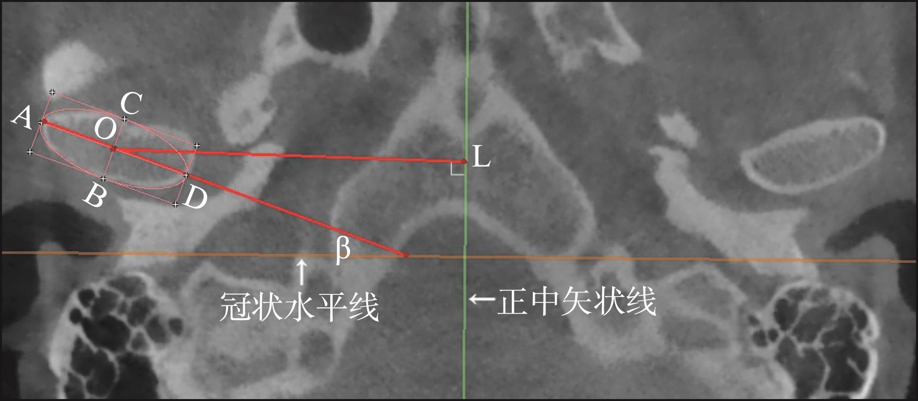
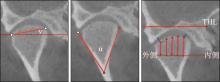
Fig 2
Example of corrected coronal measurement
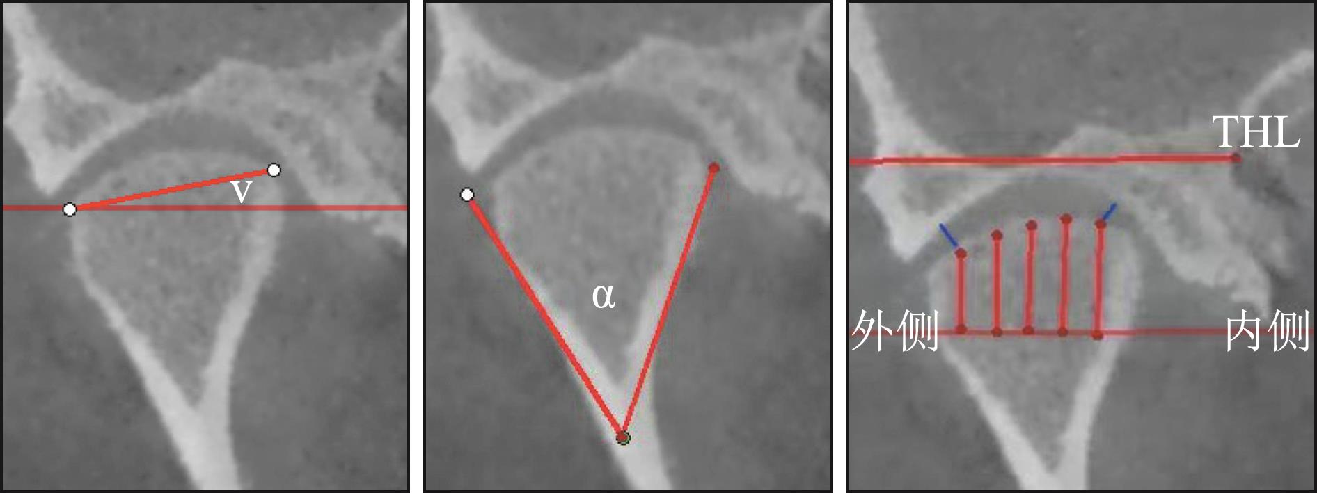
Fig 3
Example of corrected sagittal measurement
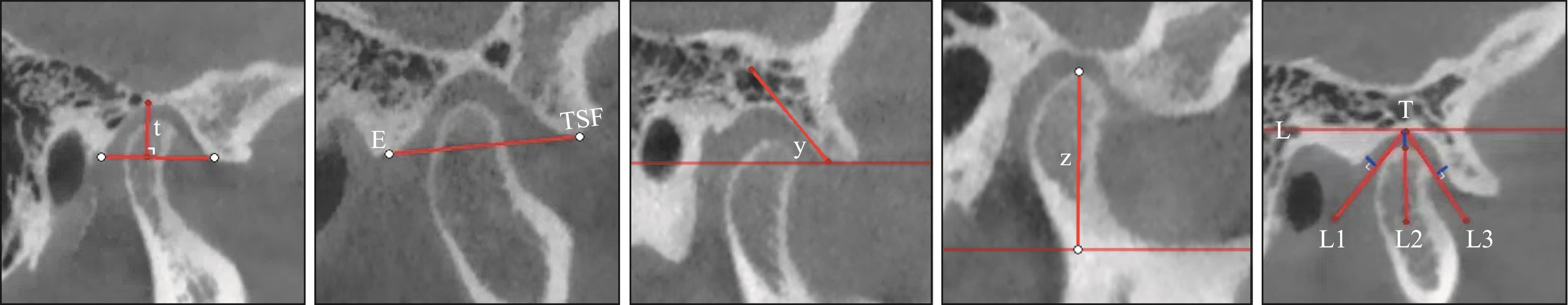
Fig 4
Effect of SS application

Tab 1
Ipsilateral measurement data at initial and follow-up visits in patients without chewing side preference
| 指标 | 右侧 | 左侧 | ||||||
|---|---|---|---|---|---|---|---|---|
| 初诊 | 复诊 | t值 | P值 | 初诊 | 复诊 | t值 | P值 | |
| 髁突内外径/mm | 17.64±3.01 | 18.84±3.08 | -1.463 | 0.154 | 18.39±1.87 | 18.09±2.04 | 0.761 | 0.453 |
| 髁突前后径/mm | 8.13±1.96 | 8.37±2.42 | -0.414 | 0.682 | 8.15±2.60 | 7.89±1.27 | 0.498 | 0.622 |
| 半径值/mm | 54.56±8.35 | 54.33±5.37 | 0.121 | 0.905 | 53.98±8.33 | 54.19±3.20 | -0.131 | 0.897 |
| 髁突水平角/° | 29.49±14.63 | 28.52±11.84 | 0.295 | 0.770 | 29.71±14.66 | 25.83±9.34 | 1.384 | 0.176 |
| 关节内间隙/mm | 2.68±1.26 | 2.78±1.18 | -0.347 | 0.731 | 3.17±1.54 | 2.75±0.93 | 1.354 | 0.186 |
| 关节外间隙/mm | 3.92±2.10 | 3.25±1.24 | 1.473 | 0.151 | 3.55±2.10 | 3.14±1.02 | 0.952 | 0.349 |
| 髁突垂直角/° | 18.33±9.10 | 19.59±9.09 | -0.490 | 0.628 | 18.41±9.11 | 19.66±9.26 | -0.476 | 0.637 |
| 髁突受力角/° | 49.26±14.51 | 53.43±12.49 | -1.450 | 0.157 | 52.11±14.43 | 56.3±7.01 | -1.431 | 0.163 |
| 关节窝宽度/mm | 22.85±4.91 | 23.07±4.38 | -0.178 | 0.860 | 23.09±2.78 | 23.16±2.58 | -0.105 | 0.917 |
| 关节窝深度/mm | 9.12±3.18 | 7.63±3.10 | 1.847 | 0.075 | 8.15±3.09 | 8.38±3.19 | -0.286 | 0.777 |
| 关节结节斜度/° | 54.95±13.94 | 55.33±15.08 | -0.104 | 0.918 | 54.96±13.97 | 51.29±15.06 | 1.028 | 0.312 |
| 关节前间隙/mm | 3.32±1.24 | 2.86±1.43 | 1.233 | 0.227 | 3.32±0.92 | 3.26±0.83 | 0.278 | 0.783 |
| 关节上间隙/mm | 3.34±0.75 | 5.15±1.64 | -5.515 | 0.000** | 4.01±2.09 | 5.06±1.65 | -2.584 | 0.015* |
| 关节后间隙/mm | 2.29±1.44 | 3.36±2.03 | -2.361 | 0.025* | 2.42±1.47 | 3.33±1.62 | -2.176 | 0.038* |
| 髁突高度/mm | 17.86±4.28 | 18.17±4.10 | -1.003 | 0.324 | 16.8±4.70 | 16.8±4.70 | -1.000 | 0.325 |
Tab 2
Bilateral measurement data at initial and follow-up visits in patients without chewing side preference n=31,xˉ±s
| 指标 | 初诊 | 复诊 | |||||
|---|---|---|---|---|---|---|---|
| 均值 | t值 | P值 | 均值 | t值 | P值 | ||
| 髁突内外径/mm | 左侧 | 18.39±1.87 | 1.175 | 0.245 | 18.09±2.04 | -1.121 | 0.267 |
| 右侧 | 17.64±3.01 | 18.84±3.08 | |||||
| 髁突前后径/mm | 左侧 | 8.15±2.60 | 0.031 | 0.975 | 7.89±1.27 | -0.983 | 0.329 |
| 右侧 | 8.13±1.96 | 8.37±2.42 | |||||
| 半径值/mm | 左侧 | 53.98±8.33 | -0.272 | 0.787 | 54.19±3.20 | -0.123 | 0.903 |
| 右侧 | 54.56±8.35 | 54.33±5.37 | |||||
| 髁突水平角/° | 左侧 | 29.71±14.66 | 0.060 | 0.953 | 25.83±9.34 | -0.992 | 0.325 |
| 右侧 | 29.49±14.63 | 28.52±11.84 | |||||
| 关节内间隙/mm | 左侧 | 3.17±1.54 | 1.378 | 0.173 | 2.75±0.93 | -0.105 | 0.917 |
| 右侧 | 2.68±1.26 | 2.78±1.18 | |||||
| 关节外间隙/mm | 左侧 | 3.55±2.10 | -0.686 | 0.496 | 3.14±1.02 | -0.369 | 0.714 |
| 右侧 | 3.92±2.10 | 3.25±1.24 | |||||
| 髁突垂直角/° | 左侧 | 18.41±9.11 | 0.037 | 0.970 | 19.66±9.26 | 0.028 | 0.977 |
| 右侧 | 18.33±9.10 | 19.59±9.09 | |||||
| 髁突受力角/° | 左侧 | 52.11±14.43 | 0.775 | 0.441 | 56.3±7.01 | 1.115 | 0.269 |
| 右侧 | 49.26±14.51 | 53.43±12.49 | |||||
| 关节窝宽度/mm | 左侧 | 23.09±2.78 | 0.243 | 0.809 | 23.16±2.58 | 0.101 | 0.919 |
| 右侧 | 22.85±4.91 | 23.07±4.38 | |||||
| 关节窝深度/mm | 左侧 | 8.15±3.09 | -1.227 | 0.225 | 8.38±3.19 | 0.932 | 0.355 |
| 右侧 | 9.12±3.18 | 7.63±3.10 | |||||
| 关节结节斜度/° | 左侧 | 54.96±13.97 | 0.003 | 0.998 | 51.29±15.06 | -1.053 | 0.297 |
| 右侧 | 54.95±13.94 | 55.33±15.08 | |||||
| 关节前间隙/mm | 左侧 | 3.32±0.92 | 0.027 | 0.979 | 3.26±0.83 | 1.360 | 0.179 |
| 右侧 | 3.32±1.24 | 2.86±1.43 | |||||
| 关节上间隙/mm | 左侧 | 4.01±2.09 | 1.690 | 0.096 | 5.06±1.65 | -0.204 | 0.839 |
| 右侧 | 3.34±0.75 | 5.15±1.64 | |||||
| 关节后间隙/mm | 左侧 | 2.42±1.47 | 0.351 | 0.727 | 3.33±1.62 | -0.064 | 0.949 |
| 右侧 | 2.29±1.44 | 3.36±2.03 | |||||
| 髁突高度/mm | 左侧 | 17.05±4.56 | -0.982 | 0.330 | 17.05±4.56 | -0.983 | 0.329 |
| 右侧 | 18.13±4.06 | 18.13±4.07 | |||||
Tab 3
Habitual and non-habitual side measurement data at initial and follow-up visits in patients with chewing side preference
| 指标 | 习惯侧 | 非习惯侧 | ||||||
|---|---|---|---|---|---|---|---|---|
| 初诊 | 复诊 | t值 | P值 | 初诊 | 复诊 | t值 | P值 | |
| 髁突内外径/mm | 18.41±2.38 | 19.29±2.39 | -2.311 | 0.026* | 18.53±2.43 | 19.12±2.35 | -2.357 | 0.023* |
| 髁突前后径/mm | 7.94±1.47 | 8.32±1.38 | -2.057 | 0.046* | 7.69±1.48 | 7.94±1.09 | -2.092 | 0.043* |
| 半径值/mm | 52.34±4.47 | 53.05±3.09 | -0.938 | 0.353 | 52.89±3.86 | 53.11±4.18 | -0.303 | 0.763 |
| 髁突水平角/° | 25.59±8.14 | 23.13±1.55 | 1.983 | 0.054 | 25.09±8.04 | 23.23±5.79 | 1.435 | 0.159 |
| 关节内间隙/mm | 2.82±0.47 | 3.16±0.85 | -2.300 | 0.026* | 2.53±0.95 | 2.79±1.03 | -2.032 | 0.049* |
| 关节外间隙/mm | 2.79±0.49 | 2.71±0.84 | 0.590 | 0.558 | 2.98±1.00 | 2.63±1.06 | 1.679 | 0.101 |
| 髁突垂直角/° | 10.37±3.41 | 11.66±4.98 | -1.509 | 0.139 | 11.63±2.97 | 11.19±3.16 | 0.706 | 0.484 |
| 髁突受力角/° | 54.09±7.74 | 53.34±7.17 | 0.445 | 0.659 | 56.64±11.81 | 55.42±6.02 | 0.577 | 0.567 |
| 关节窝宽度/mm | 22.76±2.89 | 23.09±3.73 | -0.451 | 0.654 | 22.41±2.66 | 21.97±2.15 | 0.932 | 0.357 |
| 关节窝深度/mm | 10.56±2.82 | 10.38±1.31 | 0.423 | 0.675 | 10.49±1.24 | 10.31±1.13 | 0.723 | 0.473 |
| 关节结节斜度/° | 45.52±6.92 | 46.03±6.69 | -2.567 | 0.014* | 44.86±5.69 | 44.87±6.53 | -0.010 | 0.992 |
| 关节前间隙/mm | 2.33±0.59 | 2.52±0.94 | -1.060 | 0.295 | 2.46±1.25 | 2.42±0.81 | 0.174 | 0.863 |
| 关节上间隙/mm | 3.60±0.67 | 4.14±1.45 | -2.289 | 0.027* | 3.15±1.47 | 3.83±1.12 | -2.226 | 0.031* |
| 关节后间隙/mm | 2.95±1.15 | 3.83±1.78 | -2.480 | 0.017* | 2.83±0.92 | 3.06±0.83 | -2.126 | 0.039* |
| 髁突高度/mm | 18.56±4.92 | 18.58±4.91 | -2.223 | 0.032* | 18.46±2.45 | 18.56±2.46 | 0.510 | 0.613 |
Tab 4
Measurement data of habitual and non-habi-tual sides in patients with chewing side prefe-rence
| 指标 | 位置 | 均值 | t值 | P值 |
|---|---|---|---|---|
| 髁突内外径/mm | 非习惯侧 | 19.64±2.29 | 0.008 | 0.994 |
| 习惯侧 | 19.63±2.08 | |||
| 髁突前后径/mm | 非习惯侧 | 8.13±1.34 | -1.565 | 0.121 |
| 习惯侧 | 8.6±1.44 | |||
| 半径值/mm | 非习惯侧 | 53.11±4.18 | 0.076 | 0.939 |
| 习惯侧 | 53.05±3.09 | |||
| 髁突水平角/° | 非习惯侧 | 23.23±5.79 | 0.107 | 0.915 |
| 习惯侧 | 23.13±1.55 | |||
| 关节内间隙/mm | 非习惯侧 | 2.79±1.03 | -2.195 | 0.031* |
| 习惯侧 | 3.23±0.81 | |||
| 关节外间隙/mm | 非习惯侧 | 2.63±1.06 | -0.391 | 0.697 |
| 习惯侧 | 2.71±0.84 | |||
| 髁突垂直角/° | 非习惯侧 | 11.19±3.16 | -0.521 | 0.603 |
| 习惯侧 | 11.66±4.98 | |||
| 髁突受力角/° | 非习惯侧 | 55.42±6.02 | 1.453 | 0.150 |
| 习惯侧 | 53.34±7.17 | |||
| 关节窝宽度/mm | 非习惯侧 | 21.97±2.15 | -1.713 | 0.090 |
| 习惯侧 | 23.09±3.73 | |||
| 关节窝深度/mm | 非习惯侧 | 10.31±1.13 | -0.247 | 0.805 |
| 习惯侧 | 10.38±1.31 | |||
| 关节结节斜度/° | 非习惯侧 | 44.87±6.53 | -0.948 | 0.346 |
| 习惯侧 | 46.24±6.86 | |||
| 关节前间隙/mm | 非习惯侧 | 2.42±0.81 | -0.514 | 0.609 |
| 习惯侧 | 2.52±0.94 | |||
| 关节上间隙/mm | 非习惯侧 | 4.37±1.17 | 0.632 | 0.529 |
| 习惯侧 | 4.19±1.47 | |||
| 关节后间隙/mm | 非习惯侧 | 3.06±0.83 | -2.596 | 0.011* |
| 习惯侧 | 3.83±1.78 | |||
| 髁突高度/mm | 非习惯侧 | 16.88±2.45 | -2.029 | 0.047* |
| 习惯侧 | 18.58±4.91 |
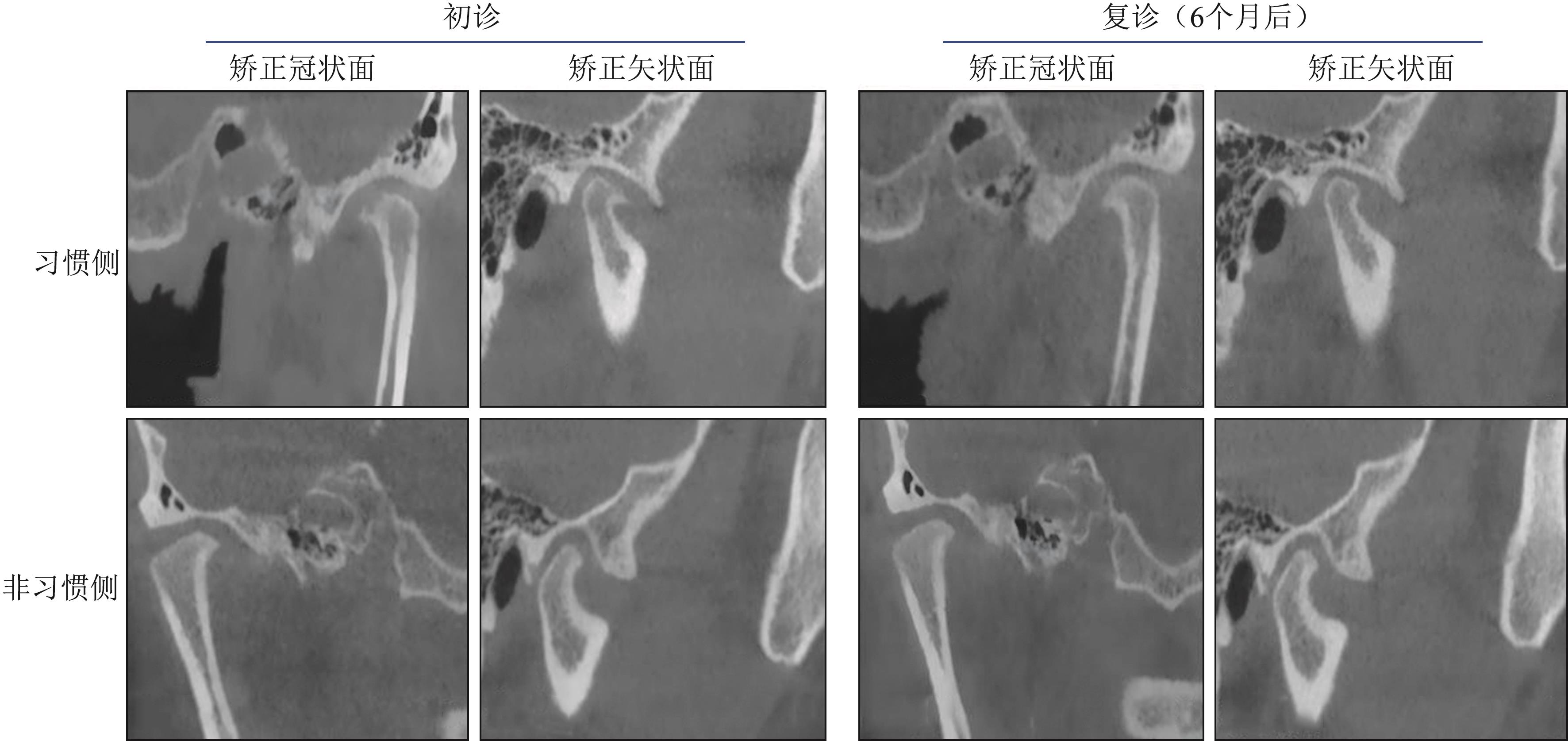
Fig 5
Bony changes of the temporomandibular joint before and after SS treatment
| [1] | Badel T, Zadravec D, Bašić Kes V, et al. Orofacial paindiagnostic and therapeutic challenges[J]. Acta Clin Croat, 2019, 58(): 82-89. |
| [2] | 孟娟红, 甘业华, 马绪臣. 颞下颌关节骨关节炎发病的分子机制及相关治疗的实验研究[J]. 北京大学学报(医学版), 2013, 45(1): 5-8. |
| Meng JH, Gan YH, Ma XC. Molecular mechanisms and related experimental therapeutic studies of temporomandibular joint osteoarthritis[J]. J Peking Univ (Health Sci), 2013, 45(1): 5-8. | |
| [3] | 傅开元, 雷杰. 颞下颌关节紊乱病的分类、诊断及治疗进展[J]. 口腔医学, 2024, 44(1): 6-10. |
| Fu KY, Lei J. Advances in classification, diagnosis and treatment of temporomandibular disorders[J]. Stomatology, 2024, 44(1): 6-10. | |
| [4] | Su N, Liu Y, Yang X, et al. Association of malocclusion, self-reported bruxism and chewing-side preference with oral health-related quality of life in patients with temporomandibular joint osteoarthritis[J]. Int Dent J, 2018, 68(2): 97-104. |
| [5] | Ma J, Wang J, Huang D, et al. A comparative study of condyle position in temporomandibular disorder patients with chewing side preference using cone-beam compu-ted tomography[J]. J Oral Rehabil, 2022, 49(2): 265-271. |
| [6] | 姜华, 刘洪臣. 偏侧咀嚼对口颌系统的影响[J]. 中华老年口腔医学杂志, 2006, 4(4): 235-237, 234. |
| Jiang H, Liu HC. Effects of unilateral mastication on the stomatognathic system[J]. Chin J Geriatr Dent, 2006, 4(4): 235-237, 234. | |
| [7] | Shu JH, Yao J, Zhang YL, et al. The influence of bilate-ral sagittal split ramus osteotomy on the stress distributions in the temporomandibular joints of the patients with facial asymmetry under symmetric occlusions[J]. Medicine (Baltimore), 2018, 97(25): e11204. |
| [8] | Jiang H, Yin H, Wang L, et al. Memory impairment of chewing-side reference mice is associated with 5-HT-BDNF signal pathway[J]. Mol Cell Biochem, 2021, 476(1): 303-310. |
| [9] | Alkhutari AS, Alyahya A, Rodrigues Conti PC, et al. Is the therapeutic effect of occlusal stabilization appliances more than just placebo effect in the management of painful temporomandibular disorders? A network meta-ana-lysis of randomized clinical trials[J]. J Prosthet Dent, 2021, 126(1): 24-32. |
| [10] | Mapelli A, Zanandrea Machado BC, Giglio LD, et al. Reorganization of muscle activity in patients with chro-nic temporomandibular disorders[J]. Arch Oral Biol, 2016, 72(1): 164-171. |
| [11] | Lee YH, Lee KM, Auh QS, et al. Magnetic resonance imaging-based prediction of the relationship between whiplash injury and temporomandibular disorders[J]. Front Neurol, 2017, 8: 725. |
| [12] | Almashraqi AA, Ahmed EA, Mohamed NS, et al. Eva-luation of the effects of chronic qat chewing on lateral pterygoid muscle using MRI[J]. Cranio, 2022, 40(2): 135-143. |
| [13] | Schiffman E, Ohrbach R, Truelove E, et al. Diagnostic criteria for emporomandibular disorders (DC/TMD) for clinical and research applications: recommendations of the International RDC/TMD Consortium Network and Orofacial Pain Special Interest Groupdagger[J]. J Oral Facial Pain Headache, 2014, 28(1): 6-27. |
| [14] | 安靖, 陈洁, 杨文静. 偏侧咀嚼患者颞下颌关节盘位置及临床症状的影响[J]. 口腔颌面修复学杂志, 2014, 15(5): 285-288. |
| An J, Chen J, Yang WJ. Effects of unilateral mastication on TMJ disc position and clinical symptoms[J]. Chin J Prosthodont, 2014, 15(5): 285-288. | |
| [15] | 傅开元, 胡敏, 余强, 等. 颞下颌关节紊乱病锥形束CT检查规范及诊断标准的专家共识[J]. 中华口腔医学杂志, 2020, 55(9): 613-616. |
| Fu KY, Hu M, Yu Q, et al. Experts consensus on cone-beam CT examination specification and diagnostic criteria of temporomandibular disorders[J]. Chin J Stomatol, 2020, 55(9): 613-616. | |
| [16] | Kazazoglu E, Heath MR, Müller F. A simple test for determination of the preferred chewing side[J]. J Oral Rehabil, 1994, 21(6): 723. |
| [17] | Diernberger S, Bernhardt O, Schwahn C, et al. Self-reported chewing side preference and its associations with occlusal, temporomandibular and prosthodontic factors: results from the population-based Study of Health in Pomerania (SHIP-0) [J]. J Oral Rehabil, 2008, 35(8): 613-620. |
| [18] | Ikeda K, Kawamura A. Assessment of optimal condylar position with limited cone-beam computed tomography[J]. Am J Orthod Dentofacial Orthop, 2009, 135(4): 495-501. |
| [19] | Kamelchuk LS, Grace MG, Major PW. Post-imaging temporomandibular joint space analysis[J]. Cranio, 1996, 14(1): 23-29. |
| [20] | 陈鹏, 张晓娟, 魏博, 等. 关节穿刺术应用于老年颞下颌关节不可复性盘前移位的临床研究[J]. 中华老年口腔医学杂志, 2022, 20(3): 162-166. |
| Chen P, Zhang XJ, Wei B, et al. Clinical study of the application of arthrocentesis in the elderly temporomandi-bular joint with irreversible anterior[J]. Chin J Geriatr Dent, 2022, 20(3): 162-166. | |
| [21] | 姜婷, 李健, 许桐楷, 等. 咬合板的临床应用特点[J]. 口腔颌面修复学杂志, 2022, 23(1): 1-10. |
| Jiang T, Li J, Xu TK, et al. Clinical application of occlusal splints[J]. Chin J Prosthodont, 2022, 23(1): 1-10. | |
| [22] | 胡敏. 颞下颌关节骨关节炎: 认识与挑战[J]. 中华口腔医学杂志, 2022, 57(7): 665-673. |
| Hu M. Temporomandibular joint osteoarthritis: understanding and challenges[J]. Chin J Stomatol, 2022, 57(7): 665-673. | |
| [23] | Song H, Lee JY, Huh KH, et al. Long-term changes of temporomandibular joint osteoarthritis on computed tomography[J]. Sci Rep, 2020, 10(1): 6731. |
| [24] | 李晓星, 马博文, 徐鑫宇, 等. 伴偏侧咀嚼颞下颌关节骨关节炎患者1年随访的CBCT研究[J]. 口腔颌面修复学杂志, 2025, 26(1): 42-49. |
| Li XX, Ma BW, Xu XY, et al. CBCT study of patients with temporomandibular joint osteoarthritis accompanied by lateral chewing for 1 year follow-up[J]. Chin J Prosthodont, 2025, 26(1): 42-49. | |
| [25] | Zhang Y, Zhang H, Liu R, et al. The efficacy of treatments for temporomandibular disorders with occlusal splints versus other conservative therapies: a meta-analysis of randomized controlled trials[J]. Oral Surg Oral Med Oral Pathol Oral Radiol, 2025, 139(5): 509-520. |
| [26] | Shu J, Xiong X, Chong DY, et al. The relations between the stress in temporomandibular joints and the deviated distances for mandibular asymmetric patients[J]. Proc Inst Mech Eng H, 2021, 235(1): 109-116. |
| [27] | Silva MAG, Pantoja LLQ, Dutra-Horstmann KL, et al. Prevalence of degenerative disease in temporomandibular disorder patients with disc displacement: a systema-tic review and meta-analysis[J]. J Craniomaxillofac Surg, 2020, 48(10): 942-955. |
| [28] | Derwich M, Mitus-Kenig M, Pawlowska E. Temporomandibular joints’ morphology and osteoarthritic chan-ges in cone-beam computed tomography images in patients with and without reciprocal clicking—A case control study[J]. Int J Environ Res Public Health, 2020, 17(10): 3428. |
| [29] | Poluha RL, Cunha CO, Bonjardim LR, et al. Temporomandibular joint morphology does not influence the presence of arthralgia in patients with disk displacement with reduction: a magnetic resonance imaging-based stu-dy[J]. Oral Surg Oral Med Oral Pathol Oral Radiol, 2020, 129(2): 149-157. |
| [30] | Pinto GNS, Grossmann E, Iwaki Filho L, et al. Correlation between joint effusion and morphology of the articular disc within the temporomandibular joint as viewed in the sagittal plane in patients with chronic disc displacement with reduction: a retrospective analytical study from magnetic resonance imaging[J]. Cranio, 2021, 39(2): 119-124. |
| [31] | Gaggl A, Enzinger S, Bürger H, et al. The condylar capping for microvascular temporomandibular joint reconstruction: a preliminary study[J]. Microsurgery, 2023, 43(4): 331-338. |
| [32] | Liu YS, Yap AU, Lei J, et al. Association between hypoplastic condyles and temporomandibular joint disc displacements: a cone beam computed tomography and magnetic resonance imaging metrical analysis[J]. Int J Oral Maxillofac Surg, 2020, 49(7): 932-939. |
| [33] | Abdelrehem A, Hu YK, Yang C, et al. Arthroscopic versus open disc repositioning and suturing techniques for the treatment of temporomandibular joint anterior disc displacement: 3-year follow-up study[J]. Int J Oral Maxillofac Surg, 2021, 50(10): 1351-1360. |
| [34] | Ok SM, Jeong SH, Ahn YW, et al. Effect of stabilization splint therapy on glenoid fossa remodeling in temporomandibular joint osteoarthritis[J]. J Prosthodont Res, 2016, 60(4): 301-307. |
| [35] | Bedran LM, Dos Santos AASMD. Changes in temporomandibular joint anatomy, changes in condylar translation, and their relationship with disc displacement: magnetic resonance imaging study[J]. Radiol Bras, 2019, 52(2): 85-91. |
| [1] | Fu Chengyi, Pan Xiao, Liu Shu, Lin Zitong. Root fracture in endodontically versus nonendodontically treated posterior teeth: the cone beam computed tomography characteristics in Chinese population [J]. West China Journal of Stomatology, 2026, 44(1): 113-119. |
| [2] | Xu Laijun, Zhang Jianying, Huang Zihua, Ou Yuemei, Wang Xiangzhu. Microscopic root canal treatment of fused mandibular molar with seven root canals: a case report [J]. West China Journal of Stomatology, 2025, 43(3): 431-435. |
| [3] | Feng Bo, Li Fengxiang, Du Qitao. Analysis on the relationship between Schneiderian membrane thickening in the posterior maxillary region and periapical lesions by cone beam computed tomography [J]. West China Journal of Stomatology, 2024, 42(5): 644-651. |
| [4] | Yang Hu, Shi Ruiwen, Liu Yue, Shi Yilin, Zhang Shengben, Lan Jing. Analysis of factors affecting bone volume changes after immediate implantation in the maxillary central incisor [J]. West China Journal of Stomatology, 2024, 42(5): 660-666. |
| [5] | Zhu Jie, Liu Ya. Morphological study of distal furcation in mandibular first molars based on cone beam computed tomography images in Beijing population [J]. West China Journal of Stomatology, 2024, 42(3): 329-333. |
| [6] | Chen Hong, Zhang Ronghua, Zhao Yuan. Non-surgical treatment of maxillary lateral incisor double dens invaginatus type Ⅲ with apical periodontitis [J]. West China Journal of Stomatology, 2024, 42(3): 409-414. |
| [7] | Li Chengxi, Song Weijian.. Root canal treatment of type Ⅱ and ⅢA double dens invaginatus in maxillary lateral incisor: a case report [J]. West China Journal of Stomatology, 2023, 41(2): 232-236. |
| [8] | Yuan Jing, Yu Sijing, You Meng, Zhang Qiong, Ye Ling, Gao Bo. Regenerative endodontic treatment of dens in dente in maxillary lateral incisor with immature root: a case report [J]. West China Journal of Stomatology, 2022, 40(6): 716-720. |
| [9] | Zhang Qiao, Huang Dongzong, Zhai Xiaoting, Li Hongbo, Hu Min, Liu Hongchen, Jiang Hua. Occlusal analysis of patients with chewing side preference and symptoms of temporomandibular disorders [J]. West China Journal of Stomatology, 2022, 40(4): 457-467. |
| [10] | Cai Pingping, Chen Xi, Jiang Yi, Lu Zhaojie, Lin Jie, Zheng Zhiqiang.. Comparing accuracy after guide access and microscope-assisted access for fiber post removal [J]. West China Journal of Stomatology, 2022, 40(3): 297-302. |
| [11] | Zhang Shuai, Wang Jianghong, Tian Lijie, Wang Baoli, Zhang Juan. Effect of 17β-estradiol on the proliferation of condylar chondrocytes [J]. West China Journal of Stomatology, 2021, 39(6): 651-657. |
| [12] | Yuan Zhiyao, Zou Xihong, Dai Linlin, Ao Huizhi, Li Houxuan.. Clinical analysis on the root fracture of the maxillary first molar [J]. West China Journal of Stomatology, 2021, 39(5): 555-559. |
| [13] | Xiao Mian, Hu Zhihui, Jiang Henghua, Fang Wei, Long Xing. Role of osteoclast differentiation in the occurrence of osteoarthritis of temporomandibular joint [J]. West China Journal of Stomatology, 2021, 39(4): 398-404. |
| [14] | Guo Meiling, Huang Zhen, Wang Chong, Wang Yujiang. Effect of bilateral sagittal split ramus osteotomy on temporomandibular joint symptom and condylar position in patients with skeletal class Ⅲ malocclusion by cone beam computed tomography [J]. West China Journal of Stomatology, 2020, 38(5): 519-524. |
| [15] | Zhang Ting, Chen Du, Miao Leiying, Xie Sijing, Tang Xuna. Guided endodontic access of calcified root canal by laser melting templates [J]. West China Journal of Stomatology, 2020, 38(5): 525-531. |
| Viewed | ||||||
|
Full text |
|
|||||
|
Abstract |
|
|||||
 This work is licensed under a Creative Commons Attribution 3.0 License.
