West China Journal of Stomatology ›› 2026, Vol. 44 ›› Issue (1): 54-64.doi: 10.7518/hxkq.2025.2025316
Previous Articles Next Articles
Hong Xinyu( ), Zhang Wenxuan, Qi Siyao, Yang Lan, Shen Wenxin, Liu Jian()
Received:2025-08-01
Online:2026-02-01
Published:2026-02-02
Contact:
Liu Jian
E-mail:306364947@qq.com;1052604322@qq.com
CLC Number:
Hong Xinyu, Zhang Wenxuan, Qi Siyao, Yang Lan, Shen Wenxin, Liu Jian. Follow-up study evaluating morphological changes in alveolar bone in the maxillary anterior region after extraction retraction in adult patients treated with clear aligners on the basis of cone beam computed tomography data[J]. West China Journal of Stomatology, 2026, 44(1): 54-64.
Add to citation manager EndNote|Ris|BibTeX
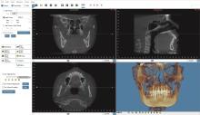
Fig 1
CBCT 3D reconstruction

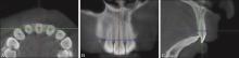
Fig 2
Acquisition of measurement plane
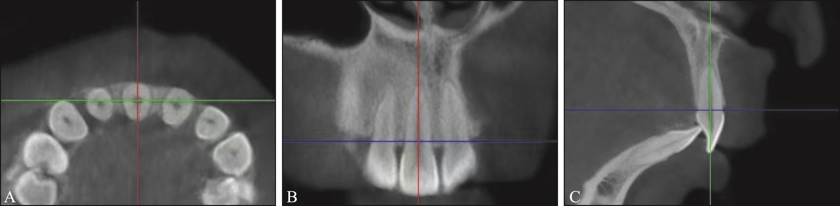
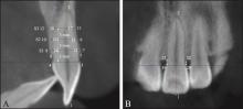
Fig 3
Sagittally and coronally measurement landmarks
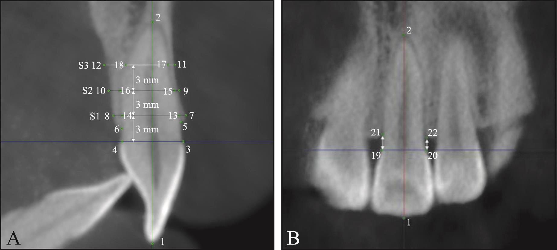
Fig 4
Preliminary registration of T1 and T2 3D images

Fig 5
Voxel-based overlap
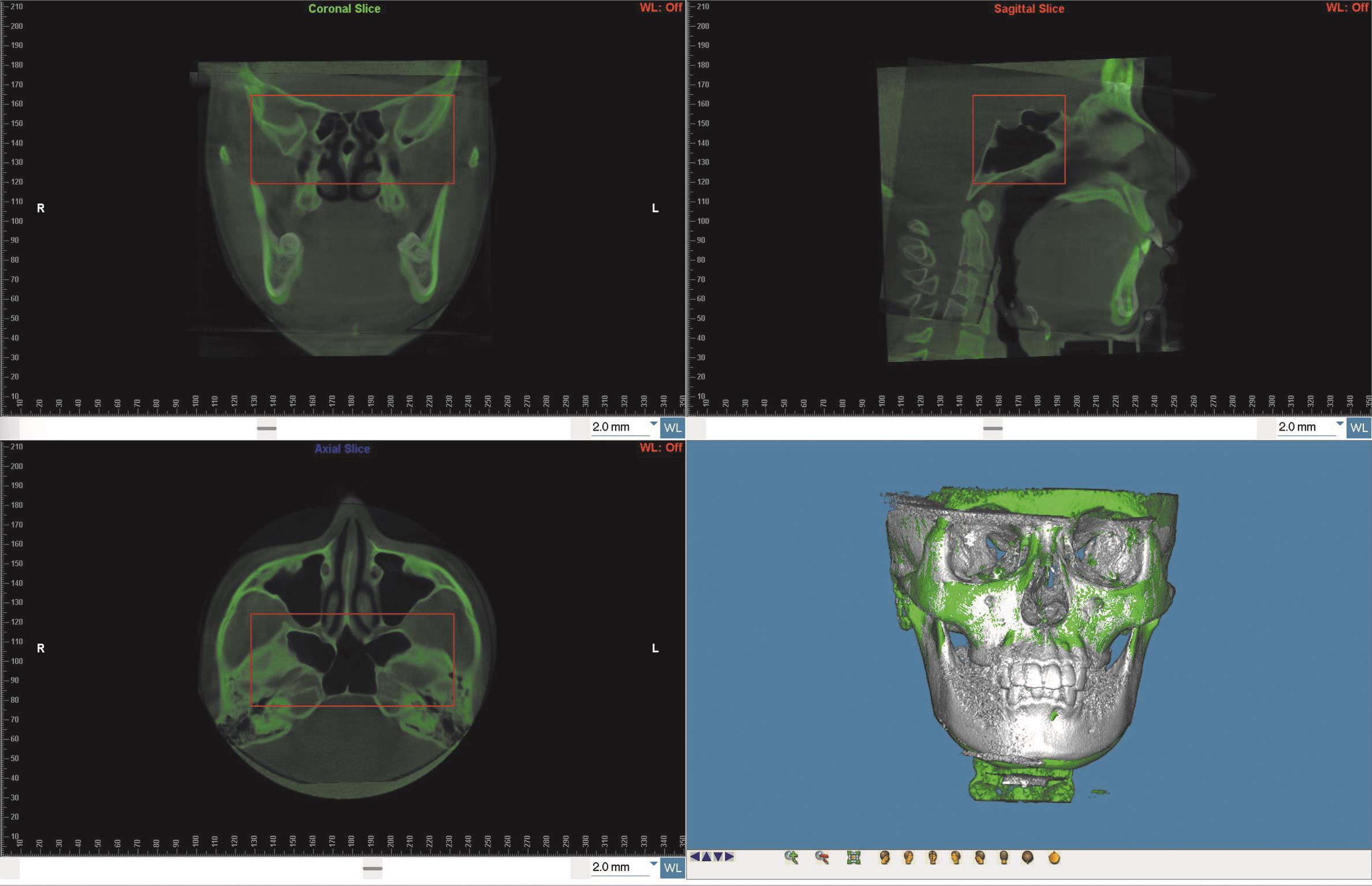
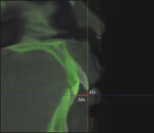
Fig 6
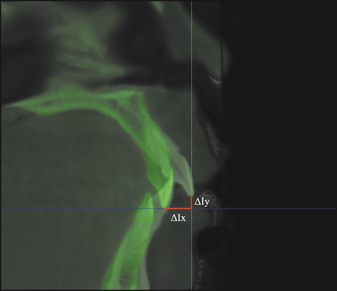
Measurement of tooth movement
Tab 1
Comparison of alveolar bone height in the maxillary anterior region pre-treatment, post-treatment, and du-ring the retention phase
| 测量项目 | T1 | T2 | T3 | P值 | ||||
|---|---|---|---|---|---|---|---|---|
| T2和T1 | T3和T2 | T3和T1 | Overall | |||||
| 上颌中切牙 | LVBL | 1.92±0.68 | 2.48±0.83 | 2.12±0.82 | 0.002b* | 0.012b* | 0.180b | 0.000b* |
| PVBL | 1.51±0.56 | 2.27±0.71 | 1.81±0.58 | 0.000b* | 0.003b* | 0.636b | 0.000b* | |
| MVBL | 1.59±0.44 | 2.23±0.87 | 1.63±0.41 | 0.023a* | 0.056a | 1.000a | 0.032a* | |
| DVBL | 1.80±0.50 | 2.08±0.47 | 2.00±0.50 | 0.186a | 1.000a | 0.522a | 0.165a | |
| 上颌侧切牙 | LVBL | 2.20±0.87 | 2.05±0.80 | 2.20±0.83 | - | - | - | 0.964b |
| PVBL | 1.42±0.37 | 1.85±0.55 | 1.76±0.50 | 0.002a* | 1.000a | 0.005a* | 0.002a* | |
| MVBL | 1.73±0.61 | 2.14±0.58 | 2.01±0.64 | 0.000b* | 0.045b* | 0.045b* | 0.000b* | |
| DVBL | 2.24±0.55 | 2.26±0.66 | 2.15±0.63 | - | - | - | 0.218b | |
| 上颌尖牙 | LVBL | 2.13±0.87 | 2.14±0.72 | 2.19±0.82 | 1.000a | 1.000a | 1.000a | 0.863a |
| PVBL | 2.10±0.98 | 2.97±1.38 | 2.83±1.36 | 0.029b* | 1.000b | 0.043b* | 0.015b* | |
| MVBL | 1.68±0.84 | 2.06±0.77 | 2.01±0.57 | 0.001b* | 1.000b | 0.001b* | 0.000b* | |
| DVBL | 1.95±0.59 | 2.19±0.80 | 2.07±0.79 | 0.218a | 0.781a | 0.827a | 0.124a | |
Tab 2
Comparison of alveolar bone thickness in the maxillary anterior region pre-treatment, post-treatment, and during the retention phase
| 测量项目 | T1 | T2 | T3 | P值 | ||||
|---|---|---|---|---|---|---|---|---|
| T2和T1 | T3和T2 | T3和T1 | Overall | |||||
| 上颌中切牙 | LTBT1 | 1.00±0.47 | 1.27±0.80 | 1.37±0.75 | 0.054a | 1.000a | 0.030a* | 0.011a |
| LTBT2 | 0.92±0.21 | 1.47±0.45 | 1.46±0.64 | 0.000b* | 0.304b | 0.002b* | 0.000b* | |
| LTBT3 | 1.06±0.32 | 1.68±0.76 | 1.60±0.85 | 0.011b* | 1.000b | 0.022b* | 0.005b* | |
| PTBT1 | 1.20±0.44 | 1.03±0.53 | 1.29±0.56 | - | - | - | 0.134b | |
| PTBT2 | 2.43±0.51 | 2.18±0.83 | 2.43±0.83 | - | - | - | 0.086b | |
| PTBT3 | 4.06±1.03 | 4.60±1.45 | 4.52±1.47 | 0.652a | 1.000a | 0.870a | 0.066a | |
| TBT1 | 8.44±0.38 | 8.90±0.66 | 9.00±0.70 | 0.015a* | 1.000a | 0.008a* | 0.001a* | |
| TBT2 | 8.82±0.66 | 9.41±0.88 | 9.37±0.96 | 0.002a* | 1.000a | 0.002a* | 0.000a | |
| TBT3 | 9.20±1.33 | 10.50±1.73 | 10.20±1.64 | 0.000a* | 0.089a | 0.000a* | 0.000a* | |
| 上颌侧切牙 | LTBT1 | 0.90±0.61 | 1.40±0.75 | 1.18±0.76 | 0.007b* | 0.066b | 0.221b | 0.007b |
| LTBT2 | 1.09±0.48 | 1.38±0.64 | 1.38±0.83 | 0.052b | 1.000b | 0.267b | 0.048b* | |
| LTBT3 | 0.92±0.86 | 1.19±0.77 | 1.15±1.03 | 0.037b* | 0.055b | 1.000b | 0.423b | |
| PTBT1 | 1.46±0.61 | 1.06±0.63 | 0.98±0.37 | 0.005b* | 1.000b | 0.002b* | 0.001b | |
| PTBT2 | 2.61±1.23 | 1.99±1.05 | 1.92±0.91 | 0.014a* | 1.000a | 0.006a* | 0.003a* | |
| PTBT3 | 4.17±2.05 | 3.97±2.00 | 3.85±1.98 | - | - | - | 0.935b | |
| TBT1 | 8.17±0.91 | 8.13±0.90 | 7.95±0.75 | - | - | - | 0.368b | |
| TBT2 | 8.74±1.60 | 8.31±1.25 | 8.24±1.04 | - | - | - | 0.576b | |
| TBT3 | 8.88±2.18 | 8.91±2.01 | 8.69±2.04 | - | - | - | 0.340b | |
| 上颌尖牙 | LTBT1 | 1.00±0.55 | 1.19±0.70 | 1.16±0.73 | 0.064a | 1.000a | 0.246a | 0.054a |
| LTBT2 | 1.02±0.32 | 1.01±0.31 | 1.12±0.40 | 1.000a | 0.112a | 0.137a | 0.048a* | |
| LTBT3 | 0.75±0.30 | 0.75±0.30 | 0.80±0.32 | - | - | - | 0.663b | |
| PTBT1 | 1.11±0.59 | 0.58±0.54 | 0.54±0.48 | 0.003b* | 1.000b | 0.001b* | 0.000b* | |
| PTBT2 | 2.26±0.72 | 1.81±0.76 | 1.66±0.63 | 0.007b* | 1.000b | 0.000b* | 0.000b* | |
| PTBT3 | 3.95±1.26 | 3.29±0.94 | 3.16±0.95 | 0.005a* | 0.957a | 0.004a* | 0.004a* | |
| TBT1 | 9.42±1.01 | 9.30±0.96 | 9.17±1.16 | 0.951a | 0.714a | 0.136a | 0.104a | |
| TBT2 | 9.86±1.23 | 9.50±1.25 | 9.43±1.10 | 0.024a* | 1.000a | 0.017a* | 0.004a* | |
| TBT3 | 10.12±1.36 | 9.46±1.35 | 9.41±1.35 | 0.001a* | 1.000a | 0.004a* | 0.002a* | |
Tab 3
Correlation of anterior alveolar bone height changes between treatment and retention pha-ses
| 测量项目 | T2-T1 | T3-T2 | r值 | P值 | |
|---|---|---|---|---|---|
| 上颌中切牙 | LVBL | 0.56±0.75 | -0.36±0.58 | -0.597b | 0.001* |
| PVBL | 0.76±0.70 | -0.46±0.57 | -0.407b | 0.039* | |
| MVBL | 0.64±0.94 | -0.60±1.01 | -0.863b | <0.001* | |
| DVBL | 0.28±0.61 | -0.08±0.70 | -0.578a | 0.010* | |
| 上颌侧切牙 | LVBL | -0.25±0.78 | 0.17±0.62 | -0.254a | 0.200 |
| PVBL | 0.43±0.60 | -0.09±0.71 | -0.691a | <0.001* | |
| MVBL | 0.41±0.55 | -0.12±0.42 | -0.142a | 0.472 | |
| DVBL | 0.03±0.57 | -0.11±0.35 | -0.154a | 0.433 | |
| 上颌尖牙 | LVBL | 0.01±0.60 | 0.05±0.69 | -0.598a | <0.001* |
| PVBL | 0.87±1.50 | -0.15±1.13 | -0.298a | 0.110 | |
| MVBL | 0.38±0.49 | -0.05±0.64 | 0.031a | 0.873 | |
| DVBL | 0.24±0.70 | -0.12±0.55 | -0.564a | 0.001* | |
Tab 4
Correlation of anterior alveolar bone thickness changes between treatment and retention pha-ses
| 测量项目 | T2-T1 | T3-T2 | r值 | P值 | |
|---|---|---|---|---|---|
| 上颌中切牙 | LTBT1 | 0.27±0.50 | 0.11±0.54 | -0.378b | 0.100b |
| LTBT2 | 0.55±0.47 | -0.01±0.40 | 0.043a | 0.856 | |
| LTBT3 | 0.62±0.90 | -0.08±0.53 | 0.078b | 0.751 | |
| PTBT1 | -0.30±0.32 | 0.29±0.44 | -0.106b | 0.719 | |
| PTBT2 | -0.25±0.65 | 0.25±0.53 | -0.558a | 0.011* | |
| PTBT3 | 0.54±0.91 | -0.08±0.48 | -0.409a | 0.082 | |
| TBT1 | 0.45±0.64 | 0.10±0.50 | -0.215a | 0.362 | |
| TBT2 | 0.59±0.65 | -0.04±0.52 | -0.503a | 0.024* | |
| TBT3 | 1.30±1.09 | -0.30±0.56 | -0.720a | 0.001* | |
| 上颌侧切牙 | LTBT1 | 0.50±0.76 | -0.22±0.46 | -0.254a | 0.202 |
| LTBT2 | 0.29±0.52 | 0.00±0.41 | -0.175a | 0.384 | |
| LTBT3 | 0.27±0.84 | -0.04±0.60 | -0.209a | 0.338 | |
| PTBT1 | -0.43±0.75 | -0.08±0.68 | -0.553b | 0.002* | |
| PTBT2 | -0.62±1.30 | -0.07±0.65 | -0.446b | 0.015* | |
| PTBT3 | -0.20±2.13 | -0.12±0.87 | -0.452a | 0.023* | |
| TBT1 | -0.04±1.06 | -0.18±0.72 | -0.454b | 0.015* | |
| TBT2 | -0.43±1.50 | -0.08±0.74 | -0.422a | 0.023* | |
| TBT3 | -0.24±1.89 | -0.22±0.98 | -0.368a | 0.070 | |
| 上颌尖牙 | LTBT1 | 0.19±0.41 | -0.03±0.42 | -0.464b | 0.011* |
| LTBT2 | -0.01±0.26 | 0.11±0.29 | -0.485a | 0.007* | |
| LTBT3 | 0.00±0.40 | 0.04±0.35 | -0.331a | 0.074 | |
| PTBT1 | -0.53±0.81 | 0.04±0.55 | -0.460a | 0.012* | |
| PTBT2 | -0.45±0.81 | 0.16±0.53 | -0.318a | 0.087 | |
| PTBT3 | -0.65±1.04 | -0.13±0.73 | -0.127b | 0.503 | |
| TBT1 | -0.12±0.64 | -0.36±0.69 | -0.471a | 0.009* | |
| TBT2 | -0.36±0.70 | 0.66±0.88 | -0.653a | <0.001* | |
| TBT3 | -0.66±0.88 | -0.05±0.66 | -0.045a | 0.814 | |
Tab 5
Correlation between retraction amount of maxillary central incisors and changes in alveolar bone height and thickness of anterior teeth during T2-T1 and T3-T2 periods
| 测量项目 | T2-T1 | T3-T2 | r1值 | P1值 | r2值 | P2值 |
|---|---|---|---|---|---|---|
| LVBL | 0.56±0.75 | -0.36±0.58 | 0.221b | 0.349 | 0.422b | 0.064 |
| PVBL | 0.76±0.70 | -0.46±0.57 | 0.091a | 0.702 | -0.160a | 0.500 |
| MVBL | 0.64±0.94 | -0.60±1.01 | -0.182b | 0.457 | 0.412a | 0.080 |
| DVBL | 0.28±0.61 | -0.08±0.70 | 0.096a | 0.694 | 0.462b | 0.030* |
| LTBT1 | 0.27±0.50 | 0.11±0.54 | -0.228a | 0.333 | -0.299b | 0.201 |
| LTBT2 | 0.55±0.47 | -0.01±0.40 | -0.350a | 0.130 | -0.151a | 0.524 |
| LTBT3 | 0.62±0.90 | -0.08±0.53 | -0.205b | 0.399 | 0.179a | 0.463 |
| PTBT1 | -0.30±0.32 | 0.29±0.44 | -0.128b | 0.664 | 0.280a | 0.333 |
| PTBT2 | -0.25±0.65 | 0.25±0.53 | 0.294a | 0.208 | -0.228b | 0.334 |
| PTBT3 | 0.54±0.91 | -0.08±0.48 | 0.682a | 0.001* | 0.005a | 0.983 |
| TBT1 | 0.45±0.64 | 0.10±0.50 | -0.333a | 0.152 | 0.314a | 0.178 |
| TBT2 | 0.59±0.65 | -0.04±0.52 | 0.060a | 0.801 | -0.307a | 0.188 |
| TBT3 | 1.30±1.09 | -0.30±0.56 | 0.043a | 0.861 | -0.109a | 0.657 |
Tab 6
Correlation between intrusion amount of maxillary central incisors and changes in alveolar bone height and thickness of anterior teeth during T2-T1 and T3-T2 periods
| 测量项目 | T2-T1 | T3-T2 | r1值 | P1值 | r2值 | P2值 |
|---|---|---|---|---|---|---|
| LVBL | 0.56±0.75 | -0.36±0.58 | 0.077b | 0.748 | 0.499b | 0.010* |
| PVBL | 0.76±0.70 | -0.46±0.57 | -0.074a | 0.755 | -0.114b | 0.633 |
| MVBL | 0.64±0.94 | -0.60±1.01 | -0.064b | 0.796 | -0.076b | 0.759 |
| DVBL | 0.28±0.61 | -0.08±0.70 | 0.063b | 0.792 | -0.037b | 0.878 |
| LTBT1 | 0.27±0.50 | 0.11±0.54 | -0.182b | 0.442 | -0.029b | 0.902 |
| LTBT2 | 0.55±0.47 | -0.01±0.40 | -0.341b | 0.142 | -0.076b | 0.750 |
| LTBT3 | 0.62±0.90 | -0.08±0.53 | -0.642b | 0.003* | -0.195b | 0.424 |
| PTBT1 | -0.30±0.32 | 0.29±0.44 | -0.135b | 0.644 | -0.611b | 0.020* |
| PTBT2 | -0.25±0.65 | 0.25±0.53 | 0.010b | 0.967 | 0.028b | 0.907 |
| PTBT3 | 0.54±0.91 | -0.08±0.48 | 0.080b | 0.745 | -0.015b | 0.950 |
| TBT1 | 0.45±0.64 | 0.10±0.50 | -0.197b | 0.469 | 0.222b | 0.927 |
| TBT2 | 0.59±0.65 | -0.04±0.52 | -0.267b | 0.255 | 0.092b | 0.700 |
| TBT3 | 1.30±1.09 | -0.30±0.56 | -0.040b | 0.870 | 0.031b | 0.901 |
| [1] | Xu Y, Xie J. Comparison of the effects of mini-implant and traditional anchorage on patients with maxillary dentoalveolar protrusion[J]. Angle Orthod, 2017, 87(2): 320-327. |
| [2] | Hahn W, Dathe H, Fialka-Fricke J, et al. Influence of thermoplastic appliance thickness on the magnitude of force delivered to a maxillary central incisor during tipping[J]. Am J Orthod Dentofacial Orthop, 2009, 136(1): 12.e1-7; discussion 12-13. |
| [3] | Lu H, Tang H, Zhou T, et al. Assessment of the periodontal health status in patients undergoing orthodontic treatment with fixed appliances and Invisalign system: a meta-analysis[J]. Medicine (Baltimore), 2018, 97(13): e0248. |
| [4] | 张瑞洁, 郭子煜, 秦文, 等. 无托槽隐形矫治器矫治错𬌗畸形与牙槽骨缺损发生的研究进展[J]. 实用口腔医学杂志, 2023, 39(4): 527-531. |
| Zhang RJ, Guo ZY, Qin W, et al. Progress on clear alig-ners in the treatment of malocclusion and the occurrence of alveolar bone defects (translation)[J]. J Pract Stomatol, 2023, 39(4): 527-531. | |
| [5] | 齐娟, 李淑芳. 无托槽隐形矫治器与固定矫治器在上颌前突拔牙病例中的疗效对比[J]. 临床口腔医学杂志, 2023, 39(3): 163-167. |
| Qi J, Li SF. Retrospective clinical study on treatment effectiveness with clear aligners and fixed appliances in extraction cases[J]. J Clin Stomatol, 2023, 39(3): 163-167. | |
| [6] | 王宇. 成人患者拔牙内收后前牙区牙槽骨变化的CBCT追踪研究[D]. 南昌: 南昌大学, 2021. |
| Wang Y. Follow-up study of changes in the anterior al-veolar bone in adult patients after retraction of anterior teeth with cone beam computed tomography[D]. Nanchang: Nanchang University, 2021. | |
| [7] | 谢飘. 青少年双颌前突患者拔牙矫治后及随访期前牙区牙槽骨变化的研究[D]. 南昌: 南昌大学, 2022. |
| Xie P. The study of anterior alveolar bone change in adolescent patients with bimaxillary protrusion involving tooth extraction after orthodontic treatment and retention phase[D]. Nanchang: Nanchang University, 2022. | |
| [8] | 王宇, 谢飘, 沈涛, 等. 成年双颌前突患者减数矫治后前牙区牙槽骨变化的锥形束CT分析[J]. 中华口腔医学杂志, 2023, 58(2): 143-150. |
| Wang Y, Xie P, Shen T, et al. Analysis of morphometric changes in the anterior alveolar bone in bimaxillary protrusion adult patients after retraction with cone-beam CT[J]. Chin J Stomatol, 2023, 58(2): 143-150. | |
| [9] | Bazina M, Cevidanes L, Ruellas A, et al. Precision and reliability of Dolphin 3-dimensional voxel-based superimposition[J]. Am J Orthod Dentofacial Orthop, 2018, 153(4): 599-606. |
| [10] | Guo R, Zhang L, Hu M, et al. Alveolar bone changes in maxillary and mandibular anterior teeth during orthodontic treatment: a systematic review and meta-analysis[J]. Orthod Craniofac Res, 2021, 24(2): 165-179. |
| [11] | 杨安迪, 毛慧敏, 雷浪. 成人双颌前突患者正畸前后上颌切牙区唇腭侧牙槽骨的变化[J]. 口腔医学研究, 2021, 37(1): 48-52. |
| Yang AD, Mao HM, Lei L. Changes of labial-palatal alveolar bone in maxillary incisor region in adult patients with bimaxillary protrusion before and after orthodontic treatment[J]. J Oral Sci Res, 2021, 37(1): 48-52. | |
| [12] | 唐娜, 赵志河, 王军, 等. 无托槽隐形矫治技术生物力学效应的有限元法研究[J]. 医用生物力学, 2010, 25(6): 399-405. |
| Tang N, Zhao ZH, Wang J, et al. Biomechanical effects of bracketless appliance technology: a finite element me-thod study[J]. J Med Biomech, 2010, 25(6): 399-405. | |
| [13] | Elhaddaoui R, Qoraich HS, Bahije L, et al. Orthodontic aligners and root resorption: a systematic review[J]. Int Orthod, 2017, 15(1): 1-12. |
| [14] | Jiang T, Wang JK, Jiang YY, et al. How well do integra-ted 3D models predict alveolar defects after treatment with clear aligners[J]. Angle Orthod, 2021, 91(3): 313-319. |
| [15] | Mulie RM, Hoeve AT. The limitations of tooth movement within the symphysis, studied with laminagraphy and standardized occlusal films[J]. J Clin Orthod, 1976, 10(12): 882-893. |
| [16] | Sarikaya S, Haydar B, Ciğer S, et al. Changes in alveolar bone thickness due to retraction of anterior teeth[J]. Am J Orthod Dentofacial Orthop, 2002, 122(1): 15-26. |
| [17] | Guo R, Li L, Lin Y, et al. Long-term bone remodeling of maxillary anterior teeth with post-treatment alveolar bone defect in adult patients with maxillary protrusion: a prospective follow-up study[J]. Prog Orthod, 2023, 24(1): 36. |
| [18] | Wang J, Zhou W, Wu Y, et al. Long-term changes in the anterior alveolar bone after orthodontic treatment with premolar extraction: a retrospective study[J]. Orthod Craniofac Res, 2022, 25(2): 174-182. |
| [19] | Almagrami I, Almashraqi AA, Almaqrami BS, et al. A quantitative three-dimensional comparative study of alveolar bone changes and apical root resorption between clear aligners and fixed orthodontic appliances[J]. Prog Orthod, 2023, 24(1): 6. |
| [20] | Gupta S, Singh VK, Pandey S, et al. 3D assessment of alveolar bone alterations in orthodontic movement among Indians[J]. Bioinformation, 2023, 19(6): 764-769. |
| [21] | 温馥嘉, 陈贵, 刘怡. 基于锥形束CT的强支抗内收上前牙病例牙根及牙槽骨的形态学分析[J]. 北京大学学报(医学版), 2016, 48(4): 702-708. |
| Wen FJ, Chen G, Liu Y. Morphological analysis of roots and alveolar bone changes after upper anterior retraction with maximum anchorage based on cone-beam compu-ted tomography[J]. J Peking Univ(Health Sci), 2016, 48(4): 702-708. | |
| [22] | Simon M, Keilig L, Schwarze J, et al. Forces and moments generated by removable thermoplastic aligners: incisor torque, premolar derotation, and molar distalization[J]. Am J Orthod Dentofacial Orthop, 2014, 145(6): 728-736. |
| [23] | Guo Z, Zhang R, Guo C, et al. A retrospective study of alveolar bone remodelling after anterior retraction in or-thodontic tooth extraction cases with clear aligners and fixed appliances[J]. Orthod Craniofac Res, 2024, 27(2): 220-227. |
| [24] | Nishimoto SK, Chang CH, Gendler E, et al. The effect of aging on bone formation in rats: biochemical and histological evidence for decreased bone formation capacity[J]. Calcif Tissue Int, 1985, 37(6): 617-624. |
| [25] | Szulc P, Seeman E, Delmas PD. Biochemical measurements of bone turnover in children and adolescents[J]. Osteoporos Int, 2000, 11(4): 281-294. |
| [26] | Misawa-Kageyama Y, Kageyama T, Moriyama K, et al. Histomorphometric study on the effects of age on orthodontic tooth movement and alveolar bone turnover in rats[J]. Eur J Oral Sci, 2007, 115(2): 124-130. |
| [27] | Li L, Chen Y, Wang J, et al. Long-term morphometric changes in the anterior alveolar bone in adolescents and adults after space closure: a retrospective study[J]. Orthod Craniofac Res, 2023, 26(4): 618-631. |
| [28] | 刘映鸿, 周泽渊, 赵奎, 等. 青少年患者前牙内收过程中牙槽骨改建情况的锥形束CT研究[J]. 华西口腔医学杂志, 2016, 34(1): 78-84. |
| Liu YH, Zhou ZY, Zhao K, et al. Morphometric evaluation of changes in the alveolar bone of adolescents with bimaxillary protrusion via cone beam computed tomography[J]. West China J Stomatol, 2016, 34(1): 78-84. | |
| [29] | Li X, Li M, Lu J, et al. Age-related effects on osteoclastic activities after orthodontic tooth movement[J]. Bone Joint Res, 2016, 5(10): 492-499. |
| [30] | Remmelink HJ, van der Molen AL. Effects of anteroposterior incisor repositioning on the root and cortical plate: a follow-up study[J]. J Clin Orthod, 1984, 18(1): 42-49. |
| [31] | Bae SM, Kim HJ, Kyung HM. Long-term changes of the anterior palatal alveolar bone after treatment with bial-veolar protrusion, evaluated with computed tomography[J]. Am J Orthod Dentofacial Orthop, 2018, 153(1): 108-117. |
| [1] | Lei Xiao, Jin Fang. Advancements in biomechanics and orthodontic strategies for clear aligner therapy [J]. West China Journal of Stomatology, 2026, 44(1): 17-28. |
| [2] | Chen Song. Research advances in clear aligner [J]. West China Journal of Stomatology, 2026, 44(1): 29-33. |
| [3] | Lai Wenli. Discussion on the application of clear aligner treatment in multidisciplinary complex cases [J]. West China Journal of Stomatology, 2026, 44(1): 9-16. |
| [4] | Tang Zhenxing, Qian Yuran, Ren Ruiting, Song Wanzhong, Li Yu. Integration of autonomous maximal smile 3D image with digital 3D dental model and investigation of its accuracy [J]. West China Journal of Stomatology, 2024, 42(3): 334-339. |
| [5] | Kang Fujia, Yu Lei, Zhang Qi, Li Xinpeng, Hu Zhiqiang, Zhu Xianchun.. Three-dimensional finite element study of mandibular first molar distalization with clear aligner [J]. West China Journal of Stomatology, 2023, 41(4): 405-413. |
| [6] | Yu Lei, Li Ziwei, Kang Fujia, Wang Songqing, Xie Zunxuan, Zhu Xianchun.. Mandibular advancement with clear aligners and functional appliances in the treatment of skeletal ClassⅡmalocclusion: a systematic review and meta-analysis [J]. West China Journal of Stomatology, 2023, 41(3): 305-314. |
| [7] | Li Bo, Xu Yimeng, Shi Ruiying, Hu Yirong, Liu Siying, Gu Zexu.. Accuracy of progress assessment with clear aligners [J]. West China Journal of Stomatology, 2022, 40(6): 698-703. |
| [8] | Shi Zean, Xia Kai, Luo Liangyu, Zhao Zhihe, Liu Jun.. Three-dimensional finite element analysis of upper anterior teeth retraction and intrusion using clear aligners and mini-implants [J]. West China Journal of Stomatology, 2022, 40(5): 589-596. |
| [9] | Li Zhou,Yanmin Wang,Lan Zhang,Jie Yao. Functional clear aligner treatment of class Ⅱ malocclusion in teenagers [J]. West China Journal of Stomatology, 2019, 37(3): 236-241. |
| [10] | Yulan Wang,Tiejun Wang,Zhonghao Liu. Changes in root and alveolar bone before and after treatment by retracting the upper incisors [J]. West China Journal of Stomatology, 2018, 36(6): 638-645. |
| [11] | Jie Yuan, Yong Wen, Haiyun Huang, Xin Xu, Baoqi Jiang. Relationship among gingival thickness, underlying alveolar bone thickness, and sagittal root position in the maxillary anterior [J]. West China Journal of Stomatology, 2018, 36(4): 389-393. |
| [12] | Liu Yinghong, Zhou Zeyuan, Zhao Kui, Tang Caomin, Wang Jun. Morphometric evaluation of changes in the alveolar bone of adolescents with bimaxillary protrusion via cone beam computed tomography [J]. West China Journal of Stomatology, 2016, 34(1): 78-84. |
| [13] | DAI Jiayin, ZHANG Miao-miao, SUN Miao, NI Hui. Treating high angle bimaxillary protrusion with three kinds of extraction method:A clinical study [J]. West China Journal of Stomatology, 2009, 27(03): 268-271. |
| [14] | XIE Yong-jian, WANG Da-wei, GIN Jii-e-wei, LU Xirthua, HE Xv-shun. Changes of Cranio-facial Hard issue after Orthodontic Treatment in Bimabllary Protrusive Patients [J]. West China Journal of Stomatology, 2004, 22(05): 408-410. |
| Viewed | ||||||
|
Full text |
|
|||||
|
Abstract |
|
|||||
 This work is licensed under a Creative Commons Attribution 3.0 License.
