West China Journal of Stomatology ›› 2026, Vol. 44 ›› Issue (1): 137-143.doi: 10.7518/hxkq.2025.2025123
• Clinical Research • Previous Articles Next Articles
Shan Xiaoxia1,2( ), Yang Qiaoyun1,2, Yang Xinghua2, Yang Yuxin2, Ma Ya’nan2, Zhao Ziang2, Li Ruimin1,2()
Received:2025-04-01
Online:2026-02-01
Published:2026-02-02
Contact:
Li Ruimin
E-mail:2643211099@qq.com;lrm0819@nyfy.com.cn
Supported by:CLC Number:
Shan Xiaoxia, Yang Qiaoyun, Yang Xinghua, Yang Yuxin, Ma Ya’nan, Zhao Ziang, Li Ruimin. Comparative study on the treatment of molar-incisor hypomineralization[J]. West China Journal of Stomatology, 2026, 44(1): 137-143.
Add to citation manager EndNote|Ris|BibTeX

Fig 1
Preoperative and postoperative photos of infiltrative resin treatment in mild MIH incisor plaque
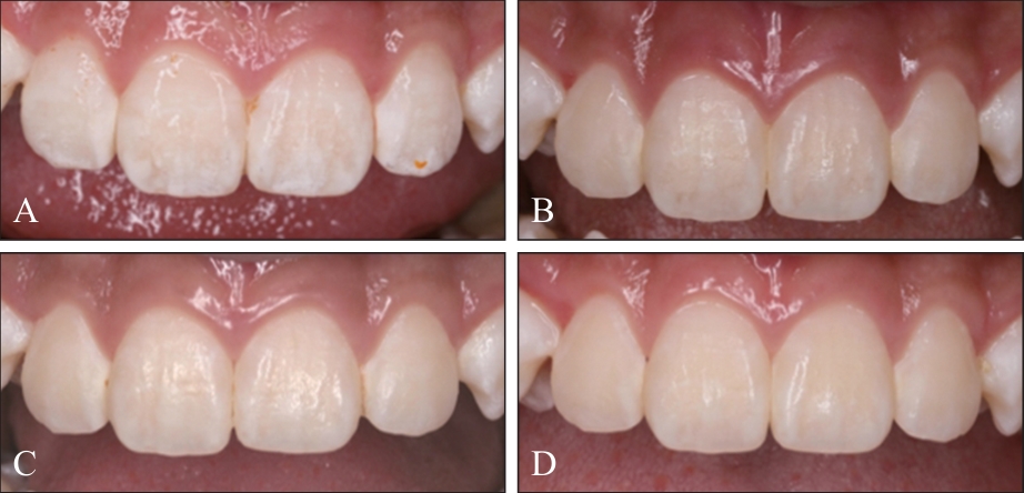
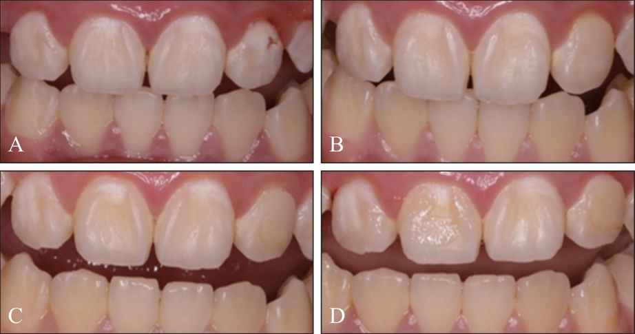
Fig 2
Preoperative and postoperative photos of fluoride treatment in mild MIH incisor plaque
Tab 1
Comparison of plaque lesion area of two treatment methods for mild MIH incisor plaque
| 时间 | 渗透树脂 | 涂氟 | P值 |
|---|---|---|---|
| P值 | <0.001 | 0.644 | |
| 术前 | 0.10(0.07,0.13) | 0.11(0.08,0.14) | 0.169 |
| 术后1月 | 0.05(0.03,0.07) | 0.10(0.08,0.14) | <0.001 |
| 术后3月 | 0.03(0.02,0.05) | 0.10(0.08,0.13) | <0.001 |
| 术后6月 | 0.03(0.02,0.04) | 0.10(0.08,0.13) | <0.001 |
Tab 2
Results of color parameter in plaque of two treatment methods for mild MIH incisor plaque
| 治疗方法 | 参数 | 术前 | 术后1月 | 术后3月 | 术后6月 | P值 |
|---|---|---|---|---|---|---|
| 渗透树脂 | L | 77.98±0.45 | 69.98±1.12 | 69.76±1.15 | 69.84±1.19 | <0.001 |
| a | 1.80±0.30 | 0.96±0.20 | 1.07±0.15 | 0.81±0.20 | - | |
| b | 25.32±0.92 | 22.93±0.87 | 23.60±0.93 | 23.16±0.83 | - | |
| ΔE | 7.15(6.27,10.65) | 3.99(2.37,8.35) | 4.50(2.91,8.88) | 4.07(2.24,9.17) | 0.001 | |
| WID | 8.93(2.68,14.67) | 8.13(2.46,14.50) | 6.96(4.16,11.84) | 8.85(2.63,14.47) | 0.926 | |
| 涂氟 | L | 76.37±0.88 | 72.34±1.25 | 73.11±1.30 | 73.68±1.37 | 0.105 |
| a | 2.38±0.17 | 2.34±0.16 | 2.30±0.15 | 2.30±0.15 | - | |
| b | 24.26±0.72 | 23.95±0.62 | 23.80±0.47 | 23.80±0.46 | - | |
| ΔE | 6.54(3.94,9.95) | 4.19(2.79,6.82) | 4.73(2.88,7.64) | 4.43(2.40,8.41) | 0.001 | |
| WID | 6.81±1.12 | 5.18±1.22 | 5.84±1.07 | 6.12±1.09 | 0.782 |
Tab 3
Statistical analysis of color parameter in plaque of two treatment methods for mild MIH incisor plaque
| 时间 | P值(渗透树脂-涂氟) | ||
|---|---|---|---|
| L | ΔE | WID | |
| 术前 | 0.103 | 0.141 | 0.393 |
| 术后1月 | 0.163 | 0.766 | 0.119 |
| 术后3月 | 0.057 | 0.443 | 0.270 |
| 术后6月 | 0.037 | 0.795 | 0.203 |
Tab 4
Comparison of the effectiveness of two pit and fissure sealant methods for mild MIH first permanent molar
| 治疗方法 | 项目 | 术后1月 | 术后3月 | 术后6月 |
|---|---|---|---|---|
| 磷酸酸蚀窝沟封闭 | 患龋 | 0/0 | 0/0 | 4/10.81 |
| 封闭剂完好 | 36/97.30 | 33/89.19 | 33/89.19 | |
| 封闭剂部分保留 | 1/2.70 | 2/5.40 | 1/2.70 | |
| 封闭剂保留 | 37/100 | 35/94.59 | 34/91.89 | |
| 封闭剂脱落 | 0/0 | 2/5.40 | 3/8.11 | |
| 窝沟封闭成功 | 36/97.30 | 33/89.19 | 29/78.38 | |
| 免酸蚀窝沟封闭 | 患龋 | 0/0 | 1/3.23 | 2/6.45 |
| 封闭剂完好 | 25/80.65 | 22/70.97 | 12/38.1* | |
| 封闭剂部分保留 | 0/0 | 0/0 | 0/0 | |
| 封闭剂保留 | 25/80.65* | 22/70.97* | 12/38.1* | |
| 封闭剂脱落 | 6/19.35* | 9/29.03* | 19/61.29* | |
| 窝沟封闭成功 | 25/80.65 | 21/67.74* | 10/32.26* |
| [1] | Weerheijm KL, Jälevik B, Alaluusua S. Molar-incisor hypomineralisation[J]. Caries Res, 2001, 35(5): 390-391. |
| [2] | Saitoh M, Shintani S. Molar incisor hypomineralisation: a review and prevalence in Japan[J]. Jpn Dent Sci Rev, 2021, 57: 71-77. |
| [3] | Zhao DD, Dong B, Yu DD, et al. The prevalence of molar incisor hypomineralisation: evidence from 70 studies[J]. Int J Paediatr Dent, 2018, 28(2): 170-179. |
| [4] | 刘晏辰, 何淼. 磨牙-切牙矿化不全的临床管理[J]. 口腔医学研究, 2022, 38(12): 1115-1118. |
| Liu YC, He M. Treatment of molar-incisor hypomineralization[J]. J Oral Sci Res, 2022, 38(12): 1115-1118. | |
| [5] | Lygidakis NA, Wong F, Jälevik B, et al. Best clinical practice guidance for clinicians dealing with children presenting with molar-incisor-hypomineralisation (MIH)[J]. Eur Arch Paediatr Dent, 2010, 11(2): 75-81. |
| [6] | 司燕, 郑树国. 窝沟封闭防龋[J]. 中国实用口腔科杂志, 2012, 5(10): 582-587. |
| Si Y, Zheng SG. Pit and fissure sealant for caries prevention[J]. Chin J Pract Stomatol, 2012, 5(10): 582-587. | |
| [7] | Joiner A, Luo W. Tooth colour and whiteness: a review[J]. J Dent, 2017, 67: S3-S10. |
| [8] | Rodd HD, Abdul-Karim A, Yesudian G, et al. Seeking children’s perspectives in the management of visible enamel defects[J]. Int J Paediatr Dent, 2011, 21(2): 89-95. |
| [9] | Paris S, Soviero VM, Schuch M, et al. Pretreatment of natural caries lesions affects penetration depth of infiltrants in vitro [J]. Clin Oral Invest, 2013, 17(9): 2085-2089. |
| [10] | 朱立霞, 汤晔, 陈黎明. 渗透树脂结合皓齿美白治疗轻中度氟斑牙效果的临床研究[J]. 贵州医药, 2020, 44(9): 1451-1452. |
| Zhu LX, Tang Y, Chen LM. Clinical study on the effect of penetrating resin combined with whitening in the treatment of mild to moderate fluorosis[J]. Guizhou Med J, 2020, 44(9): 1451-1452. | |
| [11] | 王芹, 王蕊, 胥爱文. 渗透树脂和氟化物涂布对离体乳前牙白垩色斑块龋损的修复效果及两者渗透能力的比较[J]. 广西医学, 2023, 45(14): 1681-1684, 1729. |
| Wang Q, Wang R, Xu AW. Repair effect of penetrating resin versus fluoride coating on chalky plaque caries of isolated deciduous anterior teeth and their penetrating capacity: a comparative study[J]. Guangxi Med J, 2023, 45(14): 1681-1684, 1729. | |
| [12] | 兰花, 朱琳虹, 沈旭周, 等. 渗透树脂联合微研磨治疗正畸后牙面白垩色病损的临床疗效评价[J]. 牙体牙髓牙周病学杂志, 2024, 29(7): 395-399. |
| Lan H, Zhu LH, Shen XZ, et al. Clinical evaluation of resin infiltration combined with microabrasion in the treatment of post-orthodontic white spot lesions[J]. Chin J Conserv Dent, 2024, 29(7): 395-399. | |
| [13] | 谷希, 张立亚, 陈瑞雪, 等. 渗透树脂修复磨牙-切牙矿化不全的美学效果评价[J]. 口腔疾病防治, 2021, 29(10): 689-694. |
| Gu X, Zhang LY, Chen RX, et al. Esthetic evaluation of resin infiltration for the treatment of molar-incisor hypomineralization[J]. J Prevent Treat Stomatol Dis, 2021, 29(10): 689-694. | |
| [14] | Athayde GDS, Reis PPGD, Jorge RC, et al. Impact of masking hypomineralisation opacities in anterior teeth on the esthetic perception of children and parents: a randomized controlled clinical trial[J]. J Dent, 2022, 123: 104168. |
| [15] | Pérez MM, Herrera LJ, Carrillo F, et al. Whiteness difference thresholds in dentistry[J]. Dent Mater, 2019, 35(2): 292-297. |
| [16] | Rey N, Benbachir N, Bortolotto T, et al. Evaluation of the staining potential of a caries infiltrant in comparison to other products[J]. Dent Mater J, 2014, 33(1): 86-91. |
| [17] | Paris S, Meyer-Lueckel H. Masking of labial enamel white spot lesions by resin infiltration—a clinical report[J]. Quintessence Int, 2009, 40(9): 713-718. |
| [18] | 许伟森, 熊世江. 氟化物涂膜的临床应用[J]. 中华口腔医学研究杂志(电子版), 2009, 3(2): 219-221. |
| Xu WS, Xiong SJ. The clinical effects of fluoride varnish:a review[J]. Chin J Stomatol Res (Electr Edit), 2009, 3(2): 219-221. | |
| [19] | 杨刚, 林居红, 王金华, 等. 氟保护漆预防儿童乳牙龋齿的临床效果评价[J]. 华西口腔医学杂志, 2008, 26(2): 159-161. |
| Yang G, Lin JH, Wang JH, et al. Evaluation of the clinical effect of fluoride varnish in preventing caries of primary teeth[J]. West China J Stomatol, 2008, 26(2): 159-161. | |
| [20] | 孙长芸. 渗透树脂、含氟制剂及GC护牙素治疗正畸早期釉质脱矿的比较研究[D]. 济南: 山东大学, 2017. |
| Sun CY. Comparison of penetrating resin, fluoride products and GC tooth mousse in treating enamel deminera-lization caused by orthodontic thrapy[D]. Jinan: Shandong University, 2017. | |
| [21] | 潘星星, 刘伟, 华文兵. 窝沟封闭剂联合涂氟预防儿童第一恒磨牙龋病效果的临床研究[J]. 口腔材料器械杂志, 2023, 32(4): 266-270. |
| Pan XX, Liu W, Hua WB. Clinic study on the effect of pit and fissure sealant combined with fluoride on the prevention of caries in children’s first permanent molar[J]. Chin J Dent Mater Dev, 2023, 32(4): 266-270. | |
| [22] | 曲东杰. 浅析自酸蚀粘结剂对160例小学生恒牙窝沟封闭的临床疗效评估[J]. 中国伤残医学, 2013, 21(9): 63-64. |
| Qu DJ. Since the acid etch adhesives for 160 cases of primary school students permanent teeth socket channel closed clinical curative effect evaluation[J]. Chin J Trauma Disabil, 2013, 21(9): 63-64. | |
| [23] | 谭映红, 杨鸯. 免酸蚀窝沟封闭剂对第二乳磨牙窝沟封闭效果评价[J]. 临床合理用药杂志, 2018, 11(17): 153-154. |
| Tan YH, Yang Y. Evaluation of the effect of acid-free fissure sealant on the sealing effect of the second primary molar[J]. Chin J Clin Ration Drug Use, 2018, 11(17): 153-154. | |
| [24] | Voinot J, Bedez M. Pretreatments to bonding on enamel and dentin disorders: a systematic review[J]. Evid Based Dent, 2024, 25(4): 215. |
| [25] | 张笋, 夏斌, 葛立宏. 免冲洗酸蚀复合体窝沟封闭系统的实验研究[J]. 华西口腔医学杂志, 2007, 25(6): 561-563. |
| Zhang S, Xia B, Ge LH. Experimental study of compomer sealant with non-rinse conditioner used on permanent molar[J]. West China J Stomatol, 2007, 25(6): 561-563. | |
| [26] | 张笋, 秦满, 李静. 自酸蚀和磷酸酸蚀窝沟封闭术的临床比较[J]. 华西口腔医学杂志, 2008, 26(6): 630-632. |
| Zhang S, Qin M, Li J. A comparison study on the effect of self-etching adhesive and phosphoric acid fissure sea-lant in children[J]. West China J Stomatol, 2008, 26(6): 630-632. | |
| [27] | 徐英新, 李稳, 苏敏. 2种抗氧化剂恢复漂白后釉质粘接强度的研究[J]. 华西口腔医学杂志, 2021, 39(4): 453-457. |
| Xu YX, Li W, Su M. Use of two kinds of antioxidants to restore the bond strength of bleached enamel[J]. West China J Stomatol, 2021, 39(4): 453-457. | |
| [28] | Kumar H, Palamara JEA, Burrow MF, et al. An investigation into the effect of a resin infiltrant on the micromechanical properties of hypomineralised enamel[J]. Int J Paediatr Dent, 2017, 27(5): 399-411. |
| [1] | Chen Lingzhi, Wang Xiaqin, Zhu Kaifei, Ren Kun, Wu Zhen. Machine learning-based prediction model for caries in the first molars of 9-year-old children in Suzhou [J]. West China Journal of Stomatology, 2025, 43(6): 871-880. |
| [2] | Han Jingya, Zhang Yajun, Ji Mengzhen, Sun Jingfei, Jia Shuhan, Wang Zhifeng. Meta-analysis of sealants versus fluoride varnishes for the prevention of occlusal surface caries in children’s first permanent molars [J]. West China Journal of Stomatology, 2025, 43(3): 383-394. |
| [3] | Li Fengjuan, Wang Liru, Xu Fengming, Wang Xu, Liu Jingjing, Wang Yanxin, Zhang Shufang.. Health status of children’s first permanent molars in Henan province from 2015 to 2020 [J]. West China Journal of Stomatology, 2022, 40(5): 560-565. |
| [4] | Qin Dan,Jiang Haofeng,Shen Lu,Zhang Cai,Chai Zhaowu,Wang Jinhua. Prevalence of dental caries and associated factors among 10-12-year-old students in Chongqing [J]. West China Journal of Stomatology, 2019, 37(6): 608-614. |
| [5] | Yuxiang Tang,Jie Wu,Wantian Xu,Yun Chen,Shuxiang Yu. Clinical efficacy of the glass ionomer cement used as pit and fissure sealant with and without acid etching in primary teeth [J]. West China Journal of Stomatology, 2018, 36(6): 646-649. |
| [6] | Tu Rui1,2, Zhong Yisi1, Li Xue1, Hu Deyu1, He Tao1. Three years follow-up observation and analysis of caries status of primary and permanent teeth among 652 6-yearold children in Sichuan Province [J]. West China Journal of Stomatology, 2015, 33(1): 46-49. |
| [7] | Yu Xue, Wang Lin, Li Jie, Dong Qing. Caries status of the first permanent molar among 7-to 9-year-old children in Tangshan city and their correlation [J]. West China Journal of Stomatology, 2015, 33(1): 54-57. |
| [8] | Liu Yijie, Wang Ying, Wu Xiaonan, Wang Meng, Zhao Xiulan, Rong Wensheng. Fissure morphology and caries prevalence in the first permanent molars of children in the Dalian development area [J]. West China Journal of Stomatology, 2013, 31(6): 578-582. |
| [9] | Liu Jianzhong, Li Xue, Hu Deyu, Zhang Ximu, He Songlin. Caries status of primary and permanent teeth among 6-year-old children in Sichuan Province and their correlation [J]. West China Journal of Stomatology, 2012, 30(2): 214-216. |
| [10] | CHENG Rui- bo1, TAO Wei1, ZHANG Ying1, CHENG Min2, LI Yan3. Analysis of the fir st permanent molar car ies epidemiological investigation in area of nor theast China [J]. West China Journal of Stomatology, 2008, 26(01): 73-76. |
| Viewed | ||||||
|
Full text |
|
|||||
|
Abstract |
|
|||||
 This work is licensed under a Creative Commons Attribution 3.0 License.
