| 1 |
Guo X, Yu Y, Gao S, et al. Biodegradation of dental resin-based composite—A potential factor affecting the bonding effect: a narrative review[J]. Biomedicines, 2022, 10(9): 2313.
|
| 2 |
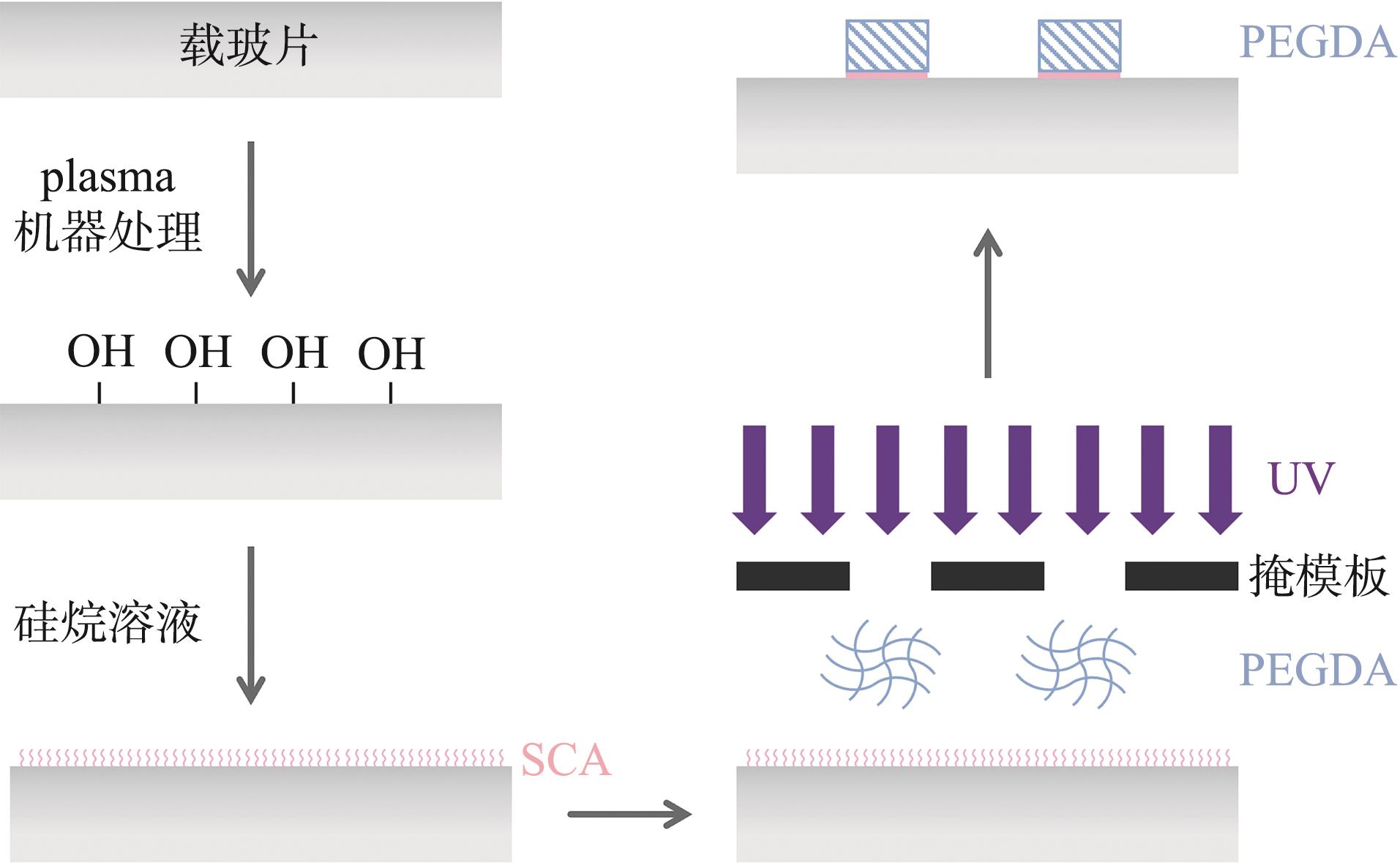
Azaryan E, Emadian Razavi F, Hanafi-Bojd MY, et al. Dentin regeneration based on tooth tissue engineering: a review[J]. Biotechnol Prog, 2023, 39(2): e3319.
|
| 3 |
Shah P, Aghazadeh M, Rajasingh S, et al. Stem cells in regenerative dentistry: current understanding and future directions[J]. J Oral Biosci, 2024, 66(2): 288-299.
|
| 4 |
Chang B, Svoboda KKH, Liu X. Cell polarization: from epithelial cells to odontoblasts[J]. Eur J Cell Biol, 2019, 98(1): 1-11.
|
| 5 |
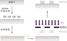
Rust K, Wodarz A. Transcriptional control of apical-ba-sal polarity regulators[J]. Int J Mol Sci, 2021, 22(22): 12340.
|
| 6 |
Li Y, Jiang W, Zhou X, et al. Advances in regulating cellular behavior using micropatterns[J]. Yale J Biol Med, 2023, 96(4): 527-547.
|
| 7 |
Tonucci FM, Hidalgo F, Ferretti A, et al. Centrosomal AKAP350 and CIP4 act in concert to define the polarized localization of the centrosome and golgi in migratory cells[J]. J Cell Sci, 2015, 128(17): 3277-3289.
|
| 8 |
Chang B, Ma C, Liu X. Nanofibrous tubular three-dimensional platform for single dental pulp stem cell polarization[J]. ACS Appl Mater Interfaces, 2020, 12(49): 54481-54488.
|
| 9 |
Fujino S, Hamano S, Tomokiyo A, et al. Dopamine is involved in reparative dentin formation through odontoblastic differentiation of dental pulp stem cells[J]. Sci Rep, 2023, 13(1): 5668.
|
| 10 |
Omi M, Kulkarni AK, Raichur A, et al. BMP-Smad signaling regulates postnatal crown dentinogenesis in mou-se molar[J]. JBMR Plus, 2019, 4(2): e10249.
|
| 11 |
Pan H, Yang Y, Xu H, et al. The odontoblastic differen-tiation of dental mesenchymal stem cells: molecular re-gulation mechanism and related genetic syndromes[J]. Front Cell Dev Biol, 2023, 11: 1174579.
|
| 12 |
刘艺萍, 王珏, 田子璐, 等. 支架微观形貌和力学性能对管状牙本质再生的影响[J]. 华西口腔医学杂志, 2020, 38(3): 314-318.
|
|
Liu YP, Wang J, Tian ZL, et al. Effects of scaffold microstructure and mechanical properties on regeneration of tubular dentin[J]. West China J Stomatol, 2020, 38(3): 314-318.
|
| 13 |
Hakim Khalili M, Zhang R, Wilson S, et al. Additive manufacturing and physicomechanical characteristics of PEGDA hydrogels: recent advances and perspective for tissue engineering[J]. Polymers (Basel), 2023, 15(10): 2341.
|
| 14 |
Park D, Kim D, Park SJ, et al. Micropattern-based nerve guidance conduit with hundreds of microchannels and stem cell recruitment for nerve regeneration[J]. NPJ Regen Med, 2022, 7(1): 62.
|
| 15 |
Bril M, Saberi A, Jorba I, et al. Shape-morphing photoresponsive hydrogels reveal dynamic topographical conditioning of fibroblasts[J]. Adv Sci (Weinh), 2023, 10(31): e2303136.
|
| 16 |
Gomes ER, Jani S, Gundersen GG. Nuclear movement regulated by Cdc42, MRCK, myosin, and actin flow establishes MTOC polarization in migrating cells[J]. Cell, 2005, 121(3): 451-463.
|
| 17 |
Fructuoso M, Legrand M, Mousson A, et al. FAK regulates dynein localisation and cell polarity in migrating mouse fibroblasts[J]. Biol Cell, 2020, 112(2): 53-72.
|
| 18 |
Zhang J, Alisafaei F, Nikolić M, et al. Nuclear mechanics within intact cells is regulated by cytoskeletal network and internal nanostructures[J]. Small, 2020, 16(18): e-1907688.
|
| 19 |
Maninová M, Klímová Z, Parsons JT, et al. The reorientation of cell nucleus promotes the establishment of front-rear polarity in migrating fibroblasts[J]. J Mol Biol, 2013, 425(11): 2039-2055.
|
| 20 |
McBeath R, Pirone DM, Nelson CM, et al. Cell shape, cytoskeletal tension, and RhoA regulate stem cell lineage commitment[J]. Dev Cell, 2004, 6(4): 483-495.
|
| 21 |
João SM, Arana-Chavez VE. Tight junctions in differentiating ameloblasts and odontoblasts differentially express ZO-1, occludin, and claudin-1 in early odontogenesis of rat molars[J]. Anat Rec A Discov Mol Cell Evol Biol, 2004, 277(2): 338-343.
|
| 22 |
Chang B, Ma C, Feng J, et al. Dental pulp stem cell polarization: effects of biophysical factors[J]. J Dent Res, 2021, 100(10): 1153-1160.
|

 ), Yu Nianzuo3,4(
 This work is licensed under a Creative Commons Attribution 3.0 License.