West China Journal of Stomatology ›› 2023, Vol. 41 ›› Issue (4): 426-433.doi: 10.7518/hxkq.2023.2023089
• Clinical Research • Previous Articles Next Articles
Li Luxin( ), Liu Honghong(), Chen Jia, Zhang Zhihong, Sang Xiao, Zhang Lili, Wang Yuantian.
Received:2023-03-22
Revised:2023-05-24
Online:2023-08-01
Published:2023-07-21
Contact:
Liu Honghong
E-mail:1215313741@qq.com;66302784@qq.com
Supported by:CLC Number:
Li Luxin, Liu Honghong, Chen Jia, Zhang Zhihong, Sang Xiao, Zhang Lili, Wang Yuantian.. Feasibility analysis of digital method for measuring supracrestal tissue height crest around implant[J]. West China Journal of Stomatology, 2023, 41(4): 426-433.
Add to citation manager EndNote|Ris|BibTeX
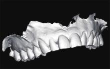
Fig 1
Pre-operative acquisition of orography image
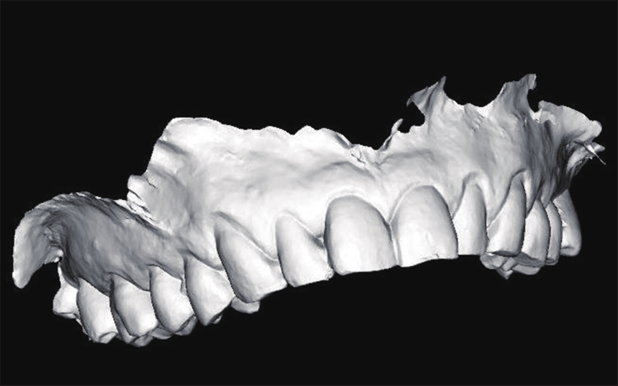
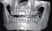
Fig 2
CBCT image acquired on the day after surgery
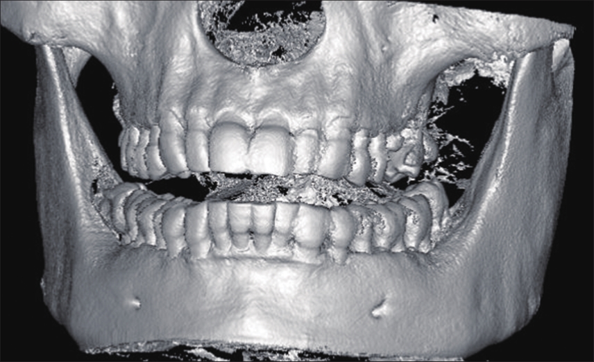
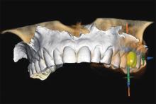
Fig 3
Fitting the CBCT image to the orography image
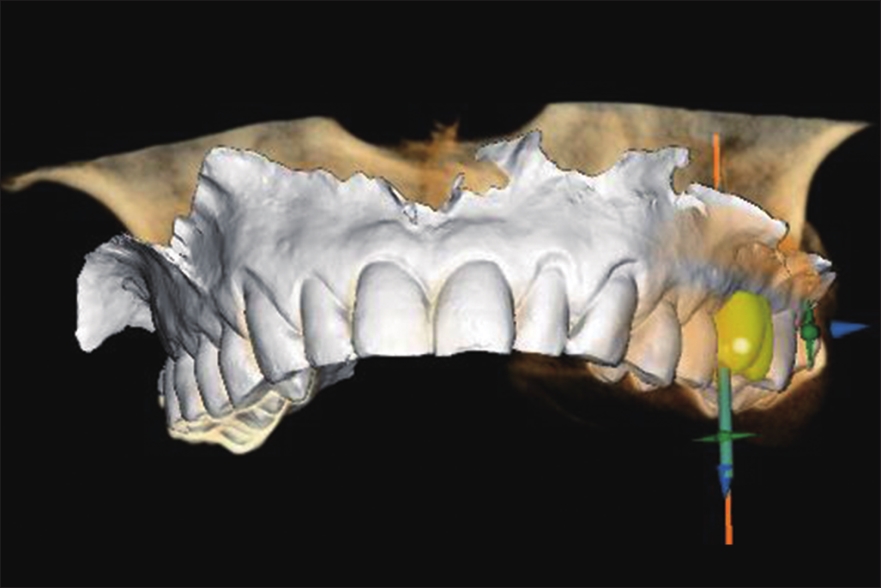
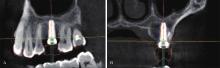
Fig 4
Placement of virtual implants overlaid with implant shadows from postoperative CBCT images
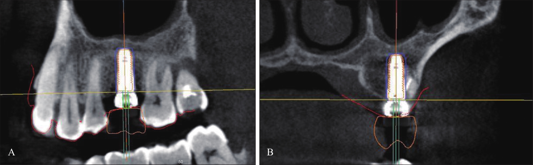
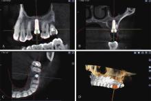
Fig 5
Cross-coordinate line centered at the center of the implant

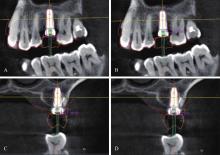
Fig 6
STH measurement

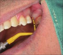
Fig 7
Intraoperative measurement of the buccal central STH using a periodontal probe after implant placement

Tab 1
Comparison of the soft tissue height on the bone crest of the proposed implantation area at 172 measurement sites by both methods
| 组别 | 颊侧 | 舌侧 | 近中 | 远中 |
|---|---|---|---|---|
| 数字化方法测量组 | 2.26±1.02 | 2.54±0.89 | 3.00±0.94 | 2.79±0.91 |
| 牙周探诊测量组 | 2.24±1.17 | 2.59±1.34 | 2.91±1.15 | 2.72±1.22 |
| t值 | -0.127 | 0.425 | -1.060 | -0.801 |
| P值 | 0.899 | 0.673 | 0.295 | 0.427 |
Tab 2
Comparative results of soft tissue height on the bone crest of the proposed implant area in 43 cases measured by both methods
| 组别 | 上牙 | 下牙 | 前牙 | 后牙 |
|---|---|---|---|---|
| 数字化方法测量组 | 3.19±1.19 | 2.41±0.80 | 3.71±1.42 | 2.56±0.90 |
| 牙周探诊测量组 | 3.15±1.33 | 2.36±0.87 | 4.00±1.73 | 2.47±0.93 |
| t值 | -0.346 | -0.850 | 1.626 | -1.460 |
| P值 | 0.735 | 0.402 | 0.203 | 0.153 |
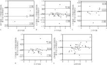
Fig 8
Consistency results of two methods of measuring

| 1 | Kao RT, Curtis DA, Kim DM, et al. American Academy of Periodontology best evidence consensus statement on modifying periodontal phenotype in preparation for orthodontic and restorative treatment[J]. J Periodontol, 2020, 91(3): 289-298. |
| 2 | Jepsen S, Caton JG, Albandar JM, et al. Periodontal manifestations of systemic diseases and developmental and acquired conditions: consensus report of workgroup 3 of the 2017 World Workshop on the Classification of Periodontal and Peri-Implant Diseases and Conditions[J]. J Periodontol, 2018, 89(): S237-S248. |
| 3 | Suárez-López Del Amo F, Lin GH, Monje A, et al. Influence of soft tissue thickness on peri-implant marginal bone loss: a systematic review and meta-analysis[J]. J Periodontol, 2016, 87(6): 690-699. |
| 4 | 张众, 孟焕新, 韩劼, 等. 软组织垂直厚度对牙周炎患者种植修复临床效果的影响[J]. 北京大学学报(医学版), 2020, 52(2): 332-338. |
| Zhang Z, Meng HX, Han J, et al. Effect of vertical soft tissue thickness on clinical manifestation of peri-implant tissue in patients with periodontitis[J]. J Peking Univ Heal Sci, 2020, 52(2): 332-338. | |
| 5 | Vervaeke S, Dierens M, Besseler J, et al. The influence of initial soft tissue thickness on peri-implant bone remodeling[J]. Clin Implant Dent Relat Res, 2014, 16(2): 238-247. |
| 6 | Chan D, Pelekos G, Ho D, et al. The depth of the implant mucosal tunnel modifies the development and resolution of experimental peri-implant mucositis: a case-control study[J]. J Clin Periodontol, 2019, 46(2): 248-255. |
| 7 | 张浩筠, 危伊萍, 韩子瑶, 等. 种植体周表型的概念及其临床应用[J]. 口腔医学, 2021, 41(2): 110-118, 132. |
| Zhang HY, Wei YP, Han ZY, et al. The concept of peri-implant phenotype and its clinical applications[J]. Stomatology, 2021, 41(2): 110-118, 132. | |
| 8 | Jeong SM, Choi BH, Kim J, et al. A 1-year prospective clinical study of soft tissue conditions and marginal bone changes around dental implants after flapless implant surgery[J]. Oral Surg Oral Med Oral Pathol Oral Radiol Endod, 2011, 111(1): 41-46. |
| 9 | Linkevicius T, Apse P, Grybauskas S, et al. The influence of soft tissue thickness on crestal bone changes around implants: a 1-year prospective controlled clinical trial[J]. Int J Oral Maxillofac Implants, 2009, 24(4): 712-719. |
| 10 | Canullo L, Camacho-Alonso F, Tallarico M, et al. Mucosa thickness and peri-implant crestal bone stability: a clinical and histologic prospective cohort trial[J]. Int J Oral Maxillofac Implants, 2017, 32(5): 675-681. |
| 11 | Zheng Z, Ao X, Xie P, et al. The biological width around implant[J]. J Prosthodont Res, 2021, 65(1): 11-18. |
| 12 | 罗昕, 冯宜, 何福明. 种植体初始生物学宽度对边缘骨水平的影响[J]. 口腔医学, 2021, 41(2): 154-158. |
| Luo X, Feng Y, He FM. Effect of the initial biological width of implant on marginal bone level[J]. Stomatology, 2021, 41(2): 154-158. | |
| 13 | Amato F, Amato G, Campriani S, et al. The role of different healing abutment sizes in tissue volume preservation of molar sockets after immediate tooth extraction and implant placement: a multicenter clinical study[J]. Int J Oral Maxillofac Implants, 2022, 37(5): 891-904. |
| 14 | Munakata M, Nagata K, Sanda M, et al. Variations in vertical mucosal thickness at edentulous ridge according to site and gender measured by cone-beam computed tomography[J]. Int J Implant Dent, 2021, 7(1): 34. |
| 15 | Sala L, Alonso-Pérez R, Agustin-Panadero R, et al. Comparative in vitro study of two methods for gingival biotype assessment[J]. J Clin Exp Dent, 2018, 10(9): e858-e863. |
| 16 | Kaya Y, Alkan Ö, Keskin S. An evaluation of the gingival biotype and the width of keratinized gingiva in the mandibular anterior region of individuals with different dental malocclusion groups and levels of crowding[J]. Korean J Orthod, 2017, 47(3): 176-185. |
| 17 | Sönmez G, Kamburoğlu K, Gülşahı A. Accuracy of high-resolution ultrasound (US) for gingival soft tissue thickness mesurement in edentulous patients prior to implant placement[J]. Dentomaxillofac Radiol, 2021, 50(5): 20200309. |
| 18 | 曹洁, 胡文杰, 张豪, 等. 基于锥形束计算机体层摄影术测量牙龈厚度[J]. 北京大学学报(医学版), 2013, 45(1): 135-139. |
| Cao J, Hu WJ, Zhang H, et al. Method and its application of gingival thickness measurement based on cone-beam computed tomography[J]. J Peking Univ (Health Sci), 2013, 45(1): 135-139. | |
| 19 | Furtak A, Leszczyńska E, Sender-Janeczek A, et al. The repeatability and reproducibility of gingival thickness measurement with an ultrasonic device[J]. Dent Med Probl, 2018, 55(3): 281-288. |
| 20 | Lau SL, Chow LK, Leung YY. A non-invasive and accurate measurement of gingival thickness using cone-beam computerized imaging for the assessment of planning immediate implant in the esthetic zone—A pig jaw mo-del[J]. Implant Dent, 2016, 25(5): 619-623. |
| 21 | Ueno D, Sato J, Igarashi C, et al. Accuracy of oral mucosal thickness measurements using spiral computed tomography[J]. J Periodontol, 2011, 82(6): 829-836. |
| 22 | Sin YW, Chang HY, Yun WH, et al. Association of gingival biotype with the results of scaling and root planing[J]. J Periodontal Implant Sci, 2013, 43(6): 283-290. |
| 23 | El Khalifa M, Abu El Sadat SM, Gaweesh YS, et al. Assessment of gingival thickness using CBCT compared to transgingival probing and its correlation with labial bone defects: a cross-sectional study[J]. Int J Oral Maxillofac Implants, 2022, 37(3): 464-472. |
| 24 | Kloukos D, Koukos G, Doulis I, et al. Gingival thickness assessment at the mandibular incisors with four methods: a cross-sectional study[J]. J Periodontol, 2018, 89(11): 1300-1309. |
| 25 | Kloukos D, Koukos G, Gkantidis N, et al. Transgingival probing: a clinical gold standard for assessing gingival thickness[J]. Quintessence Int, 2021, 52(5): 394-401. |
| [1] | Jing Bingshuai, Shi Bing, Zheng Qian, Li Chenghao.. Effectiveness of iliac cancellous bone grafting in alveolar cleft repair and analysis of factors affecting it [J]. West China Journal of Stomatology, 2023, 41(3): 284-289. |
| [2] | Yan Li, Zhou Maoqiang, Qiu Jiaxuan. Association between bone morphology and sagittal disc position in temporomandibular joints of patients with anterior disc displacement [J]. West China Journal of Stomatology, 2022, 40(4): 414-421. |
| [3] | Guo Biao, Lu Rongjian. Morphological change analysis based on cone beam CT of the upper airway for obstructive sleep apnea syndrome patients treated with oral appliance in skeletal class Ⅱ malocclusion with different vertical patterns [J]. West China Journal of Stomatology, 2020, 38(4): 419-424. |
| [4] | You Meng, Xu Laiqing, Jiang Meng, Li Na, Liu Yuanyuan, Wang Hu.. Cone beam CT radiographic diagnosis of submandibular radiopaque sialolithiasis [J]. West China Journal of Stomatology, 2014, 32(5): 459-463. |
| [5] | Lei Qiaoling, Zhou Li, Lei Lei, Wang Yanmin. Comparison of mesiodistal tooth angulations determined through traditional panoramic radiographs and cone beam CT panoramic images [J]. West China Journal of Stomatology, 2014, 32(4): 331-335. |
| [6] | Feng Chi, Li Conghua, Zeng Xing-qi, Wang Qinhua, Zheng Qian, Qiu Ye. Investigation of accuracy of premolar length measured by cone beam CT in vivo [J]. West China Journal of Stomatology, 2014, 32(1): 36-39. |
| [7] | Huang Hong1,2, Liu Peng2,3, Li Xiaodong1,2, Pei Zhongqiu1,2, Yang Xiaozhu1,2, Bai Shi1,2, Huang Yuanding1,2.. Mandibular incisive canal by cone beam CT [J]. West China Journal of Stomatology, 2013, 31(5): 479-482. |
| [8] | Huang Rui, Liu Peng, Xiao Maode, Zhou Zhi.. A comparative study on apexification using different kinds of materials in dogs [J]. West China Journal of Stomatology, 2013, 31(4): 377-380,384. |
| [9] | Wang Hongwei1, Qi Suqing1, Wang Jianguo2, Cai Zhifang2, Li Chuang3.. Detection to changes in hyoid and tongue positions, and pharyngeal airway following mandibular setback surgery by cone beam CT [J]. West China Journal of Stomatology, 2012, 30(6): 650-654. |
| [10] | Tong Fangli, Liu Wei, Chen Zhusu, Liu Ziqiang, Zeng Xiongqun.. Repair of root fracture in maxillary second premolar with MTA:A case report [J]. West China Journal of Stomatology, 2012, 30(5): 552-553. |
| [11] | Liao Rui1, Sun Miaogen1, Gu Yajun1, Wang Renfei2, Liu Min2. Clinical application of cone beam CT in the treatment of jaw bone cyst [J]. West China Journal of Stomatology, 2012, 30(3): 262-266. |
| [12] | Xie Zhiwei1,2, Li Guoju2,3, Guo Jing1,2. Anthropometric analysis of the mandible morphology in young females with different vertical skeletal pattern [J]. West China Journal of Stomatology, 2012, 30(3): 299-303. |
| [13] | Zhang Jiangshan1, Li Xueyan2, Zhang Yanzhen3. Application research of cone beam CT in dowel preparation for the mandibular first molar [J]. West China Journal of Stomatology, 2012, 30(2): 179-182. |
| Viewed | ||||||
|
Full text |
|
|||||
|
Abstract |
|
|||||
 This work is licensed under a Creative Commons Attribution 3.0 License.
