| 1 |
Berglundh T, Armitage G, Araujo MG, et al. Peri-implant diseases and conditions: consensus report of workgroup 4 of the 2017 World Workshop on the Classification of Periodontal and Peri-Implant Diseases and Conditions[J]. J Periodontol, 2018, 89(): S313-S318.
|
| 2 |
Astolfi V, Ríos-Carrasco B, Gil-Mur FJ, et al. Incidence of peri-implantitis and relationship with different conditions: a retrospective study[J]. Int J Environ Res Public Health, 2022, 19(7): 4147.
|
| 3 |
Quaranta A, Lim ZW, Tang J, et al. The impact of resi-dual subgingival cement on biological complications a-round dental implants: a systematic review[J]. Implant Dent, 2017, 26(3): 465-474.
|
| 4 |
Fu JH, Wang HL. Breaking the wave of peri-implantitis[J]. Periodontol 2000, 2020, 84(1): 145-160.
|
| 5 |
童子安, 姒蜜思. 种植体表面菌斑去污方式的体外研究进展[J]. 国际口腔医学杂志, 2020, 47(5): 589-594.
|
|
Tong ZA, Si MS. Advances in the decontamination of plaque on implant surface in vitro [J]. Int J Stomatol, 2020, 47(5): 589-594.
|
| 6 |
Steiger-Ronay V, Merlini A, Wiedemeier DB, et al. Location of unaccessible implant surface areas during debridement in simulated peri-implantitis therapy[J]. BMC Oral Health, 2017, 17(1): 137.
|
| 7 |
苗磊, 潘亚萍. 口腔显微技术发展与应用[J]. 中国实用口腔科杂志, 2014, 7(4): 196-199.
|
|
Miao L, Pan YP. Development and application of microscopy in oral treatment[J]. Chin J Pract Stomatol, 2014, 7(4): 196-199.
|
| 8 |
Cha JK, Paeng K, Jung UW, et al. The effect of five mechanical instrumentation protocols on implant surface topography and roughness: a scanning electron microsco-pe and confocal laser scanning microscope analysis[J]. Clin Oral Implants Res, 2019, 30(6): 578-587.
|
| 9 |
La Monaca G, Pranno N, Annibali S, et al. Clinical and radiographic outcomes of a surgical reconstructive approach in the treatment of peri-implantitis lesions: a 5-year prospective case series[J]. Clin Oral Implants Res, 2018, 29(10): 1025-1037.
|
| 10 |
Ronay V, Merlini A, Attin T, et al. In vitro cleaning potential of three implant debridement methods. Simulation of the non-surgical approach[J]. Clin Oral Implants Res, 2017, 28(2): 151-155.
|
| 11 |
John G, Becker J, Schwarz F. Rotating titanium brush for plaque removal from rough titanium surfaces: an in vitro study[J]. Clin Oral Implants Res, 2014, 25(7): 838-842.
|
| 12 |
Batalha VC, Bueno RA, Fronchetti Junior E, et al. Dental implants surface in vitro decontamination protocols[J]. Eur J Dent, 2021, 15(3): 407-411.
|
| 13 |
高跃跃, 李厚轩. 不同去污方法对种植体表面处理的体外研究[J]. 中国实用口腔科杂志, 2022, 15(1): 41-46.
|
|
Gao YY, Li HX. In vitro research of different deconta-mination methods for contaminated implant surfaces[J]. Chin J Pract Stomatol, 2022, 15(1): 41-46.
|
| 14 |
Monje A, Amerio E, Cha JK, et al. Strategies for implant surface decontamination in peri-implantitis therapy[J]. Int J Oral Implantol (Berl), 2022, 15(3): 213-248.
|
| 15 |
Sahrmann P, Ronay V, Sener B, et al. Cleaning potential of glycine air-flow application in an in vitro peri-implantitis model[J]. Clin Oral Implants Res, 2013, 24(6): 666-670.
|
| 16 |
Orsini E, Giavaresi G, Trirè A, et al. Dental implant thread pitch and its influence on the osseointegration process: an in vivo comparison study[J]. Int J Oral Maxillofac Implants, 2012, 27(2): 383-392.
|
| 17 |
Sanz-Martín I, Paeng K, Park H, et al. Significance of implant design on the efficacy of different peri-implantitis decontamination protocols[J]. Clin Oral Investig, 2021, 25(6): 3589-3597.
|

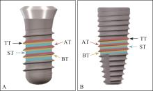
 ), 李静雯, 蒋立姗, 崔雯洁, 赵阳, 李厚轩(