West China Journal of Stomatology ›› 2026, Vol. 44 ›› Issue (1): 113-119.doi: 10.7518/hxkq.2025.2025057
• Clinical Research • Previous Articles Next Articles
Fu Chengyi1( ), Pan Xiao1, Liu Shu2, Lin Zitong2()
Received:2025-02-17
Online:2026-02-01
Published:2026-02-02
Contact:
Lin Zitong
E-mail:000fcy@163.com;linzitong710@163.com
Supported by:CLC Number:
Fu Chengyi, Pan Xiao, Liu Shu, Lin Zitong. Root fracture in endodontically versus nonendodontically treated posterior teeth: the cone beam computed tomography characteristics in Chinese population[J]. West China Journal of Stomatology, 2026, 44(1): 113-119.
Add to citation manager EndNote|Ris|BibTeX
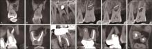
Fig 1
The orientation of fracture lines
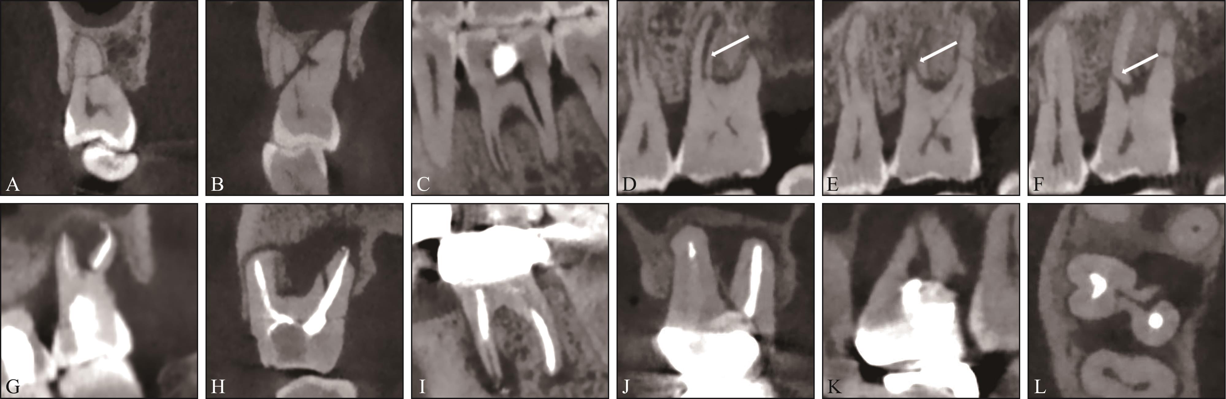
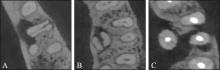
Fig 2
Direction of vertical root fracture

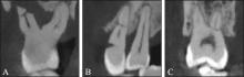
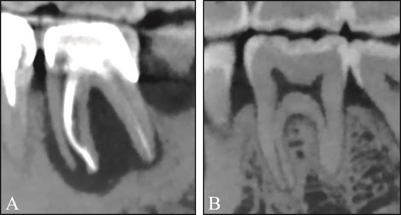
Fig 3
Location of horizontal root fracture
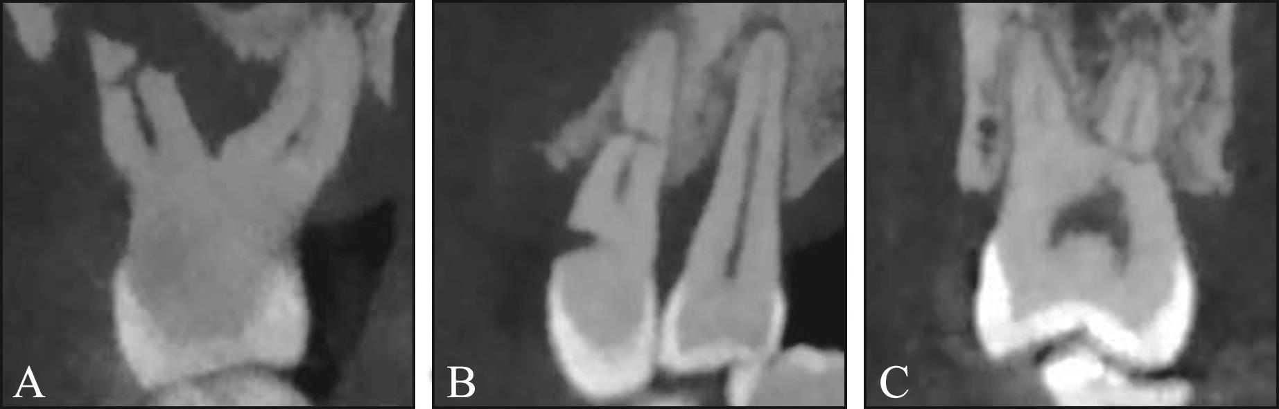
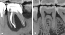
Fig 4
Bone loss around fractured roots
Tab 1
Proportion of single tooth and multiple teeth with root fracture of nonendodontically treated patients and endodontically treated patients
| 类别 | 单发性根折 | 多发性根折 |
|---|---|---|
| 非根管治疗 | 161/86.6% | 25/13.4% |
| 根管治疗 | 113/98.3% | 2/1.7% |
Tab 2
Age distribution of nonendodontically treated patients and endodontically treated patients
| 类别 | 年龄分组 | 平均年龄/岁 | |||
|---|---|---|---|---|---|
| 10~30岁 | 31~50岁 | 51~70岁 | 71~90岁 | ||
| 合计 | 8 | 90 | 162 | 41 | 56.8±13.2 |
| 非根管治疗 | 4 | 43 | 114 | 25 | 58.1±12.2 |
| 根管治疗 | 4 | 47 | 48 | 16 | 54.7±14.4 |
Tab 3
Gender distribution of nonendodontically trea-ted patients and endodontically treated patients
| 类别 | 男 | 女 |
|---|---|---|
| 非根管治疗 | 125/67.2% | 61/32.8% |
| 根管治疗 | 65/56.3% | 50/43.7% |
Tab 4
The tooth distribution in nonendodontically treated group and endodontically treated group
| 牙位 | 非根管治疗 | 根管治疗 |
|---|---|---|
| 上颌前磨牙 | 21/9.6% | 24/19.8% |
| 上颌磨牙 | 87/39.7% | 39/32.2% |
| 下颌前磨牙 | 10/4.6% | 9/7.4% |
| 下颌磨牙 | 101/46.1% | 49/40.5% |
Tab 5
Root distribution in nonendodontically treated group and endodontically treated group
| 牙根位 | 非根管治疗 | 根管治疗 | |
|---|---|---|---|
| 上颌前磨牙 | 21/10.0% | 24/20.3% | |
| 下颌前磨牙 | 10/4.8% | 9/7.6% | |
| 上颌磨牙 | 近颊根 | 28/13.4% | 18/15.3% |
| 远颊根 | 15/7.2% | 4/3.4% | |
| 腭根 | 48/23.0% | 16/13.6% | |
| 下颌磨牙 | 近中根 | 76/36.4% | 38/32.2% |
| 远中根 | 11/5.3% | 9/7.6% | |
| 合计 | 209/100% | 118/100% | |
Tab 6
Orientation of root fractures in nonendodontically treated group and endodontically treated group
| 根折类型 | 非根管治疗 | 根管治疗 |
|---|---|---|
| 合计 | 227/100% | 123/100% |
| 横折 | 57/25.1% | 8/6.5% |
| 斜折 | 56/24.7% | 20/16.2% |
| 纵折 | 99/43.6% | 93/75.6% |
| 不规则折裂 | 15/6.6% | 2/1.6% |
Tab 7
Orientation of root fractures in different roots of posterior teeth
| 牙根 | 横折 | 斜折 | 纵折 | 不规则折裂 | 合计 | |
|---|---|---|---|---|---|---|
| 上颌前磨牙 | 8(7/1) | 13(9/4) | 22(3/19) | 2(2/0) | 45(21/24) | |
| 下颌前磨牙 | 2(2/0) | 8(7/1) | 7(0/7) | 2(1/1) | 19(10/9) | |
| 上颌磨牙 | 近颊根 | 20(17/3) | 4(1/3) | 18(7/11) | 4(3/1) | 46(28/18) |
| 远颊根 | 15(13/2) | 3(2/1) | 1(0/1) | 0(0/0) | 19(15/4) | |
| 腭根 | 17(16/1) | 25(17/8) | 18(11/7) | 4(4/0) | 64(48/16) | |
| 下颌磨牙 | 近中根 | 2(1/1) | 9(7/2) | 100(65/35) | 3(3/0) | 114(76/38) |
| 远中根 | 1(1/0) | 4(3/1) | 15(7/8) | 0(0/0) | 20(11/9) | |
| 合计 | 65(57/8) | 66(46/20) | 181(93/88) | 15(13/2) | 327(209/118) | |
Tab 8
Directions of vertical root fractures in nonendodontically treated group and endodontically treated group
| 牙根纵折方向 | 非根管治疗 | 根管治疗 |
|---|---|---|
| 合计 | 93/100% | 88/100% |
| 近远中向 | 8/8.6% | 6/6.8% |
| 颊舌向 | 80/86.0% | 74/84.1% |
| 斜向 | 5/5.4% | 8/9.1% |
Tab 9
Directions of vertical root fractures in different roots of posterior teeth
| 牙根 | 近远中向 | 颊舌向 | 斜向 | |
|---|---|---|---|---|
| 前磨牙 | 2(0/2) | 26(3/23) | 1(0/1) | |
| 上颌磨牙 | 近颊根 | 0(0/0) | 18(7/11) | 0(0/0) |
| 远颊根 | 0(0/0) | 0(0/0) | 1(0/1) | |
| 腭根 | 11(8/3) | 2(0/2) | 5(3/2) | |
| 下颌磨牙 | 近中根 | 1(0/1) | 95(63/32) | 4(2/2) |
| 远中根 | 0(0/0) | 13(7/6) | 2(0/2) | |
| 合计 | 14(8/6) | 154(80/74) | 13(5/8) | |
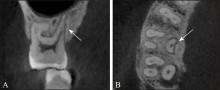
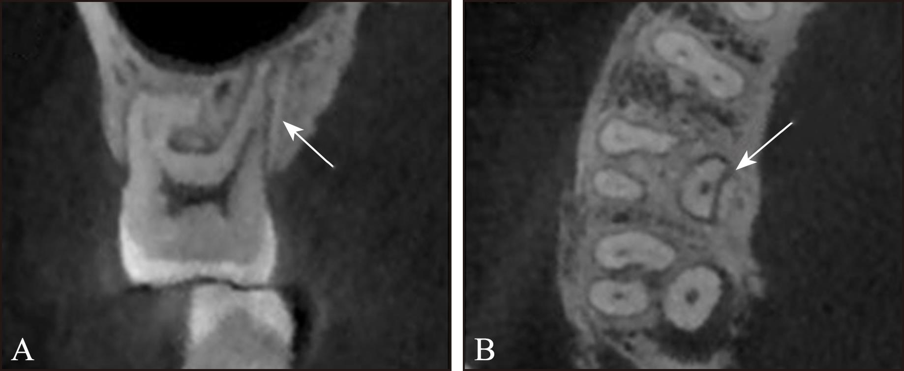
Fig 5
Vertical root fracture of palatal root of maxillary first molar, the fracture line was in a mesio-distal direction
Tab 10
Location of horizontal root fractures in nonendodontically treated group and endodontically treated group
| 横折位置 | 非根管治疗 | 根管治疗 |
|---|---|---|
| 合计 | 57/100% | 8/100% |
| 根颈1/3区 | 37/64.9% | 8/100% |
| 根中1/3区 | 14/24.5% | 0/0% |
| 根尖1/3区 | 6/10.5% | 0/0% |
Tab 11
Bone loss around fractured roots in nonendo-dontically treated group and endodontically treated group
| 根折线周围骨吸收 | 非根管治疗 | 根管治疗 |
|---|---|---|
| 合计 | 227/100% | 123/100% |
| 骨吸收 | 134/59.0% | 113/91.8% |
| 无明显骨吸收 | 93/41.0% | 10/8.1% |
| [1] | Pan X, Tang R, Gao AT, et al. Cross-sectional study of posterior tooth root fractures in 2015 and 2019 in a Chinese population[J]. Clin Oral Invest, 2022, 26(10): 6151-6157. |
| [2] | Yeh CJ. Fatigue root fracture: a spontaneous root fracture in non-endodontically treated teeth[J]. Br Dent J, 1997, 182(7): 261-266. |
| [3] | Versluis A, Messer HH, Pintado MR. Changes in compaction stress distributions in roots resulting from canal preparation[J]. Int Endod J, 2006, 39(12): 931-939. |
| [4] | Mizuhashi F, Ogura I, Sugawara Y, et al. Diagnosis of root fractures using cone-beam computed tomography: difference of vertical and horizontal root fracture[J]. Oral Radiol, 2021, 37(2): 305-310. |
| [5] | Chan CP, Lin CP, Tseng SC, et al. Vertical root fracture in endodontically versus nonendodontically treated tee-th: a survey of 315 cases in Chinese patients[J]. Oral Surg Oral Med Oral Pathol Oral Radiol Endod, 1999, 87(4): 504-507. |
| [6] | Liao WC, Chen CH, Pan YH, et al. Horizontal root fracture in posterior teeth without dental trauma: a diseased condition with special characteristics[J]. J Formos Med Assoc, 2022, 121(9): 1625-1635. |
| [7] | Tsai YL, Liao WC, Wang CY, et al. Horizontal root fractures in posterior teeth without dental trauma: tooth/root distribution and clinical characteristics[J]. Int Endod J, 2017, 50(9): 830-835. |
| [8] | Liao WC, Chen CH, Pan YH, et al. Vertical root fracture in non-endodontically and endodontically treated teeth: current understanding and future challenge[J]. J Pers Med, 2021, 11(12): 1375. |
| [9] | Sheikhi M, Ghazizadeh M, Aminian M, et al. Accuracy of digital image enhancement in detection of vertical and horizontal root fracture[J]. Dent Res J, 2020, 17(4): 266-272. |
| [10] | Xu HP, Zheng QH, Shao YF, et al. The effects of ageing on the biomechanical properties of root dentine and fracture[J]. J Dent, 2014, 42(3): 305-311. |
| [11] | Liao WC, Tsai YL, Wang CY, et al. Clinical and radiographic characteristics of vertical root fractures in endodontically and nonendodontically treated teeth[J]. J Endod, 2017, 43(5): 687-693. |
| [12] | Lim MJ, Kim JA, Choi Y, et al. Differentiating spontaneous vertical root fracture in endodontically treated tooth[J]. Eur J Dent, 2017, 11(1): 122-125. |
| [13] | Wang P, Su LY. Clinical observation in 2 representative cases of vertical root fracture in nonendodontically trea-ted teeth[J]. Oral Surg Oral Med Oral Pathol Oral Radiol Endod, 2009, 107(4): e39-e42. |
| [14] | Tamse A, Fuss Z, Lustig J, et al. An evaluation of en-dodontically treated vertically fractured teeth[J]. J Endod, 1999, 25(7): 506-508. |
| [15] | Tsesis I, Rosen E, Tamse A, et al. Diagnosis of vertical root fractures in endodontically treated teeth based on clinical and radiographic indices: a systematic review[J]. J Endod, 2010, 36(9): 1455-1458. |
| [16] | Patel S, Bhuva B, Bose R. Present status and future directions: vertical root fractures in root filled teeth[J]. Int Endod J, 2022, 55(S3): 804-826. |
| [17] | Zhou YF, Hu ZY, Hu YN, et al. Patterns of stress distribution of endodontically treated molar under different types of loading using finite element models: the exploring of mechanism of vertical root fracture[J]. J Mech Behav Biomed Mater, 2023, 144: 105947. |
| [18] | Wang P, Lv H, Sun HT, et al. Horizontal root fractures in posterior teeth: a case series[J]. Dent Traumatol, 2011, 27(2): 152-155. |
| [19] | Zhang L, Wang TM, Cao Y, et al. In vivo detection of subtle vertical root fracture in endodontically treated teeth by cone-beam computed tomography[J]. J Endod, 2019, 45(7): 856-862. |
| [20] | Ku HM, Oh YR, Lee ES, et al. Using autofluorescence to detect bacterial contamination in root fractures[J]. J Dent, 2019, 86: 27-32. |
| Viewed | ||||||
|
Full text |
|
|||||
|
Abstract |
|
|||||
 This work is licensed under a Creative Commons Attribution 3.0 License.
