West China Journal of Stomatology ›› 2024, Vol. 42 ›› Issue (2): 227-233.doi: 10.7518/hxkq.2024.2023277
• Clinical Research • Previous Articles Next Articles
Wang Siyu1( ), Zhou Zheqing1, Yuan Quan2, Yue Li1, Yang Shengtao1()
Received:2023-08-28
Revised:2023-12-07
Online:2024-04-01
Published:2024-03-26
Contact:
Yang Shengtao
E-mail:2732078280@qq.com;shengtao@scu.edu.cn
Supported by:CLC Number:
Wang Siyu, Zhou Zheqing, Yuan Quan, Yue Li, Yang Shengtao. Trueness evaluation of three intraoral scanners for the recording of maximal intercuspal position[J]. West China Journal of Stomatology, 2024, 42(2): 227-233.
Add to citation manager EndNote|Ris|BibTeX
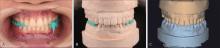
Fig 1
Acquisition of the control group’s digital casts

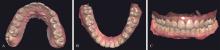
Fig 2
Maxillary and mandibular arch scans obtained by Trios 3 intraoral scanner
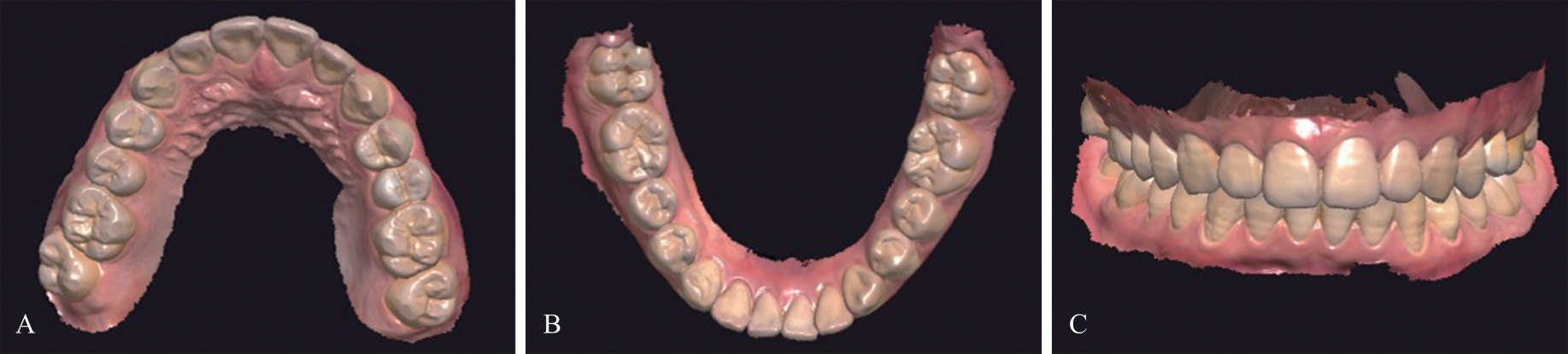
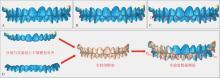
Fig 3
Distance measurements of control and experimental groups

Tab 1
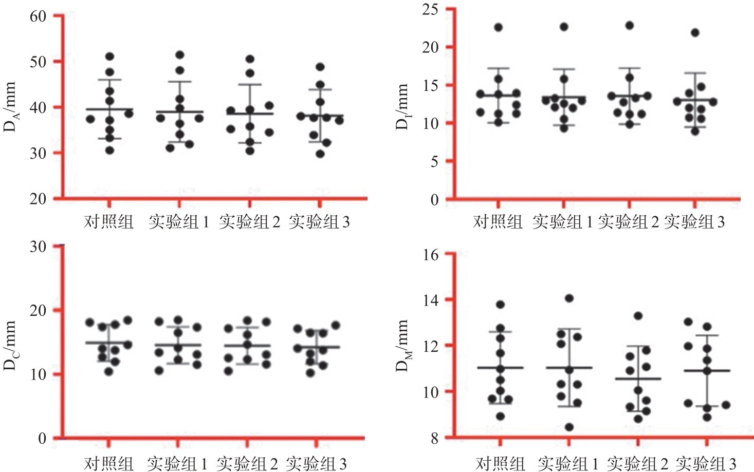
The measured values of DA, DI, DC, and DM in the control and experimental groups
| 距离 | 对照组 | 实验组1 | 实验组2 | 实验组3 |
|---|---|---|---|---|
| DI | 13.64±3.58 | 13.42±3.66 | 13.56±3.68 | 13.03±3.54 |
| DC | 14.91±2.85 | 14.55±2.87 | 14.45±2.85 | 14.23±2.59 |
| DM | 11.03±1.56 | 11.03±1.69 | 10.55±1.41 | 10.90±1.54 |
| DA | 39.58±6.40 | 38.99±6.60 | 38.57±6.36 | 38.16±5.69 |
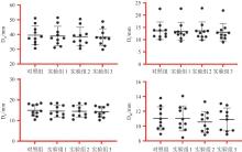
Fig 4
Scatter plots for DA, DI, DC, and DM in the control and experimental groups
Tab 2
Variance analysis of DA, DI, DC, and DM va-lues in the control and experimental groups
| 距离 | 变异来源 | SS | df | MS | F值 | P值 |
|---|---|---|---|---|---|---|
| DI | 组间变异 | 2.22 | 3 | 0.74 | 0.057 | 0.98 |
| 组内变异 | 471.60 | 36 | 13.10 | |||
| 总变异 | 473.80 | 39 | ||||
| DC | 组间变异 | 2.41 | 3 | 0.80 | 0.10 | 0.96 |
| 组内变异 | 281.20 | 36 | 7.81 | |||
| 总变异 | 283.60 | 39 | ||||
| DM | 组间变异 | 1.52 | 3 | 0.50 | 0.21 | 0.89 |
| 组内变异 | 87.31 | 36 | 2.42 | |||
| 总变异 | 88.83 | 39 | ||||
| DA | 组间变异 | 11.13 | 3 | 3.70 | 0.094 | 0.96 |
| 组内变异 | 1 418.00 | 36 | 39.39 | |||
| 总变异 | 1 429.00 | 39 |
| 1 | Tripodakis AP, Vergos VK, Tsoutsos AG. Evaluation of the accuracy of interocclusal records in relation to two recording techniques[J]. J Prosthet Dent, 1997, 77(2): 141-146. |
| 2 | Thongthammachat S, Moore BK, Barco MT, et al. Dimensional accuracy of dental casts: influence of tray material, impression material, and time[J]. J Prosthodont, 2002, 11(2): 98-108. |
| 3 | Naumovski B, Kapushevska B. Dimensional stability and acuracy of silicone-based impression materials using different impression techniques—A literature review[J]. Pril (Makedon Akad Nauk Umet Odd Med Nauki), 2017, 38(2): 131-138. |
| 4 | Al-Odinee NM, Al-Hamzi M, Al-Shami IZ, et al. Eva-luation of the quality of fixed prosthesis impressions in private laboratories in a sample from Yemen[J]. BMC Oral Health, 2020, 20(1): 304. |
| 5 | Aragón ML, Pontes LF, Bichara LM, et al. Validity and reliability of intraoral scanners compared to conventional gypsum models measurements: a systematic review[J]. Eur J Orthod, 2016, 38(4): 429-434. |
| 6 | Kong L, Li Y, Liu Z. Digital versus conventional full-arch impressions in linear and 3D accuracy: a systematic review and meta-analysis of in vivo studies[J]. Clin Oral Investig, 2022, 26(9): 5625-5642. |
| 7 | Keul C, Güth JF. Accuracy of full-arch digital impressions: an in vitro and in vivo comparison[J]. Clin Oral Investig, 2020, 24(2): 735-745. |
| 8 | Yehia A, Abo El Fadl A, El Sergany O, et al. Effect of different span lengths with different total occlusal convergences on the accuracy of intraoral scanners[J]. J Prosthodont, 2023. doi: 10.1111/jopr.13686 . |
| 9 | Chen Y, Zhai Z, Watanabe S, et al. Understanding the effect of scan spans on the accuracy of intraoral and desktop scanners[J]. J Dent, 2022, 124: 104220. |
| 10 | DeLong R, Ko CC, Anderson GC, et al. Comparing ma-ximum intercuspal contacts of virtual dental patients and mounted dental casts[J]. J Prosthet Dent, 2002, 88(6): 622-630. |
| 11 | Zimmermann M, Ender A, Attin T, et al. Accuracy of buccal scan procedures for the registration of habitual intercuspation[J]. Oper Dent, 2018, 43(6): 573-580. |
| 12 | Revilla-León M, Alonso Pérez-Barquero J, Zubizarreta-Macho Á, et al. Influence of the number of teeth and location of the virtual occlusal record on the accuracy of the maxillo-mandibular relationship obtained by using an intraoral scanner[J]. J Prosthodont, 2023, 32(3): 253-258. |
| 13 | Ren S, Morton D, Lin WS. Accuracy of virtual interocclusal records for partially edentulous patients[J]. J Prosthet Dent, 2020, 123(6): 860-865. |
| 14 | 陈玲, 陈成, 李志勇, 等. 口内扫描数字化印模对固定修复临床应用效果的Meta分析[J]. 华西口腔医学杂志, 2021, 39(3): 306-312. |
| Chen L, Chen C, Li ZY, et al. Clinical performance of intraoral digital impression for fixed prosthodontics: a Meta-analysis[J]. West China J Stomatol, 2021, 39(3): 306-312. | |
| 15 | Burzynski JA, Firestone AR, Beck FM, et al. Comparison of digital intraoral scanners and alginate impressions: time and patient satisfaction[J]. Am J Orthod Dentofacial Orthop, 2018, 153(4): 534-541. |
| 16 | 奚祺, 吴国锋. 数字化口内扫描技术的发展与应用[J]. 实用口腔医学杂志, 2021, 37(1): 136-140. |
| Xi Q, Wu GF. Development and application of digital intraoral scanning technology[J]. J Pract Stomatol, 2021, 37(1): 136-140. | |
| 17 | Revilla-León M, Kois DE, Kois JC. A guide for maximizing the accuracy of intraoral digital scans: part 2—patient factors[J]. J Esthet Restor Dent, 2023, 35(1): 241-249. |
| 18 | Revilla-León M, Kois DE, Kois JC. A guide for maximizing the accuracy of intraoral digital scans. Part 1: operator factors[J]. J Esthet Restor Dent, 2023, 35(1): 230-240. |
| 19 | Revilla-León M, Subramanian SG, Özcan M, et al. Clinical study of the influence of ambient lighting conditions on the mesh quality of an intraoral scanner[J]. J Prosthodont, 2020, 29(8): 651-655. |
| 20 | Edher F, Hannam AG, Tobias DL, et al. The accuracy of virtual interocclusal registration during intraoral scanning[J]. J Prosthet Dent, 2018, 120(6): 904-912. |
| 21 | Abdulateef S, Edher F, Hannam AG, et al. Clinical accuracy and reproducibility of virtual interocclusal records[J]. J Prosthet Dent, 2020, 124(6): 667-673. |
| 22 | Chen SY, Liang WM, Chen FN. Factors affecting the accuracy of elastometric impression materials[J]. J Dent, 2004, 32(8): 603-609. |
| 23 | Surapaneni H, Samatha YP, Shankar YR, et al. Polyvinylsiloxanes in dentistry: an overview[J]. Trends Biomater Artif Organ, 2013, 27(3): 115-123. |
| 24 | Dugal R, Railkar B, Musani S. Comparative evaluation of dimensional accuracy of different polyvinyl siloxane putty-wash impression techniques—in vitro study[J]. J Int Oral Health, 2013, 5(5): 85-94. |
| 25 | Mei J, Ma L, Chao J, et al. Three-dimensional analysis of the outcome of different scanning strategies in virtual interocclusal registration[J]. J Adv Prosthodont, 2022, 14(6): 369-378. |
| 26 | Revilla-León M, Subramanian SG, Özcan M, et al. Clinical study of the influence of ambient light scanning conditions on the accuracy (trueness and precision) of an intraoral scanner[J]. J Prosthodont, 2020, 29(2): 107-113. |
| 27 | Revilla-León M, Subramanian SG, Att W, et al. Analysis of different illuminance of the room lighting condition on the accuracy (trueness and precision) of an intraoral scanner[J]. J Prosthodont, 2021, 30(2): 157-162. |
| 28 | Revilla-León M, Agustín-Panadero R, Zeitler JM, et al. Differences in maxillomandibular relationship recorded at centric relation when using a conventional method, four intraoral scanners, and a jaw tracking system: a clinical study[J]. J Prosthet Dent, 2023. doi: 10.1016/j.prosdent.2022.12.007 . |
| [1] | Tang Zhenxing, Qian Yuran, Ren Ruiting, Song Wanzhong, Li Yu. Integration of autonomous maximal smile 3D image with digital 3D dental model and investigation of its accuracy [J]. West China Journal of Stomatology, 2024, 42(3): 334-339. |
| [2] | Zhu Zhanfeng, Yang Tingting, Chen Qinyi, Qiu Weien, Li Yongshan, Lin Yilan, Ban Yu. Concentrated growth factor and collagen as barrier materials in alveolar ridge preservation for posterior teeth: a prospective cohort study with one-year follow-up [J]. West China Journal of Stomatology, 2024, 42(3): 346-352. |
| [3] | Chen Luona, Zhang Xin, Tian Zhengyu, Wang Jian. Effect of artificial aging on optical properties of ultra-translucent zirconia ceramics of different brands [J]. West China Journal of Stomatology, 2024, 42(3): 353-358. |
| [4] | Du Qiao, Niu Guangliang. Effect of laser and coating surface treatment on the bond strength of zirconia ceramics [J]. West China Journal of Stomatology, 2024, 42(3): 359-364. |
| [5] | Wang Ruibin, Xu Mingzhang, Wang Lan, Zheng Ziyang, Deng Yunyi, Zeng Maoyun, Yuan Lingling, Peng Peizhao, Liu Qiqi, Yu Ke. Accuracy evaluation of a universal dental implant guide for simulating implantation in posterior area on dental molds [J]. West China Journal of Stomatology, 2024, 42(3): 365-371. |
| [6] | Ren Bihui, Xu Yehao, Dai Jieting, Guo Shuigen, Wei Hongwu. Experimental study on implant-abutment locking force and abutment subsidence in a pure Morse taper connection implant system [J]. West China Journal of Stomatology, 2024, 42(3): 372-381. |
| [7] | Chen Yunyi, Sun Ciji, Li Hong. Effects of flapless and flapped implantations on soft tissue: a systematic review and Meta-analysis [J]. West China Journal of Stomatology, 2024, 42(3): 382-393. |
| [8] | Guo Yanling, Li Jiaxin, Liu Xinran, Yue Yuan, Wei Na, Wang Min, Hao Liang. Case of precise full-mouth occlusal reconstruction guided by digital occlusal function analysis [J]. West China Journal of Stomatology, 2024, 42(3): 394-402. |
| [9] | Wang Jian, Yang Linxin. Clinical application principles and new developments of zirconia crown [J]. West China Journal of Stomatology, 2024, 42(2): 135-141. |
| [10] | Yu Haiyang, Zhao Junyi, Sun Manlin. Classified diagnosis and treatment scheme of oral cosmetic restoration based on aesthetic analysis (part Ⅱ): Chinese aesthetic connotation and analysis scheme [J]. West China Journal of Stomatology, 2024, 42(2): 154-162. |
| [11] | Chen Xinyi, Jiang Xiaoge, Chen Song. Site selection of micro-implant anchorages in the infrazygomatic crest in adult orthodontic patients [J]. West China Journal of Stomatology, 2024, 42(2): 207-213. |
| [12] | Gong Yanji, Liu Yang, Yin Deqiang. Effect of the application of digital technology-assisted optimization in the process of adjusting jaw position [J]. West China Journal of Stomatology, 2024, 42(2): 268-276. |
| [13] | Zhou Zheqing, Wang Siyu, Yuan Quan, Yue Li, Yang Shengtao. Evaluation of the accuracy of a fully digital method of measuring sagittal condylar inclination [J]. West China Journal of Stomatology, 2024, 42(1): 67-74. |
| [14] | Yu Haiyang, Zhao Junyi, Sun Manlin. Classified diagnosis and treatment scheme of oral cosmetic restoration based on aesthetic analysis (part Ⅰ): basic concept, decision tree and clinical pathway [J]. West China Journal of Stomatology, 2024, 42(1): 19-27. |
| [15] | Hou Weiwei, Zheng Xuhong, Chen Xiaoling, Cai Weiliang, Wang Chaoyang, Su Zhiwei, Zhao Juan. Application of digital technology in the repair of functional and aesthetic defects in patients with acid erosion and severe attrition: a case report [J]. West China Journal of Stomatology, 2024, 42(1): 111-120. |
| Viewed | ||||||
|
Full text |
|
|||||
|
Abstract |
|
|||||
 This work is licensed under a Creative Commons Attribution 3.0 License.
