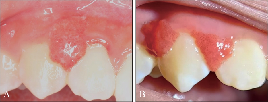
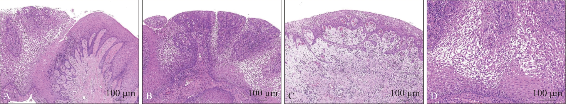
| [1] |
Darling MR, Daley TD, Wilson A, et al. Juvenile spongiotic gingivitis[J]. J Periodontol, 2007, 78(7): 1235-1240.
|
| [2] |
Chang JY, Kessler HP, Wright JM. Localized juvenile spongiotic gingival hyperplasia[J]. Oral Surg Oral Med Oral Pathol Oral Radiol Endod, 2008, 106(3): 411-418.
|
| [3] |
杨靖梅, 曾昕, 吴亚菲, 等. 青少年局限性海绵状牙龈增生1例[J]. 华西口腔医学杂志, 2024, 42(5): 667-670.
|
|
Yang JM, Zeng X, Wu YF, et al. Localized juvenile spongiotic gingival hyperplasia: a case report[J]. West China J Stomatol, 2024, 42(5): 667-670.
|
| [4] |
Kalogirou EM, Chatzidimitriou K, Tosios KI, et al. Localized juvenile spongiotic gingival hyperplasia: report of two cases[J]. J Clin Pediatr Dent, 2017,41(3): 228-231.
|
| [5] |
Solomon LW, Trahan WR, Snow JE.Localized juvenile spongiotic gingival hyperplasia:a report of 3 cases[J]. Pediatr Dent, 2013, 35(4): 360-363.
|
| [6] |
Wang MZ, Jordan RC. Localized juvenile spongiotic gingival hyperplasia: a report of 27 cases[J]. J Cutan Pathol, 2019, 46(11): 839-843.
|
| [7] |
Argyris PP, Nelson AC, Papanakou S, et al. Localized juvenile spongiotic gingival hyperplasia featuring unusual p16INK4A labeling and negative human papillomavirus status by polymerase chain reaction[J]. J Oral Pathol Med, 2015, 44(1): 37-44.
|
| [8] |
Moragonzalez D. Localized “juvenile” spongiotic gingival hyperplasia in adults; investigation of possible viral etiology and comparison with pyogenic granuloma[D]. North Carolina: University of North Carolina at Chapel Hill, 2014: 1-52.
|
| [9] |
Vargo RJ, Bilodeau EA. Reappraising localized juvenile spongiotic gingival hyperplasia[J]. J Am Dent Assoc, 2019, 150(2): 147-153.
|
| [10] |
Allon I, Lammert KM, Iwase R, et al. Localized juvenile spongiotic gingival hyperplasia possibly originates from the junctional gingival epithelium—an immunohistoche-mical study[J]. Histopathology, 2016, 68(4): 549-555.
|
| [11] |
Theofilou VI, Pettas E, Georgaki M, et al. Localized juvenile spongiotic gingival hyperplasia: microscopic variations and proposed change to nomenclature[J]. Oral Surg Oral Med Oral Pathol Oral Radiol, 2021, 131(3): 329-338.
|
| [12] |
de Freitas Silva BS, Sant’Ana SSS, Watanabe S, et al. Multifocal red bands of the marginal gingiva[J]. Oral Surg Oral Med Oral Pathol Oral Radiol, 2015, 119(1): 3-7.
|
| [13] |
Nogueira VK, Fernandes D, Navarro CM, et al. Cryotherapy for localized juvenile spongiotic gingival hyperplasia: preliminary findings on two cases[J]. Int J Paediatr Dent, 2017, 27(3): 231-235.
|
| [14] |
Fernandes DT, Wright JM, Lopes SMP, et al. Localized juvenile spongiotic gingival hyperplasia: a report of four cases and literature review[J]. Clin Adv Periodontics, 2018, 8(1): 17-21.
|
| [15] |
Silveira HA, Toral-Rizo VH, Lara-Carrillo E, et al. Spongiotic hyperplasia of the oral mucosa: case series and immunohistochemical analysis[J]. Oral Maxillofac Surg, 2022, 26(2): 333-337.
|
| [16] |
Vieira DL, Leite AF, Figueiredo PTS, et al. A conservative approach for localized spongiotic gingivitis hyperplasia using photodynamic therapy:a case report and review of the literature[J]. Photobiomodul Photomed Laser Surg, 2019, 37(1): 57-61.
|
| [17] |
Lafuente-Ibáñez de Mendoza I, Alberdi-Navarro J, Ma-richalar-Mendia X, et al. Characterization of juvenile spongiotic gingival hyperplasia as an entity of odontogenic origin[J]. J Periodontol, 2019, 90(12): 1490-1495.
|
| [18] |
MacNeill SR, Rokos JW, Umaki MR, et al. Conservative treatment of localized juvenile spongiotic gingival hyperplasia[J]. Clin Adv Periodontics, 2011, 1(3): 199-204.
|
| [19] |
Roberts EP, Schuster GM, Haub S. Case report of spongiotic gingivitis in an adult male treated with novel 9,300-nanometer carbon dioxide laser low-energy ablation[J]. J Am Dent Assoc, 2022, 153(1): 67-73.
|
| [20] |
Goldberg AS, Fatahzadeh M, Bortnik V, et al. Novel treatment approach of localized juvenile spongiotic gingival hyperplasia with use of Nd:YAG laser[J]. Clin Adv Periodontics, 2023, 13(4): 253-257.
|
| [21] |
Decani S, Lodi G, Sardella A, et al. Localised juvenile spongiotic gingival hyperplasia: a case of spontaneous resolution and a literature review[J]. Eur J Paediatr Dent, 2021, 22(2): 159-162.
|
| [22] |
Deseta M, Baldwin D, Siddik D, et al. Conservative management of juvenile spongiotic gingivitis: a series of ten cases[J]. Br Dent J, 2020, 229(5): 287-291.
|
| [23] |
Siamantas I, Kalogirou EM, Tosios KI, et al. Spongiotic gingival hyperplasia synchronously involving multiple sites: case report and review of the literature[J]. Head Neck Pathol, 2018, 12(4): 517-521.
|
| [24] |
Rezende KM, Moraes Pde C, Oliveira LB, et al. Cryosurgery as an effective alternative for treatment of oral lesions in children[J]. Braz Dent J, 2014, 25(4): 352-356.
|
| [25] |
Prażmo EJ, Kwaśny M, Łapiński M, et al. Photodynamic therapy as a promising method used in the treatment of oral diseases[J]. Adv Clin Exp Med, 2016, 25(4): 799-807.
|

 ), Cao Rongfang1,2, Yao Rui1,2, Shang Jianwei2,3(
 This work is licensed under a Creative Commons Attribution 3.0 License.