| 1 |
Valenti M, Schmitz JH, Cortellini D, et al. A diagnostically and digitally driven tooth preparation protocol by using a patient monitoring tool with an intraoral scanner[J]. J Prosthet Dent, 2023, 129(1): 7-13.
|
| 2 |
Badar SB, Ghafoor R, Hameed MH, et al. Assessment of iatrogenic damage to adjacent teeth during crown and bridge preparation[J]. Indian J Dent Res, 2019, 30(1): 107-111.
|
| 3 |
Li J, Moon HS, Kim JH, et al. Accuracy of impression-making methods in edentulous arches: an in vitro study encompassing conventional and digital methods[J]. J Pro-sthet Dent, 2022, 128(3): 479-486.
|
| 4 |
Li C, Zou B, Xin W, et al. Investigation on the effectiveness of digital scanning combined with reverse enginee-ring technology in demonstrating full crown tooth preparation[J]. Int J Prosthodont, 2024: 1-22.
|
| 5 |
王勇. 口内数字印模技术[J]. 口腔医学, 2015, 35(9): 705-709, 743.
|
|
Wang Y. Intraoral digital impression technique[J]. Stomatology, 2015, 35(9): 705-709, 743.
|
| 6 |
杨日桃, 张晓峥. 265件钴铬烤瓷单冠返工的原因分析[J]. 临床口腔医学杂志, 2012, 28(12): 736-737.
|
|
Yang RT, Zhang XZ. Cause analysis on reprocessing of 265 cobalt-chromium PFM single crowns[J]. J Clin Stomatol, 2012, 28(12): 736-737.
|
| 7 |
卢嘉仪, 赵君仪, 高静, 等. 1 312件单冠预备体数字化模型的关键预备质量指标的分析研究[J]. 华西口腔医学杂志, 2022, 40(1): 52-60.
|
|
Lu JY, Zhao JY, Gao J, et al. Investigation on the quality analysis of 1 312 single crown digital models[J]. West China J Stomatol, 2022, 40(1): 52-60.
|
| 8 |
Shahmoradi M, Wan B, Zhang Z, et al. Monolithic crowns fracture analysis: the effect of material properties, cusp angle and crown thickness[J]. Dent Mater, 2020, 36(8): 1038-1051.
|
| 9 |
李忠义, 白鹤飞, 王勇, 等. 牙体预备定量引导技术的研究现状[J]. 中华口腔医学杂志, 2018, 53(2): 137-140.
|
|
Li ZY, Bai HF, Wang Y, et al. Research status of tooth preparation quantitative guide technique[J]. Chin J Stomatol, 2018, 53(2): 137-140.
|
| 10 |
赵铱民. 口腔修复学[M]. 8版. 北京: 人民卫生出版社, 2020.
|
|
Zhao YM. Prosthodontics[M]. 8th ed. Beijing: People’s Medical Publishing House, 2020.
|
| 11 |
罗天, 李俊颖, 于海洋. 制备高精度牙预备体肩台的临床路径和预备方法[J]. 华西口腔医学杂志, 2020, 38(6): 712-717.
|
|
Luo T, Li JY, Yu HY. Clinical pathway and preparation method of high-precision tooth shoulder platform[J]. West China J Stomatol, 2020, 38(6): 712-717.
|
| 12 |
UrliĆ I, Demoli N, Pavan J, et al. Measuring tooth vibrations induced during cavity preparation with time-ave-raged holography and its influence on near vision acuity in dentists[J]. Dent Mater J, 2021, 40(1): 123-128.
|
| 13 |
Okawa T, Abe S, Nakano M, et al. Evaluation of the measurement precision and accuracy in the dental CAD/CAM system[J]. Dent Mater J, 2020, 39(5): 784-791.
|
| 14 |
萧宁, 孙玉春, 赵一姣, 等. 三种数字化分析算法测量咬合接触分布及面积的对比研究[J]. 北京大学学报(医学版), 2020, 52(1): 144-151.
|
|
Xiao N, Sun YC, Zhao YJ, et al. Preliminary study on three digital analysis methods for analyzing the distribution and area of occlusal contacts[J]. J Peking Univ: Health Sci, 2020, 52(1): 144-151.
|
| 15 |
Tekin YH, Hayran Y. Fracture resistance and marginal fit of the zirconia crowns with varied occlusal thickness[J]. J Adv Prosthodont, 2020, 12(5): 283-290.
|
| 16 |
Zimmermann M, Ender A, Mehl A. Influence of CAD/CAM fabrication and sintering procedures on the fracture load of full-contour monolithic zirconia crowns as a function of material thickness[J]. Oper Dent, 2020, 45(2): 219-226.
|
| 17 |
皮昕.口腔解剖生理学[M]. 8版.北京: 人民卫生出版社, 2020.
|
|
Pi X. Oral anatomy and physiology[M]. 8th ed. Beijing: People’s Medical Publishing House, 2020.
|
| 18 |
Veldt EA, Vermaire JH, van Houtem CMHH, et al. A study to determine possible success variables in the treatment of gag reflex patients[J]. Ned Tijdschr Tandheelkd, 2018, 125(2): 101-107.
|
| 19 |
Cheng Y, Zhou YF, Ding YP, et al. Cleaning the palate and tongue without nausea: a mixed methods study exploring the appropriate depth and direction of oral care[J]. BMC Oral Health, 2021, 21(1): 67.
|
| 20 |
Markerink H, van Geffen GJ, Bruhn J. Topical anaesthesia using a soft mist spray device allows comfortable awake visualisation of the airway via self-videolaryngoscopy in volunteers[J]. Medicina (Kaunas), 2024, 60(1): 176.
|
| 21 |
Amine M, Wahid HO, Fahi S, et al. Assessment of convergence angle of tooth preparations for complete crowns among dental students: typodont vs simulator[J]. Int J Dent, 2022, 2022: 7615892.
|

 ), Nie Rongrong3(
 This work is licensed under a Creative Commons Attribution 3.0 License.
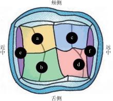

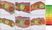

 面分区
面分区 面分区
面分区