West China Journal of Stomatology ›› 2018, Vol. 36 ›› Issue (5): 576-579.doi: 10.7518/hxkq.2018.05.021
Di Wu( ),Jin Zhang,Wei Wang,Bing-chang Xin()
Received:2018-03-08
Revised:2018-07-06
Online:2018-10-01
Published:2018-10-18
CLC Number:
Di Wu,Jin Zhang,Wei Wang,Bing-chang Xin. Mandibular first molar with C-shaped root canal system: a two-case report[J]. West China Journal of Stomatology, 2018, 36(5): 576-579.
Add to citation manager EndNote|Ris|BibTeX
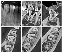
Fig 1
X-ray and CBCT of the case 1 before treatment


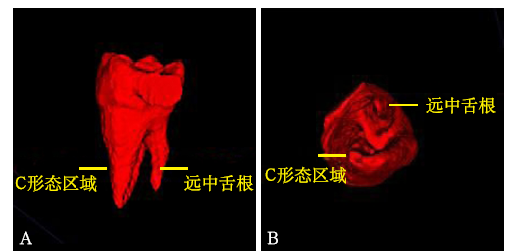
Fig 2
3D reconstruction of case 1 before treatment
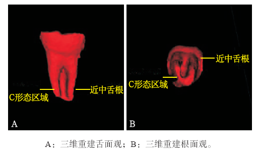
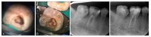
Fig 3
The treatment of case 1

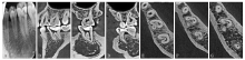
Fig 4
X-ray and CBCT of the case 2 before treatment
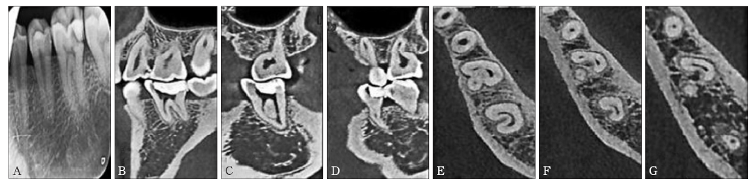

Fig 5
3D reconstruction of case 2 before treatment
| [1] | 高荣寰 . 下颌第一恒磨牙5根管2例[J]. 北京口腔医学, 2008,16(6):321. |
| Gao RH . Mandibular first molar of five root canals: two cases report[J]. Beijing J Stomatol, 2008,16(6):321. | |
| [2] |
王瑶, 郑广宁, 郑庆华 , 等. 下颌恒磨牙根管系统的锥形束CT研究[J]. 泸州医学院学报, 2011,34(5):616-619.
doi: 10.3969/j.issn.1000-2669.2011.05.038 URL |
|
Wang Y, Zheng GN, Zheng QH , et al. A cone-beam com-puted tomography study of mandibular permanent molar root canal system morphology[J]. J Luzhou Med Coll, 2011,34(5):616-619.
doi: 10.3969/j.issn.1000-2669.2011.05.038 URL |
|
| [3] |
姜楠, 周洲, 周耀 , 等. 华东地区中国人下颌第一恒磨牙根管形态的锥形束CT分析[J]. 南京医科大学学报(自然科学版), 2013,33(5):693-697.
doi: 10.7655/NYDXBNS20130529 URL |
|
Jiang N, Zhou Z, Zhou Y , et al. Use of cone-beam computed tomography to evaluate root and canal morphology of man-dibular first molars in Chinese individuals[J]. Acta Univ Med Nanjing (Nat Sci), 2013,33(5):693-697.
doi: 10.7655/NYDXBNS20130529 URL |
|
| [4] |
姚娜, 贾立辉, 张晓东 , 等. 辽沈地区人群下颌第一磨牙牙根及根管解剖的CBCT观察[J]. 临床口腔医学杂志, 2014,30(4):205-207.
doi: 10.3969/j.issn.1003-1634.2014.04.005 URL |
|
Yao N, Jia LH, Zhang XD , et al. Observation of the roots and canals morphology of mandibular first permanent molars in Liaoning province by cone-beam computed tomography[J]. J Clin Stomatol, 2014,30(4):205-207.
doi: 10.3969/j.issn.1003-1634.2014.04.005 URL |
|
| [5] | Demirbuga S, Sekerci AE, Dinçer AN , et al. Use of cone-beam computed tomography to evaluate root and canal mor-phology of mandibular first and second molars in Turkish individuals[J]. Med Oral Patol Oral Cir Bucal, 2013,18(4):e737-e744. |
| [6] |
李明霞, 王光平, 杨江华 . 青少年下颌第一恒磨牙牙根及根管的锥形束 CT 研究[J]. 实用口腔医学杂志, 2015,31(3):397-400.
doi: 10.3969/j.issn.1001-3733.2015.03.022 URL |
|
Li MX, Wang GP, Yang JH . Investigation of root and root canal of mandibular first permanent molars in adolescents by cone-beam computed tomography[J]. J Pract Stomatol, 2015,31(3):397-400.
doi: 10.3969/j.issn.1001-3733.2015.03.022 URL |
|
| [7] |
王俊, 刘青梅, 程琳 . 双侧下颌第一恒磨牙融合根伴C形根管1例[J]. 国际口腔医学杂志, 2015,42(3):263-264.
doi: 10.7518/gjkq.2015.03.004 URL |
|
Wang J, Liu QM, Cheng L . Bilateral mandibular permanent first molars with fused roots and C-shaped root canal[J]. Int J Stomatol, 2015,42(3):263-264.
doi: 10.7518/gjkq.2015.03.004 URL |
|
| [8] |
Manning SA . Root canal anatomy of mandibular second molars. Part Ⅱ. C-shaped canals[J]. Int Endod J, 1990,23(1):40-45.
doi: 10.1111/j.1365-2591.1990.tb00801.x URL pmid: 2391179 |
| [9] |
Fan B, Cheung GS, Fan MW , et al. C-shaped canal system in mandibular second molars: Part Ⅰ—Anatomical features[J]. J Endod, 2004,30(12):899-903.
doi: 10.1097/01.don.0000136207.12204.e4 URL pmid: 15564874 |
| [10] |
Fan B, Cheung GS, Fan MW , et al. C-shaped canal system in mandibular second molars: Part Ⅱ—Radiographic fea-tures[J]. J Endod, 2004,30(12):904-908.
doi: 10.1097/01.don.0000136206.73115.93 URL |
| [11] | 欧阳勇, 凌均棨, 陈慧芝 , 等. 下颌第二磨牙C形根管的临床研究[J]. 中山大学学报(医学科学版), 2003,24(s1):172-174. |
| Ouyang Y, Ling JQ, Chen HZ , et al. Clinical research of C-shaped root canals in mandibular second molars[J]. J Sun Yat-Sen Univ (Med Sci), 2003,24(s1):172-174. | |
| [12] |
Al-Qudah AA, Awawdeh LA . Root and canal morphology of mandibular first and second molar teeth in a Jordanian population[J]. Int Endod J, 2009,42(9):775-784.
doi: 10.1111/j.1365-2591.2009.01578.x URL pmid: 19549153 |
| [13] | 喻刚, 叶玲, 黄定明 , 等. 成都地区378颗中国人下颌第一恒磨牙远舌根的临床研究[J]. 实用口腔医学杂志, 2012,28(4):498-501. |
| Yu G, Ye L, Huang DM , et al. Clinical study of radix ento-molaris in permanent mandibular first molars in Chinese individuals in Chengdu[J]. J Pract Stomatol, 2012,28(4):498-501. | |
| [14] |
李颖超, 刘荣森, 石校伟 . 老年人下颌磨牙牙根及根管的锥形束CT研究[J]. 中华老年口腔医学杂志, 2009,7(4):206-209.
doi: 10.3969/j.issn.1672-2973.2009.04.005 URL |
|
Li YC, Liu RS, Shi XW . Investigation of root and root canal of mandibular molar in the elderly by cone beam computer tomography[J]. Chin J Geriatr Dent, 2009,7(4):206-209.
doi: 10.3969/j.issn.1672-2973.2009.04.005 URL |
|
| [15] | 范兵, 吴昊 . C形根管的形态特点及处理方法[J]. 中国实用口腔科杂志, 2011,4(9):521-524. |
| Fan B, Wu H . The anatomical characteristics of C-shaped canal and its treatment[J]. Chin J Pract Stomatol, 2011,4(9):521-524. |
| [1] | Cai Meijuan, Xiang Shaowen, Xie Chengjie, Ouyang Chuhong, Tong Fangli. Intentional replantation for the retreatment of mandibular second molar: a case report [J]. West China Journal of Stomatology, 2023, 41(4): 471-477. |
| [2] | Li Chengxi, Song Weijian.. Root canal treatment of type Ⅱ and ⅢA double dens invaginatus in maxillary lateral incisor: a case report [J]. West China Journal of Stomatology, 2023, 41(2): 232-236. |
| [3] | Su Wenqi, Shi Jiahong, Cheng Yan, Lei Lang, Li Houxuan. Periodontal treatment of furcation involvement at the mandibular first molar with a follow-up of 27 years [J]. West China Journal of Stomatology, 2021, 39(3): 347-354. |
| [4] | Zhai Xiaoyang,Zhang Jingya,Zhang Sanke,Jiang Chuanjing,Qiu Xiaoxia. Finite-element analysis of mandibular first molar with two marginal designs of endocrown for the repair of different defects [J]. West China Journal of Stomatology, 2019, 37(5): 480-484. |
| [5] | Xu Weizhe,Song Dongzhe,Tan Xuelian,Zhang Lan,Huang Dingming. Vital pulp preservation treatment in mandibular right first molar with vertical root fractures: a case report [J]. West China Journal of Stomatology, 2019, 37(5): 563-567. |
| [6] | Liu Tongxi,Zheng Zhiguo,Yang Jian. Apical barrier technology to treat chronic apical periodontitis caused by type Ⅱ dens invaginatus: a case report [J]. West China Journal of Stomatology, 2019, 37(5): 568-570. |
| [7] | Weilin Qiang,Yuxuan Li,Gang Liu,Lihong Zhang,Jingyang Zhang,Wenjin Cao. Three-year retrospective clinical evaluation of pulp-less molars with defects of varying degree repaired by cast ceramic onlays of three marginal types [J]. West China Journal of Stomatology, 2018, 36(5): 493-497. |
| [8] | Yang Meng, Degang. Sun. Maxillary first premolar with four canals: a case report [J]. West China Journal of Stomatology, 2018, 36(2): 229-231. |
| [9] | Lili Yang, Yan Zhang, Shijun Zhao, Shuai Zhang, Na Wang, Jie Xu, Shue Hu, Zhiyuan Xu. Clinical application of cone beam computed tomography combined with micro-ultrasound technique in treating three mesial canals in mandibular first molars [J]. West China Journal of Stomatology, 2017, 35(4): 384-388. |
| [10] | Tan Baochun, Xiao Jianping, Yan Fuhua, Huan Hong. Clinical comparative study on the efficacy of periodontal endodontic therapy and periodontal treatment alone for advanced periodontitis [J]. West China Journal of Stomatology, 2016, 34(6): 600-605. |
| [11] | Cui Pingping, Wang Xiaoya, Yu Jian, Sun Qinfeng.. Maxillary first molar with two distobuccal root canals: a case report [J]. West China Journal of Stomatology, 2016, 34(5): 539-540. |
| [12] | Wu Di, Wu Hongbin.. Maxillary first molar with twin-root canal in palatal side: two case reports [J]. West China Journal of Stomatology, 2015, 33(3): 329-330. |
| [13] | Yu Qing, Yang Yang, Chang Bei.. The technology of apical infection control [J]. West China Journal of Stomatology, 2014, 32(5): 427-431. |
| [14] | Xie Kexian, Wang Xiao, Li Yuangao, Zhang Ping. Root canal treatment of two-rooted three-canal maxillary first premolar: a case report [J]. West China Journal of Stomatology, 2013, 31(6): 641-643. |
| [15] | Yu Gang1, Ye Ling2, Huang Dingming2. Clinical investigation of radix entomolaris in mandibular first molars [J]. West China Journal of Stomatology, 2012, 30(3): 259-261. |
| Viewed | ||||||
|
Full text |
|
|||||
|
Abstract |
|
|||||
 This work is licensed under a Creative Commons Attribution 3.0 License.
