| 1 |
Sanchez-Larsen A, Sopelana D, Diaz-Maroto I, et al. Assessment of efficacy and safety of eslicarbazepine acetate for the treatment of trigeminal neuralgia[J]. Eur J Pain, 2018, 22(6): 1080-1087.
|
| 2 |
Zhao H, Wang XH, Zhang Y, et al. Management of primary bilateral trigeminal neuralgia with microvascular decompression: 13-case series[J]. World Neurosurg, 2018, 109: e724-e730.
|
| 3 |
Tsai YH, Yuan R, Patel D, et al. Altered structure and functional connection in patients with classical trigeminal neuralgia[J]. Hum Brain Mapp, 2018, 39(2): 609-621.
|
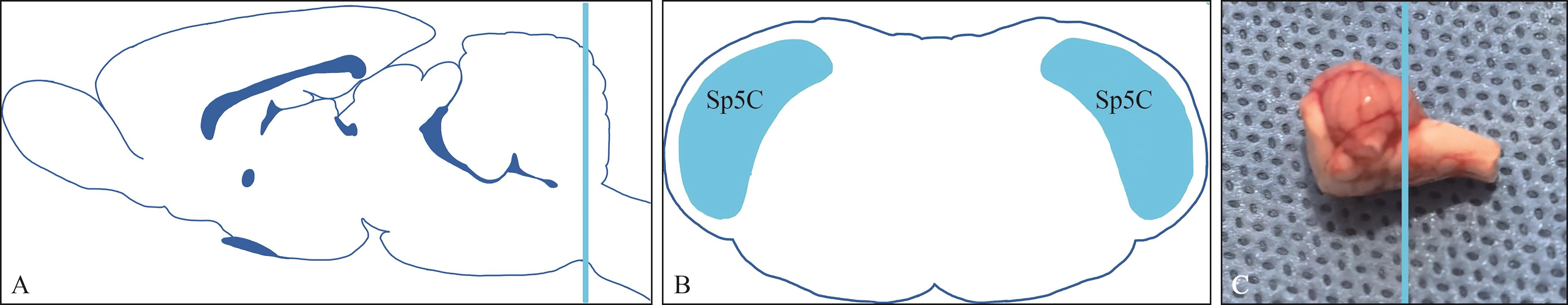
| 4 |
Kobayashi M, Nakaya Y. Anatomical aspects of corticotrigeminal projections to the medullary dorsal horn[J]. J Oral Sci, 2020, 62(2): 144-146.
|
| 5 |
Giordano KR, Denman CR, Dubisch PS, et al. An update on the rod microglia variant in experimental and clinical brain injury and disease[J]. Brain Commun, 2021, 3(1): fcaa227.
|
| 6 |
Graeber MB. Changing face of microglia[J]. Science, 2010, 330(6005): 783-788.
|
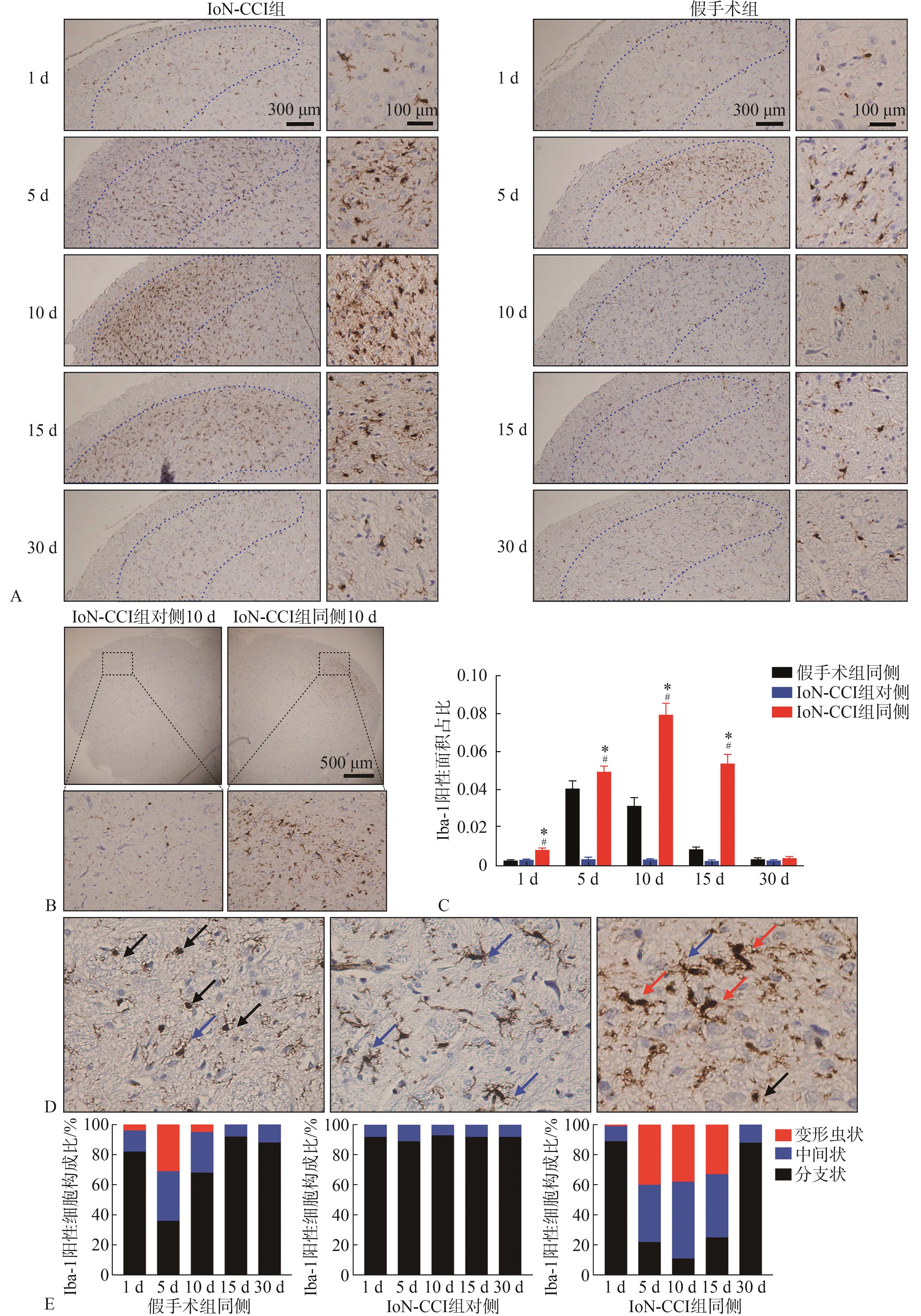
| 7 |
Young K, Morrison H. Quantifying microglia morpho-logy from photomicrographs of immunohistochemistry prepared tissue using imageJ[J]. J Vis Exp, 2018(136): 57648.
|
| 8 |
Pomilio C, Gorojod RM, Riudavets M, et al. Microglial autophagy is impaired by prolonged exposure to β-amyloid peptides: evidence from experimental models and Alzheimer’s disease patients[J]. Geroscience, 2020, 42(2): 613-632.
|
| 9 |
Bathla G, Hegde AN. The trigeminal nerve: an illustrated review of its imaging anatomy and pathology[J]. Clin Radiol, 2013, 68(2): 203-213.
|
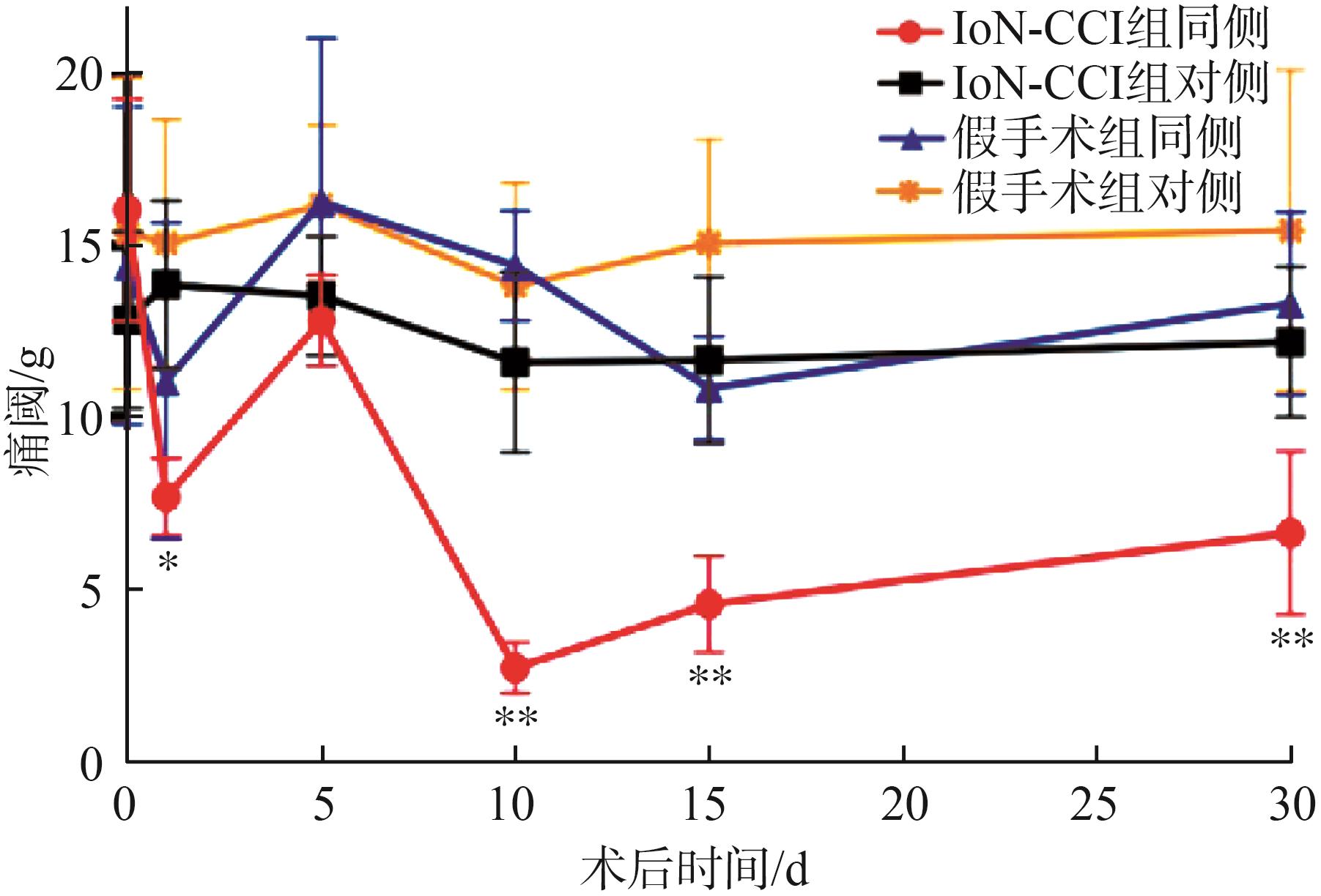
| 10 |
Zuo X, Ling JX, Xu GY, et al. Operant behavioral responses to orofacial cold stimuli in rats with chronic constrictive trigeminal nerve injury: effects of menthol and capsazepine[J]. Mol Pain, 2013, 9: 28.
|
| 11 |
Fink BR, Aasheim G, Kish SJ, et al. Neurokinetics of lidocaine in the infraorbital nerve of the rat in vivo: relation to sensory block[J]. Anesthesiology, 1975, 42(6): 731-736.
|
| 12 |
Henry MA, Fairchild DD, Patil MJ, et al. Effect of a novel, orally active matrix metalloproteinase-2 and -9 inhibitor in spinal and trigeminal rat models of neuropa-thic pain[J]. J Oral Facial Pain Headache, 2015, 29(3): 286-296.
|
| 13 |
Ding WH, You ZR, Shen SQ, et al. An improved rodent model of trigeminal neuropathic pain by unilateral chro-nic constriction injury of distal infraorbital nerve[J]. J Pain, 2017, 18(8): 899-907.
|
| 14 |
Dingle A, Zeng WF, Ness JP, et al. Strategies for interfa-cing with the trigeminal nerves in rodents for bioelectric medicine[J]. J Neurosci Methods, 2019, 324: 108321.
|
| 15 |
Imamura Y, Kawamoto H, Nakanishi O. Characterization of heat-hyperalgesia in an experimental trigeminal neuropathy in rats[J]. Exp Brain Res, 1997, 116(1): 97-103.
|
| 16 |
樊林花, 李丹, 樊平花, 等. 清洁级SD大鼠体重和主要脏器系数正常参考值研究及相关性分析[J]. 中国卫生检验杂志, 2012, 22(4): 750-752.
|
|
Fan LH, Li D, Fan PH, et al. Study and correlation analysis of normal reference range of body weight and the main organs coefficient of clean SD rat[J]. Chin J Heal Lab Tech, 2012, 22(4): 750-752.
|
| 17 |
Xia L, Liu MX, Zhong J, et al. Pain threshold monitoring during chronic constriction injury of the infraorbital nerve in rats[J]. Br J Neurosurg, 2019, 33(4): 409-412.
|
| 18 |
Ananthan S, Benoliel R. Chronic orofacial pain[J]. J Neural Transm (Vienna), 2020, 127(4): 575-588.
|
| 19 |
Yatziv SL, Devor M. Suppression of neuropathic pain by selective silencing of dorsal root ganglion ectopia using nonblocking concentrations of lidocaine[J]. Pain, 2019, 160(9): 2105-2114.
|
| 20 |
Catterall WA, Lenaeus MJ, Gamal El-Din TM. Structure and pharmacology of voltage-gated sodium and calcium channels[J]. Annu Rev Pharmacol Toxicol, 2020, 60: 133-154.
|
| 21 |
Goodwin G, McMahon SB. The physiological function of different voltage-gated sodium channels in pain[J]. Nat Rev Neurosci, 2021, 22(5): 263-274.
|
| 22 |
Inoue K, Tsuda M. Microglia in neuropathic pain: cellular and molecular mechanisms and therapeutic potential[J]. Nat Rev Neurosci, 2018, 19(3): 138-152.
|

 ), Zhang Jingqi, Lai Wenli.(
 This work is licensed under a Creative Commons Attribution 3.0 License.