West China Journal of Stomatology ›› 2022, Vol. 40 ›› Issue (5): 582-588.doi: 10.7518/hxkq.2022.05.012
Previous Articles Next Articles
Li Yaqi( ), Wang Ziqian, Liu Jiaqi, Yan Zhebin, Xiao Chuqiao, Wang Jun, Xiong Xin.()
Received:2022-02-15
Revised:2022-07-13
Online:2022-10-01
Published:2022-10-17
Contact:
Xiong Xin.
E-mail:liyaqi@stu.scu.edu.cn;drxiongxin@scu.edu.cn
Supported by:CLC Number:
Li Yaqi, Wang Ziqian, Liu Jiaqi, Yan Zhebin, Xiao Chuqiao, Wang Jun, Xiong Xin.. Morphometric evaluation of sella turcica and cranial base in patients with congenital absence of teeth[J]. West China Journal of Stomatology, 2022, 40(5): 582-588.
Add to citation manager EndNote|Ris|BibTeX
Tab 1
Distribution of age and sex in each group
| 组别 | 患者人数 | 年龄/岁 | 男性(n/%) | 女性(n/%) |
|---|---|---|---|---|
| 合计 | 322 | 15.46±4.66 | 159/49.4 | 163/50.6 |
| 对照组 | 112 | 15.19±4.49 | 51/45.5 | 61/54.5 |
| 试验Ⅰ组 | 104 | 15.79±4.93 | 51/49.0 | 53/51.0 |
| 试验Ⅱ组 | 106 | 15.42±4.59 | 57/53.8 | 49/46.2 |

Fig 1
Number of missing teeth in the study group
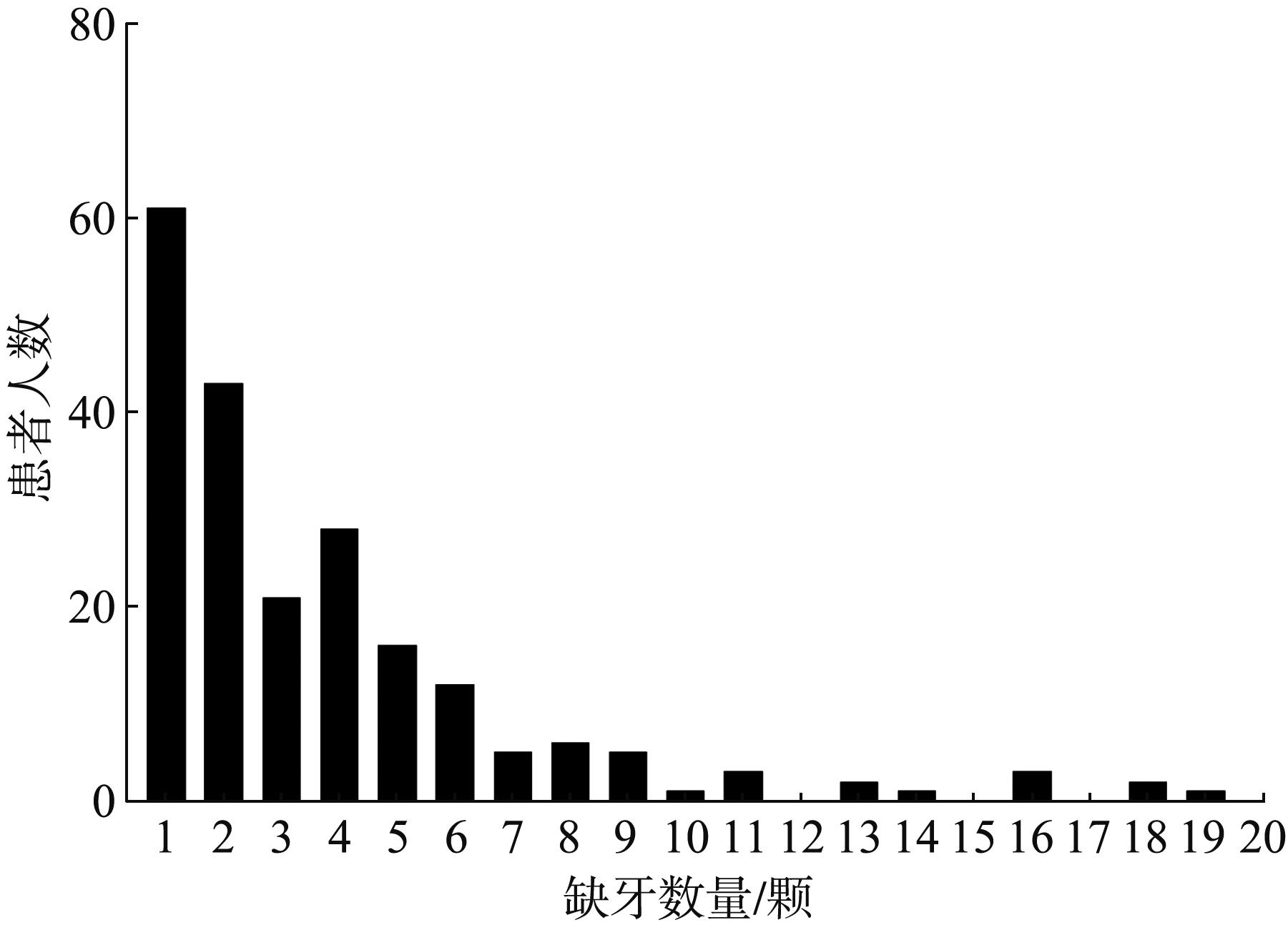

Fig 2
Distribution of missing teeth in the study group
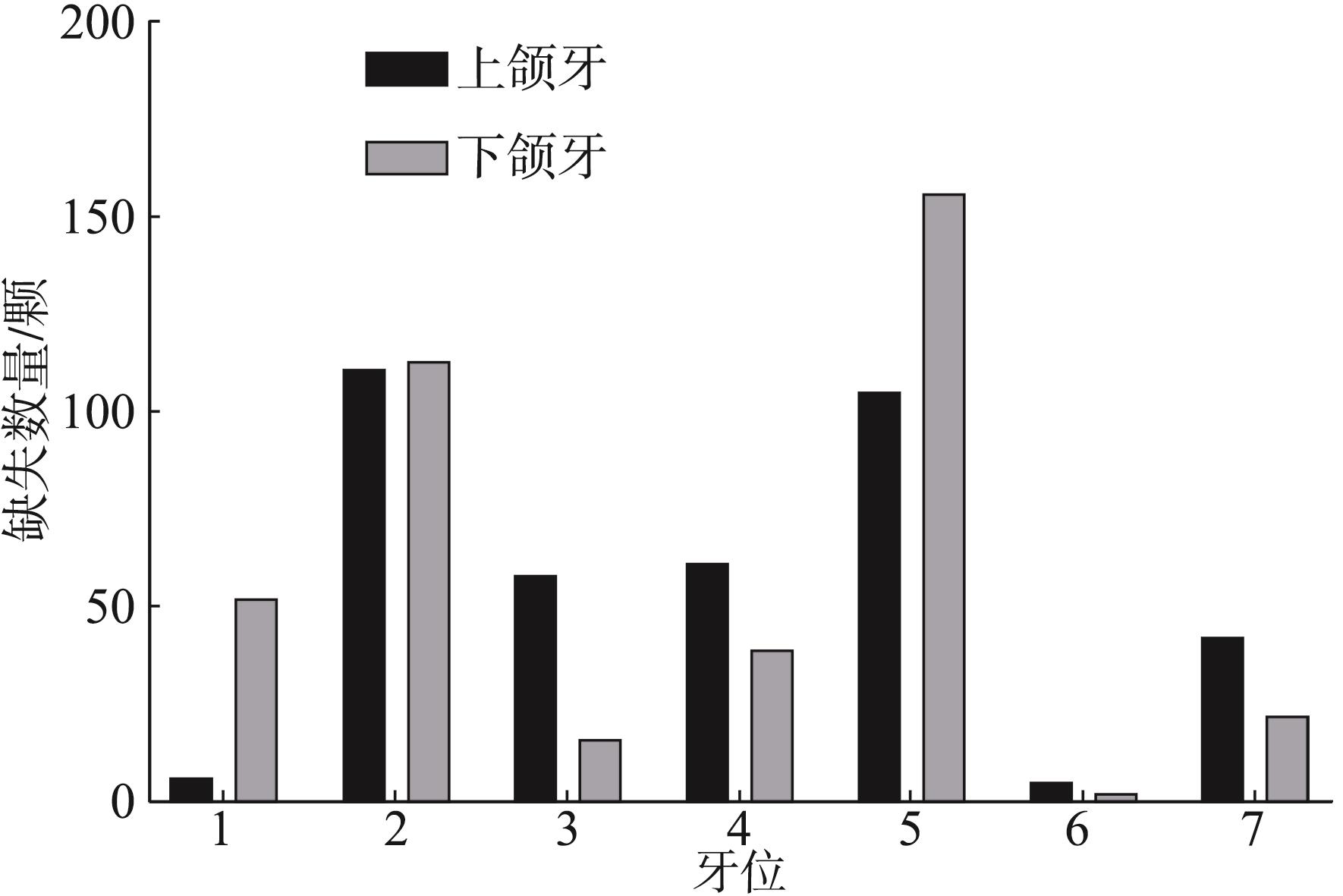
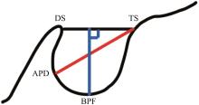
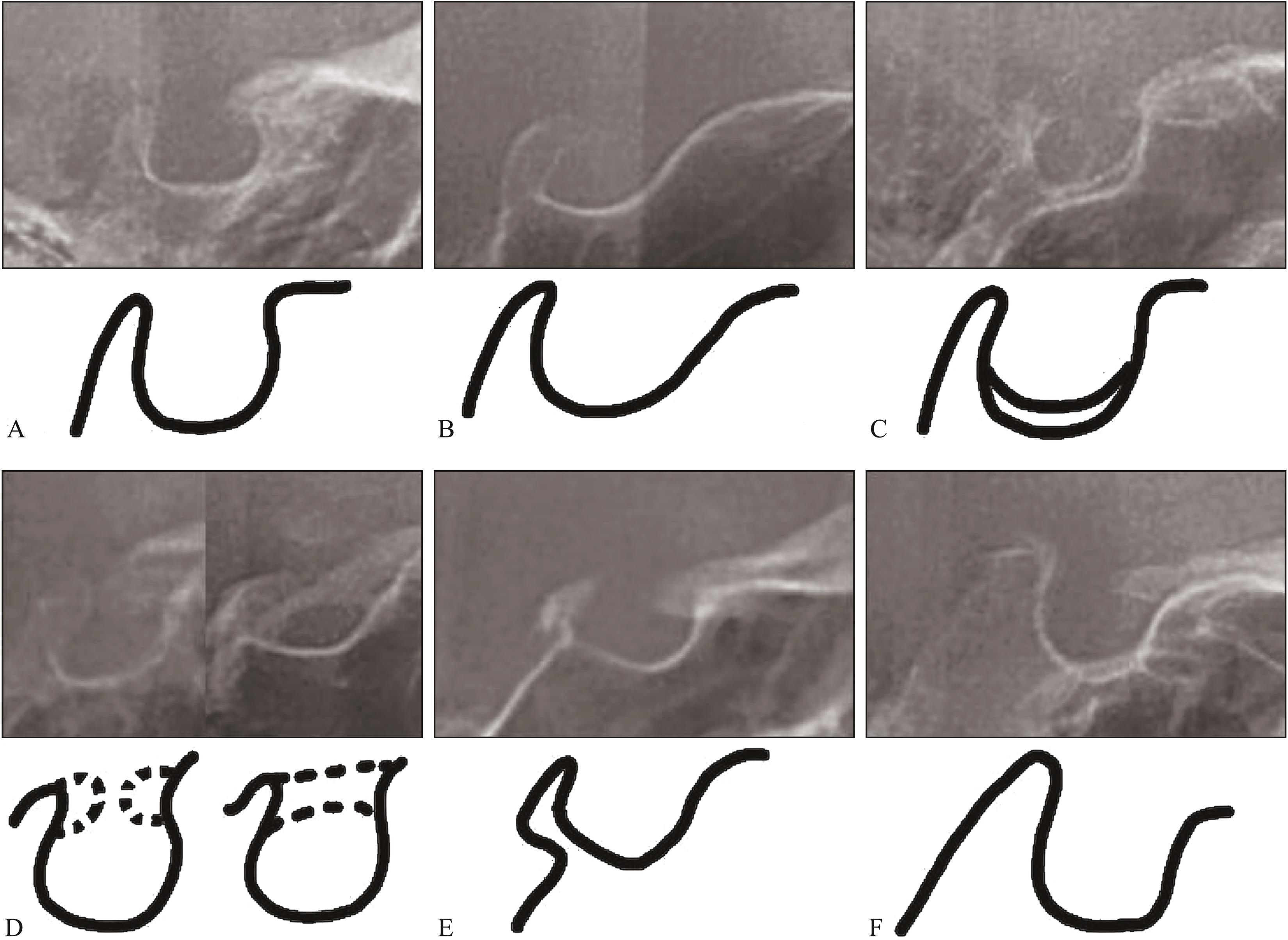
Fig 3
Normal sella turcica morphology and mark points and re-ference lines used for measuring sella turcica size
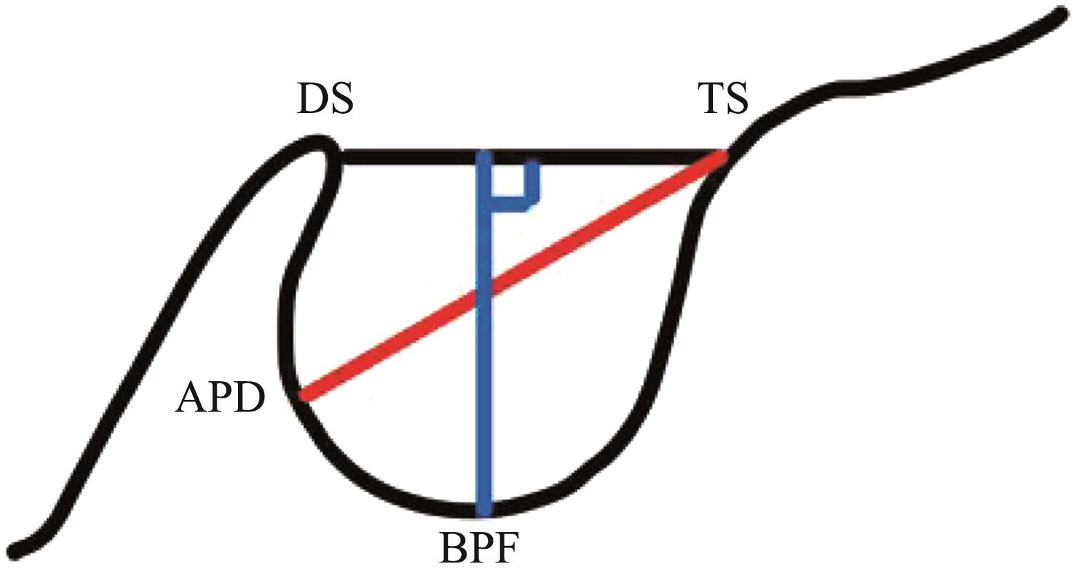
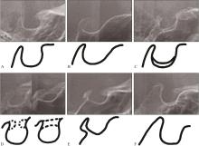
Fig 4
The shape of sella turcica
Tab 2
Cephalometric landmarks used in the study
| 标志点 | 定义 |
|---|---|
| 鼻根点(N) | 鼻额缝的最前点 |
| 蝶鞍点(S) | 蝶鞍影像的中心 |
| 颅底点(Ba) | 枕骨大孔前缘的中点 |
| 关节点(Ar) | 颅底下缘与下颌髁突颈后缘的交点 |
| Sphenoidale点(Se) | 蝶骨大翼和颅前窝的交点 |
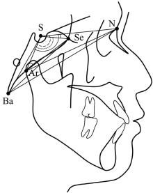
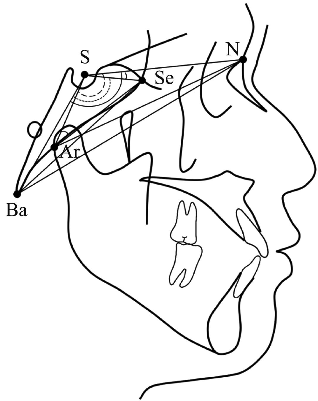
Fig 5
Linear and angular measurements related to cranial base
Tab 3
Comparative analysis of cephalometric parameters in each group
| 项目 | 对照组 | 试验Ⅰ组 | 试验Ⅱ组 | P值 | |||
|---|---|---|---|---|---|---|---|
| 均值±标准差 | 最小值~最大值 | 均值±标准差 | 最小值~最大值 | 均值±标准差 | 最小值~最大值 | ||
| 蝶鞍 | |||||||
| 长度/mm | 8.58±1.42 | 5.83~12.19 | 8.78±1.44 | 5.55~13.47 | 8.84±1.49 | 4.58~12.93 | 0.379a |
| 深度/mm | 7.03±1.10 | 3.36~10.00 | 7.01±1.13 | 4.44~9.83 | 6.88±1.10 | 4.51~9.80 | 0.553a |
| 直径/mm | 11.02±1.26 | 8.26~13.81 | 10.98±1.23 | 7.40~14.65 | 10.85±1.42 | 8.20~15.65 | 0.615a |
| 前颅底 | |||||||
| S-N/mm | 62.92±4.08 | 46.35~73.69 | 63.23±3.72 | 55.64~74.28 | 63.12±3.96 | 50.95~73.59 | 0.922b |
| S-Se/mm | 24.46±2.41 | 18.79~29.65 | 24.91±2.45 | 20.27~32.15 | 24.68±2.59 | 19.72~30.88 | 0.416a |
| ∠N-S-Se/° | 0.91±3.33 | -9.95~9.09 | 0.78±3.82 | -11.15~10.76 | 1.02±3.94 | -10.06~10.07 | 0.898a |
| 后颅底 | |||||||
| S-Ba/mm | 45.99±3.69 | 37.31~53.14 | 45.47±3.55 | 39.30~54.11 | 45.65±3.37 | 36.63~54.91 | 0.543a |
| S-Ar/mm | 33.91±3.68 | 25.08~43.09 | 33.78±3.77 | 26.49~44.12 | 33.73±3.59 | 26.76~41.75 | 0.935a |
| Se-Ar/mm | 51.29±4.38 | 41.31~66.18 | 51.67±4.26 | 40.93~67.97 | 51.41±3.85 | 42.33~59.00 | 0.789a |
| Se-Ba/mm | 64.08±4.31 | 55.14~76.71 | 63.99±3.88 | 56.68~78.04 | 64.00±3.80 | 55.00~72.30 | 0.829b |
| ∠Se-S-Ba/° | 128.40±5.15 | 115.12~140.58 | 128.77±6.79 | 113.19~153.91 | 128.74±5.65 | 115.43~146.29 | 0.864b |
| ∠Se-Ar-S/° | 23.83±3.18 | 17.99~34.38 | 23.94±3.89 | 10.97~36.11 | 23.86±3.41 | 17.03~34.32 | 0.898b |
| 全颅底 | |||||||
| N-Ba/mm | 98.64±6.27 | 76.97~114.74 | 98.52±5.79 | 86.25~115.36 | 98.69±5.59 | 86.14~109.26 | 0.976a |
| N-Ar/mm | 86.22±6.21 | 67.73~104.16 | 86.58±5.94 | 74.57~103.32 | 86.48±5.67 | 74.48~99.06 | 0.899a |
| ∠N-S-Ba/° | 129.31±4.17 | 119.81~140.04 | 129.55±5.49 | 118.09~149.68 | 129.76±4.46 | 118.10~140.65 | 0.783a |
| ∠N-S-Ar/° | 123.12±4.52 | 112.76~136.17 | 123.66±5.61 | 111.46~145.92 | 123.73±4.49 | 112.15~134.67 | 0.466b |
Tab 4
Morphology of sella turcica in each group
| 组别 | 正常蝶鞍 | 前壁倾斜 | 鞍底双重轮廓 | 鞍桥 | 后部不规则 | 金字塔鞍背 |
|---|---|---|---|---|---|---|
| 对照组 | 63/56.3%a | 6/5.4% | 5/4.5% | 25/22.3%c | 9/8.0% | 4/3.6% |
| 试验Ⅰ组 | 39/37.5%b | 8/7.7% | 5/4.8% | 40/38.5%d | 5/4.8% | 7/6.7% |
| 试验Ⅱ组 | 42/39.6%b | 7/6.6% | 4/3.8% | 41/38.7%d | 7/6.6% | 5/4.7% |
| χ2值 | 9.33 | 0.48 | 0.22 | 8.74 | 0.92 | 1.16 |
| P值 | 0.009** | 0.785 | 0.944 | 0.013* | 0.630 | 0.560 |
| 1 | Khalaf K, Miskelly J, Voge E, et al. Prevalence of hypodontia and associated factors: a systematic review and Meta-analysis[J]. J Orthod, 2014, 41(4): 299-316. |
| 2 | Kjær I. Mechanism of human tooth eruption: review article including a new theory for future studies on the eruption process[J]. Scientifica (Cairo), 2014, 2014: 341905. |
| 3 | Becktor JP, Einersen S, Kjaer I. A sella turcica bridge in subjects with severe craniofacial deviations[J]. Eur J Orthod, 2000, 22(1): 69-74. |
| 4 | 刘军, 杨洁, 李铎, 等. 第二前磨牙先天缺失与蝶鞍的相关性研究[J]. 中国实用口腔科杂志, 2019, 12(2): 92-95. |
| Liu J, Yang J, Li D, et al. Study on the relationship between sella and the congenital absence of second premolar[J]. Chin J Pract Stomatol, 2019, 12(2): 92-95. | |
| 5 | Alqahtani H. Association between sella turcica bridging and congenitally missing maxillary lateral incisors[J]. J Dent Sci, 2020, 15(1): 59-64. |
| 6 | Canigur Bavbek N, Arslan Avan B. Morphometric evaluation of cranial base and sella turcica in patients with bilateral agenesis of maxillary lateral incisors[J]. Odonto-logy, 2021, 109(3): 701-709. |
| 7 | 李亚其, 王梓千, 刘家琦, 等. 蝶鞍与颈椎成熟度的相关性研究[J]. 口腔医学研究, 2022, 38(4): 367-371. |
| Li YQ, Wang ZQ, Liu JQ, et al. Study on correlation between sella turcica and cervical vertebral maturation[J]. J Oral Sci Res, 2022, 38(4): 367-371. | |
| 8 | Silverman FN. Roentgen standards fo-size of the pitui-tary fossa from infancy through adolescence[J]. Am J Roentgenol Radium Ther Nucl Med, 1957, 78(3): 451-460. |
| 9 | Axelsson S, Storhaug K, Kjaer I. Post-natal size and morphology of the sella turcica. longitudinal cephalometric standards for Norwegians between 6 and 21 years of age[J]. Eur J Orthod, 2004, 26(6): 597-604. |
| 10 | Leonardi R, Barbato E, Vichi M, et al. A sella turcica bridge in subjects with dental anomalies[J]. Eur J Orthod, 2006, 28(6): 580-585. |
| 11 | Proff P, Will F, Bokan I, et al. Cranial base features in skeletal Class Ⅲ patients[J]. Angle Orthod, 2008, 78(3): 433-439. |
| 12 | 李蓝, 张博文, 赵志河. 牙缺失基因的研究进展[J]. 华西口腔医学杂志, 2013, 31(4): 436-439. |
| Li L, Zhang BW, Zhao ZH. Research progress on gene involved in tooth agenesis[J]. West China J Stomatol, 2013, 31(4): 436-439. | |
| 13 | Nieminen P, Arte S, Tanner D, et al. Identification of a nonsense mutation in the PAX9 gene in molar oligodontia[J]. Eur J Hum Genet, 2001, 9(10): 743-746. |
| 14 | Vieira AR, Modesto A, Meira R, et al. Interferon regulatory factor 6 (IRF6) and fibroblast growth factor receptor 1 (FGFR1) contribute to human tooth agenesis[J]. Am J Med Genet A, 2007, 143A(6): 538-545. |
| 15 | Vieira AR, Meira R, Modesto A, et al. MSX1, PAX9, and TGFA contribute to tooth agenesis in humans[J]. J Dent Res, 2004, 83(9): 723-727. |
| 16 | Frazier-Bowers SA, Guo DC, Cavender A, et al. A novel mutation in human PAX9 causes molar oligodontia[J]. J Dent Res, 2002, 81(2): 129-133. |
| 17 | Fauzi NH, Ardini YD, Zainuddin Z, et al. A review on non-syndromic tooth agenesis associated with PAX9 mutations[J]. Jpn Dent Sci Rev, 2018, 54(1): 30-36. |
| 18 | Abdalla EM, Mostowska A, Jagodziński PP, et al. A no-vel WNT10A mutation causes non-syndromic hypodontia in an Egyptian family[J]. Arch Oral Biol, 2014, 59(7): 722-728. |
| 19 | Scribante A, Sfondrini MF, Cassani M, et al. Sella turcica bridging and dental anomalies: is there an association[J]. Int J Paediatr Dent, 2017, 27(6): 568-573. |
| 20 | Canigur Bavbek N, Dincer M. Dimensions and morphologic variations of sella turcica in type 1 diabetic patients[J]. Am J Orthod Dentofacial Orthop, 2014, 145(2): 179-187. |
| 21 | Korayem M, AlKofide E. Size and shape of the sella turcica in subjects with Down syndrome[J]. Orthod Craniofac Res, 2015, 18(1): 43-50. |
| 22 | Jankowski T, Jedliński M, Schmeidl K, et al. Sella turcica abnormalities, dental age and dental abnormalities in Polish children[J]. Int J Environ Res Public Health, 2021, 18(19): 10101. |
| 23 | Antonarakis GS, Huanca Ghislanzoni L, La Scala GC, et al. Sella turcica morphometrics in children with unilateral cleft lip and palate[J]. Orthod Craniofac Res, 2020, 23(4): 398-403. |
| 24 | Sato D, Endo T. Size and bridging of the sella turcica in Japanese orthodontic patients with tooth agenesis[J]. O-dontology, 2020, 108(4): 730-737. |
| 25 | Arntsen T, Kjær I, Sonnesen L. Lengths of the maxillary central incisor, the nasal bone, and the anterior cranial base in different skeletal malocclusions[J]. Acta Odontol Scand, 2009, 67(5): 265-270. |
| 26 | Endo T, Yoshino S, Ozoe R, et al. Association of advanced hypodontia and craniofacial morphology in Japanese orthodontic patients[J]. Odontology, 2004, 92(1): 48-53. |
| 27 | Kjaer I, Becktor KB, Lisson J, et al. Face, palate, and craniofacial morphology in patients with a solitary median maxillary central incisor[J]. Eur J Orthod, 2001, 23(1): 63-73. |
| 28 | Miletich I, Sharpe PT. Neural crest contribution to mammalian tooth formation[J]. Birth Defects Res C Embryo Today, 2004, 72(2): 200-212. |
| [1] | Zhang Sui, Sun Yi, Huang Changbo, He Dongning. Correlation between differences in intraoperative jumping gaps and soft tissue changes around immediate implant placement and provisionalization in the maxillary anterior region [J]. West China Journal of Stomatology, 2023, 41(6): 678-685. |
| [2] | Yan Xinmiao, Sun Taolan, Lu Yuhang, Tan Xin, Wang Zhuo, Li Miaojing. Prediction model of dental caries in 12-year-old children in Sichuan Province based on machine learning [J]. West China Journal of Stomatology, 2023, 41(6): 686-693. |
| [3] | Jiang Jianhong, Shi Xinglian, He Quanmin, Gao Li, Yang Kun, Wang Taiping, Li Zhezhen, Liu Mei. Correlation between health literacy and life quality in elderly patients with chronic periodontitis [J]. West China Journal of Stomatology, 2023, 41(6): 694-700. |
| [4] | Li Dandan, Yu Zhujun, Nie Cheng, Wang Jianli.. Analysis of drug factors in 5-aminolevulinic acid photodynamic therapy for oral potentially malignant disorder [J]. West China Journal of Stomatology, 2023, 41(6): 701-707. |
| [5] | Bi Sisi, Ren Zhanping, Li Jinfeng, Tao Yongwei, Gao Ming. Retrospective study on primary rhinoplasty for unilateral complete cleft lip nasal deformity [J]. West China Journal of Stomatology, 2023, 41(6): 708-712. |
| [6] | Dong Zhe, Xie Ying, Yang Jiegang, Fu Yuchuan, Li Jian.. Clinical efficacy of Hogan posterior pharyngeal flap in repairing velopharyngeal insufficiency secondary to cleft palate in older patients [J]. West China Journal of Stomatology, 2023, 41(6): 713-718. |
| [7] | Wu Min, Yin Heng, Chen Lixian, Shi Bing, Li Yang.. Analysis of risk factors affecting poor wound healing after primary cleft palate surgery [J]. West China Journal of Stomatology, 2023, 41(6): 719-724. |
| [8] | Han Yue, Liu Yixuan, Zhao Jihong, Zhong Haoyan. Cemento-ossifying fibroma resection combined with heterotopic canine autotransplantation: a case report [J]. West China Journal of Stomatology, 2023, 41(6): 731-736. |
| [9] | Wang Jun.. Vital pulp therapy of permanent teeth with irreversible pulpitis [J]. West China Journal of Stomatology, 2023, 41(6): 622-627. |
| [10] | Ma Yanning, Jin Zuolin.. Orthodontic program design based on aesthetic [J]. West China Journal of Stomatology, 2023, 41(6): 628-634. |
| [11] | Wang Qintao, Ma Zhiwei, Wang Jinjin.. Personal understanding of the extraction or rescue on severe periodontitis teeth [J]. West China Journal of Stomatology, 2023, 41(6): 635-640. |
| [12] | Liu Feng, Zhan Yalin, Liu Xinran.. Clinical solutions for natural abutments with preexisting defected crown margin in the aesthetic area [J]. West China Journal of Stomatology, 2023, 41(6): 641-646. |
| [13] | Wang Liangtao, Li Shan, Lu Doudou, Chen Zheng.. Structural design of gradient porous dental implant based on orthogonal test [J]. West China Journal of Stomatology, 2023, 41(6): 647-652. |
| [14] | Zhang Yanbiao, Wei Meirong, Xia Tianyong, Yin Wenting, Mao Shumei. Association between serum Galectin-3 and periodontitis in patients with type 2 diabetes mellitus [J]. West China Journal of Stomatology, 2023, 41(6): 653-661. |
| [15] | Wang Xudong, Wei Hongpu, Li Biao.. From “Empirical Surgery” to “Precision Surgery”: establishment and clinical application of precision orthognathic surgery system [J]. West China Journal of Stomatology, 2023, 41(5): 491-501. |
| Viewed | ||||||
|
Full text |
|
|||||
|
Abstract |
|
|||||
 This work is licensed under a Creative Commons Attribution 3.0 License.
