West China Journal of Stomatology ›› 2022, Vol. 40 ›› Issue (4): 414-421.doi: 10.7518/hxkq.2022.04.007
Previous Articles Next Articles
Yan Li1( ), Zhou Maoqiang2, Qiu Jiaxuan1()
Received:2022-01-24
Revised:2022-04-10
Online:2022-07-25
Published:2022-07-27
Contact:
Qiu Jiaxuan
E-mail:yanli2020628@163.com;qiujiaxuan@163.com
Supported by:CLC Number:
Yan Li, Zhou Maoqiang, Qiu Jiaxuan. Association between bone morphology and sagittal disc position in temporomandibular joints of patients with anterior disc displacement[J]. West China Journal of Stomatology, 2022, 40(4): 414-421.
Add to citation manager EndNote|Ris|BibTeX
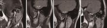
Fig 1
Diagram of classification according to the disc positions evaluated by sagittal MRI
Tab 1
Definitions of the measurements performed in this study
| 测量参数 | 定义 | |
|---|---|---|
髁突 关节窝 | 宽度 | 轴面上测量的最大内外径 |
| 长度 | 轴面上经过最大内外径中点的垂直线上髁突的最前点与最后点的距离 | |
| 体积 | 利用Mimics软件在轴向上以髁突顶点和下颌切迹最底点所在轴面为上下界,分割建立髁突模型,自动测量体积 | |
| 表面积 | 利用Mimics软件在轴向上以髁突顶点和下颌切迹最底点所在轴面为上下界,分割建立髁突模型,自动测量表面积 | |
| 高度 | 正中矢状面上髁突最顶点到经过下颌切迹最底点所在轴面的最短距离 | |
| 长度 | 正中矢状面上关节结节最低点到关节窝后部最低点的距离 | |
| 深度 | 正中矢状面上关节窝最高点至关节窝宽度线的垂直距离 | |
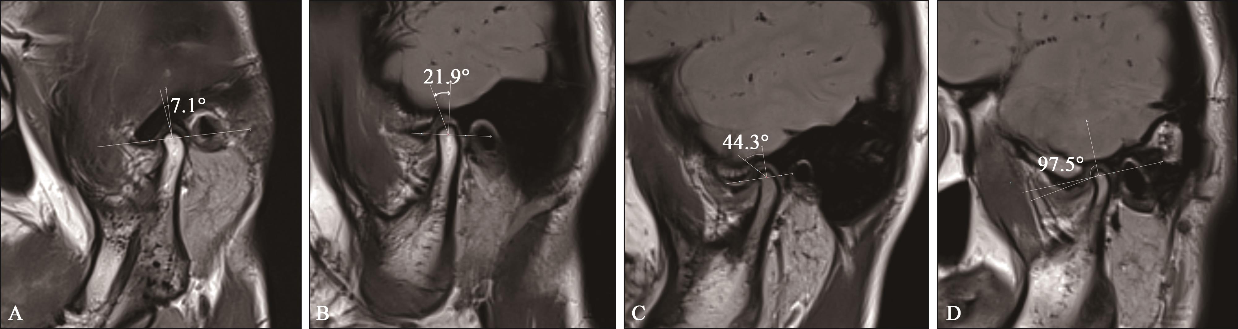
| 关节结节 | 倾斜角 | 正中矢状面上经过关节窝最高点和关节结节最低点的直线与关节窝长度所在直线所形成的夹角 |
关节间隙 | 前 | 画过关节窝最高点,与髁突前缘相切的线,过切点的垂线上切点到对应关节窝的距离 |
| 上 | 上关节间隙是从关节窝最上点到髁头最顶点的距离 | |
| 后 | 画过关节窝最高点,与髁突后缘相切的线,过切点的垂线上切点到对应关节窝的距离 | |
髁突位置 | 前 | 根据公式:(后间隙-前间隙)/(后间隙+前间隙),若比值R>12%,则为髁突前位 |
| 后 | 根据公式:(后间隙-前间隙)/(后间隙+前间隙),若比值R<-12%,则为髁突后位 | |
| 中 | 根据公式:(后间隙-前间隙)/(后间隙+前间隙),若比值R介于-12%~12%之间,为髁突中间位 | |
Fig 2
Schematic measurement of bone morphological parameters of the TMJ
| 项目 | 对照组(n=52) | 轻度组 (n=50) | 中度组(n=31) | 重度组(n=45) | P值 |
|---|---|---|---|---|---|
| 平均 | 26.69±4.66 | 26.24±4.98 | 26.00±4.67 | 24.29±4.67 | 0.081 12 |
| 男 | 26.79±5.31 | 26.20±6.60 | 24.33±4.72 | 22.45±4.11 | 0.177 21 |
| 女 | 26.81±4.28 | 26.25±4.60 | 26.40±4.66 | 24.88±4.75 | 0.367 22 |
Tab 3
Comparison of measurement of bone morphological parameters of the TMJ among groups
| 测量参数 | 对照组 | 轻度组 | 中度组 | 重度组 | P值 |
|---|---|---|---|---|---|
| 髁突 | |||||
| 宽度/mm | 19.69±2.09a | 19.07±1.57a | 18.78±2.11a | 17.32±2.46b | 0.000 12 |
| 长度/mm | 8.18±1.20a | 7.95±1.42?? | 7.16±1.40?? | 6.73±1.41? | <0.000 11 |
| 高度/mm | 19.30±3.12a | 18.10±2.75a | 17.74±2.56a | 14.86±3.38b | <0.000 11 |
| 体积/mm3 | 2 033±442.1a | 1 832±455.4a | 1 709±422.4a | 1 216±477.9b | <0.000 12 |
| 表面积/mm2 | 1 019.0±160.1a | 941.1±158.7a | 917.8±144.4a | 735.2±184.4b | <0.000 12 |
| 关节窝 | |||||
| 深度/mm | 6.33±1.16a | 6.31±1.14a | 6.50±1.03a | 5.40±1.57b | 0.000 31 |
| 长度/mm | 18.19±1.77 | 17.68±2.22 | 17.26±1.29 | 18.03±2.02 | 0.142 11 |
| 深/长比 | 0.35±0.06?? | 0.36±0.06? | 0.38±0.05? | 0.30±0.09? | <0.000 12 |
| 关节结节倾斜角/? | 29.70±4.60?? | 32.02±6.50a | 32.49±5.33a | 26.33±8.70b | <0.000 11 |
| 关节间隙 | |||||
| 前/mm | 2.33±0.62? | 2.67±0.80?? | 2.89±0.90? | 2.54±0.86?? | 0.021 02 |
| 上/mm | 3.57±0.68a | 3.52±0.87a | 3.52±0.86ab | 3.07±1.05b | 0.009 02 |
| 后/mm | 2.06±0.53ab | 1.84±0.51a | 2.01±0.60ab | 2.24±0.67b | 0.005 72 |
| (后-前)/(后+前)/% | -5.97±16.82? | -17.52±16.69? | 16.95±20.22? | -5.46±19.22? | 0.000 61 |
| 矢状髁突位置/例(%) | 0.044 43 | ||||
| 前 | 8(15.38) | 1(2.00) | 3(9.68) | 8(17.78) | |
| 后 | 21(40.39) | 31(62.00) | 20(64.51) | 18(40.00) | |
| 中 | 23(44.23) | 18(36.00) | 8(35.81) | 19(42.22) |
Tab 4
Analysis of correlation between TMJ morphological measurements and sagittal disc positions
| 测量参数 | 关节盘矢状位置 | ||
|---|---|---|---|
| r | P值 | ||
髁突 | 宽度 | -0.406 6 | <0.000 1 |
| 长度 | -0.358 4 | <0.000 1 | |
| 高度 | -0.481 2 | <0.000 1 | |
| 体积 | -0.562 8 | <0.000 1 | |
| 表面积 | -0.541 8 | <0.000 1 | |
| 关节窝 | 深度 | -0.325 2 | <0.000 1 |
| 长度 | 0.030 7 | 0.685 6 | |
| 关节结节 | 倾斜角 | -0.318 1 | <0.000 1 |
关节间隙 | 前 | 0.029 8 | 0.695 6 |
| 上 | -0.244 7 | 0.001 1 | |
| 后 | 0.187 8 | 0.012 8 | |
| 1 | de Farias JF, Melo SL, Bento PM, et al. Correlation between temporomandibular joint morphology and disc displacement by MRI[J]. Dentomaxillofac Radiol, 2015, 44(7): 20150023. |
| 2 | Chang MS, Choi JH, Yang IH, et al. Relationships between temporomandibular joint disk displacements and condylar volume[J]. Oral Surg Oral Med Oral Pathol O-ral Radiol, 2018, 125(2): 192-198. |
| 3 | Derwich M, Mitus-Kenig M, Pawlowska E. Temporomandibular joints' morphology and osteoarthritic changes in cone-beam computed tomography images in patients with and without reciprocal clicking-a case control study[J]. Int J Environ Res Public Health, 2020, 17(10): 3428. |
| 4 | Basaran M, Bozdemir E, Evrimler S. Evaluation of morphometric and volumetric measurements of temporomandibular joint structures on patients with disc displacement[J]. J Anat Soc India, 2021, 70(1): 41-47. |
| 5 | Torres MG, Crusoé-Rebello IM, Rosário M, et al. Morphometric features of the mandibular condyle and association with disk abnormalities[J]. Oral Surg Oral Med Oral Pathol Oral Radiol, 2016, 121(5): 566-572. |
| 6 | Guercio Monaco E, De Stefano AA, Hernandez-Andara A, et al. Correlation between condylar size on CT and position of the articular disc on MRI of the temporomandibular joint[J]. Cranio, 2022, 40(1): 64-71. |
| 7 | Noh KJ, Baik HS, Han SS, et al. Differences in mandibular condyle and glenoid fossa morphology in relation to vertical and sagittal skeletal patterns: a cone-beam computed tomography study[J]. Korean J Orthod, 2021, 51(2): 126-134. |
| 8 | Yalcin ED, Ararat E. Cone-beam computed tomography study of mandibular condylar morphology[J]. J Craniofac Surg, 2019, 30(8): 2621-2624. |
| 9 | Seo BY, An JS, Chang MS, et al. Changes in condylar dimensions in temporomandibular joints with disk displacement[J]. Oral Surg Oral Med Oral Pathol Oral Radiol, 2020, 129(1): 72-79. |
| 10 | Yang Z, Wang M, Ma Y, et al. Magnetic resonance imaging (MRI) evaluation for anterior disc displacement of the temporomandibular joint[J]. Med Sci Monit, 2017, 23: 712-718. |
| 11 | Cai XY, Jin JM, Yang C. Changes in disc position, disc length, and condylar height in the temporomandibular joint with anterior disc displacement: a longitudinal retrospective magnetic resonance imaging study[J]. J Oral Maxillofac Surg, 2011, 69(11): e340-e346. |
| 12 | Drace JE, Enzmann DR. Defining the normal temporomandibular joint: closed-, partially open-, and open-mouth MR imaging of asymptomatic subjects[J]. Radio-logy, 1990, 177(1): 67-71. |
| 13 | Kurita H, Ohtsuka A, Kobayashi H, et al. The relationship between the degree of disk displacement and ability to perform disk reduction[J]. Oral Surg Oral Med Oral Pathol Oral Radiol Endod, 2000, 90(1): 16-20. |
| 14 | 刘学业, 李齐明, 唐弘毅, 等. 年轻成人颞下颌关节髁突体积、表面积与关节盘矢向位置的关系[J]. 山东大学学报(医学版), 2021, 59(6): 117-121. |
| Liu XY, Li QM, Tang HY, et al. Relationship among the volume and surface area of the temporomandibular joint condyle and the sagittal position of disc in young adults[J]. J Shandong Univ (Health Sci), 2021, 59(6): 117-121. | |
| 15 | Incesu L, Taşkaya-Yilmaz N, Oğütcen-Toller M, et al. Relationship of condylar position to disc position and morphology[J]. Eur J Radiol, 2004, 51(3): 269-273. |
| 16 | Choudhary A, Ahuja US, Rathore A, et al. Association of temporomandibular joint morphology in patients with and without temporomandibular joint dysfunction: a cone-beam computed tomography based study[J]. Dent Res J (Isfahan), 2020, 17(5): 338-346. |
| 17 | Pullinger AG, Hollender L, Solberg WK, et al. A tomographic study of mandibular condyle position in an asymptomatic population[J]. J Prosthet Dent, 1985, 53(5): 706-713. |
| 18 | Alexiou K, Stamatakis H, Tsiklakis K. Evaluation of the severity of temporomandibular joint osteoarthritic changes related to age using cone beam computed tomography[J]. Dentomaxillofac Radiol, 2009, 38(3): 141-147. |
| 19 | Liu X, Xu Q, Guo J. The relationship between the size of temporomandibular joint condyle and the sagittal disc-condyle position in adults[J]. Cranio, 2021: 1-8. |
| 20 | Kurita H, Ohtsuka A, Kobayashi H, et al. Alteration of the horizontal mandibular condyle size associated with temporomandibular joint internal derangement in adult females[J]. Dentomaxillofac Radiol, 2002, 31(6): 373-378. |
| 21 | Karayol KC, Karayol SS. Morphometric Analysis of the glenoid fossa in the skull base[J]. J Craniofac Surg, 2022, 33(1): 319-321. |
| 22 | Sülün T, Cemgil T, Duc JM, et al. Morphology of the mandibular fossa and inclination of the articular eminence in patients with internal derangement and in symptom-free volunteers[J]. Oral Surg Oral Med Oral Pathol Oral Radiol Endod, 2001, 92(1): 98-107. |
| 23 | Kurita H, Ohtsuka A, Kobayashi H, et al. Is the morphology of the articular eminence of the temporomandibular joint a predisposing factor for disc displacement[J]. Dentomaxillofac Radiol, 2000, 29(3): 159-162. |
| 24 | Atkinson WB, Bates RE Jr. The effects of the angle of the articular eminence on anterior disk displacement[J]. J Prosthet Dent, 1983, 49(4): 554-555. |
| 25 | Kurita H, Ohtsuka A, Kobayashi H, et al. A study of the relationship between the position of the condylar head and displacement of the temporomandibular joint disk[J]. Dentomaxillofac Radiol, 2001, 30(3): 162-165. |
| 26 | Ahmed J, Sujir N, Shenoy N, et al. Morphological assessment of TMJ spaces, mandibular condyle, and glenoid fossa using cone beam computed tomography (CB-CT): a retrospective analysis[J]. Indian J Radiol Imaging, 2021, 31(1): 78-85. |
| Viewed | ||||||
|
Full text |
|
|||||
|
Abstract |
|
|||||
 This work is licensed under a Creative Commons Attribution 3.0 License.
