West China Journal of Stomatology ›› 2022, Vol. 40 ›› Issue (3): 314-319.doi: 10.7518/hxkq.2022.03.011
Previous Articles Next Articles
Shi Xiaoyang( ), Lin Xuefen, Ma Chi, Chen Muhan, Liu Dongxu.()
Received:2021-08-30
Revised:2022-04-10
Online:2022-06-01
Published:2022-06-01
Contact:
Liu Dongxu.
E-mail:1140212323@qq.com;liudongxu@sdu.edu.cn
Supported by:CLC Number:
Shi Xiaoyang, Lin Xuefen, Ma Chi, Chen Muhan, Liu Dongxu.. Evaluation of changes in orbital volume in adult female patients with maxillary transverse deficiency treated with a maxillary skeletal expander[J]. West China Journal of Stomatology, 2022, 40(3): 314-319.
Add to citation manager EndNote|Ris|BibTeX
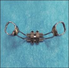
Fig 1
MSE
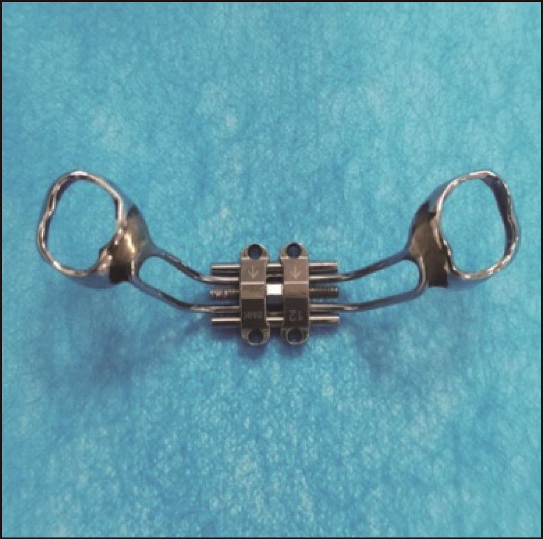
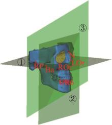
Fig 2
Head position correction
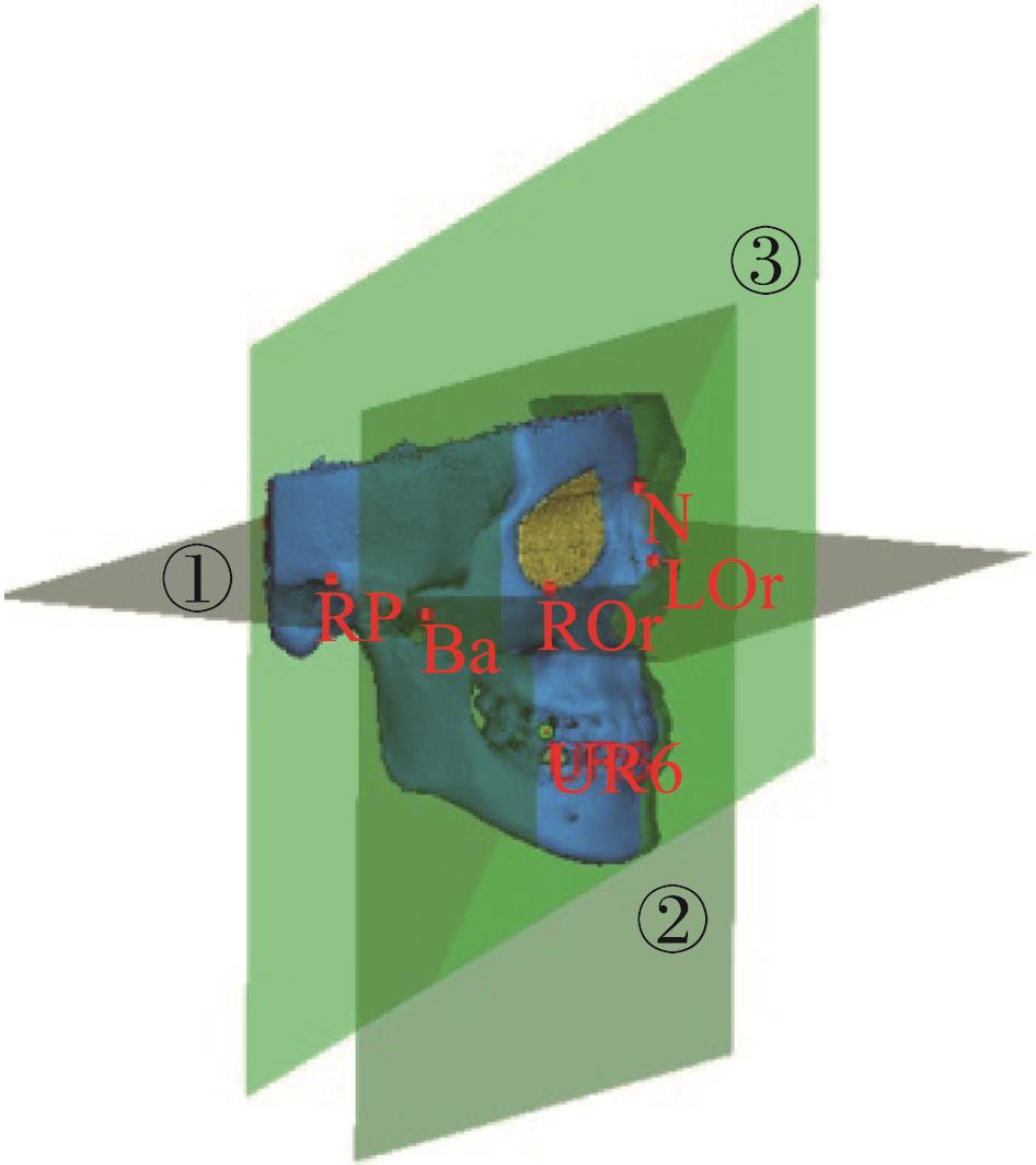

Fig 3
Periorbital mark points
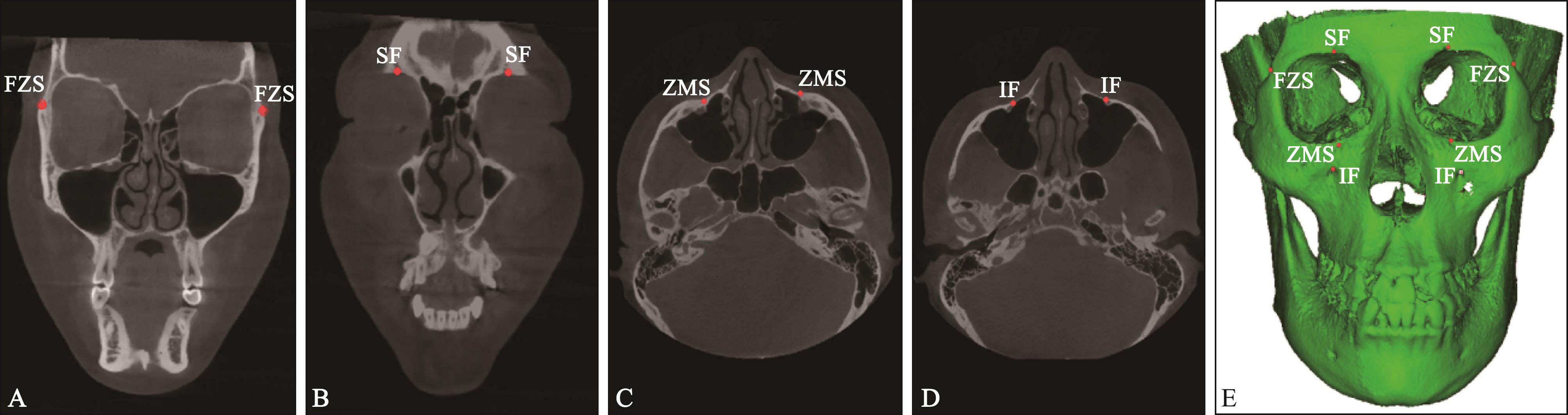
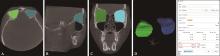
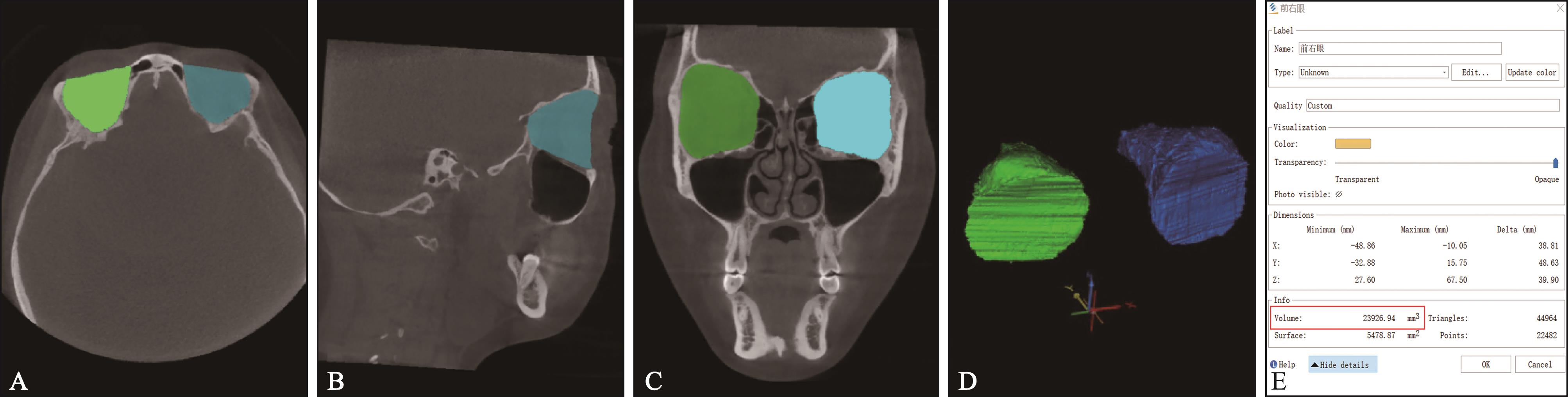
Fig 4
Orbital boundary and 3D orbital model
Tab 1
Changes in the width of the periorbital bones before and after treatment with MSE
| 测量项目/mm | T0 | T1 | 变化量(T1-T0) | P值 |
|---|---|---|---|---|
| FZS间宽度 | 100.69±4.10 | 100.84±4.01 | 0.15±0.32 | 0.052 |
| ZMS间宽度 | 50.02±6.84 | 51.71±6.76 | 1.69±0.57 | <0.01* |
| SF间宽度 | 47.87±4.98 | 48.10±4.82 | 0.23±0.52 | 0.066 |
| IF间宽度 | 51.09±4.04 | 52.80±0.97 | 1.71±0.70 | <0.01* |
| 1 | Proffit WR, White RP Jr. Who needs surgical-orthodontic treatment[J]. Int J Adult Orthodon Orthognath Surg, 1990, 5(2): 81-89. |
| 2 | Betts NJ, Vanarsdall RL, Barber HD, et al. Diagnosis and treatment of transverse maxillary deficiency[J]. Int J Adult Orthodon Orthognath Surg, 1995, 10(2): 75-96. |
| 3 | Brunelle JA, Bhat M, Lipton JA. Prevalence and distribution of selected occlusal characteristics in the US po⁃pulation, 1988-1991[J]. J Dent Res, 1996, 75(Spec): 706-713. |
| 4 | Ramires T, Maia RA, Barone JR. Nasal cavity changes and the respiratory standard after maxillary expansion[J]. Braz J Otorhinolaryngol, 2008, 74(5): 763-769. |
| 5 | Baccetti T, Franchi L, Cameron CG, et al. Treatment ti-ming for rapid maxillary expansion[J]. Angle Orthod, 2001, 71(5): 343-350. |
| 6 | Ghoneima A, Abdel-Fattah E, Hartsfield J, et al. Effects of rapid maxillary expansion on the cranial and circummaxillary sutures[J]. Am J Orthod Dentofacial Orthop, 2011, 140(4): 510-519. |
| 7 | Kiliç N, Kiki A, Oktay H. A comparison of dentoalveolar inclination treated by two palatal expanders[J]. Eur J Orthod, 2008, 30(1): 67-72. |
| 8 | Gurel HG, Memili B, Erkan M, et al. Long-term effects of rapid maxillary expansion followed by fixed applian⁃ces[J]. Angle Orthod, 2010, 80(1): 5-9. |
| 9 | Kartalian A, Gohl E, Adamian M, et al. Cone-beam computerized tomography evaluation of the maxillary dentoskeletal complex after rapid palatal expansion[J]. Am J Orthod Dentofacial Orthop, 2010, 138(4): 486-492. |
| 10 | Lagravère MO, Carey J, Heo G, et al. Transverse, vertical, and anteroposterior changes from bone-anchored ma-xillary expansion vs traditional rapid maxillary expansion: a randomized clinical trial[J]. Am J Orthod Dentofacial Orthop, 2010, 137(3): 304.e1-e12. |
| 11 | MacGinnis M, Chu H, Youssef G, et al. The effects of micro-implant assisted rapid palatal expansion (MAR-PE) on the nasomaxillary complex—a finite element me-thod (FEM) analysis[J]. Prog Orthod, 2014, 15(1): 52. |
| 12 | Cantarella D, Dominguez-Mompell R, Mallya SM, et al. Changes in the midpalatal and pterygopalatine sutures induced by micro-implant-supported skeletal expander, analyzed with a novel 3D method based on CBCT ima⁃ging[J]. Prog Orthod, 2017, 18(1): 34. |
| 13 | Colak O, Paredes NA, Elkenawy I, et al. Tomographic assessment of palatal suture opening pattern and pterygopalatine suture disarticulation in the axial plane after midfacial skeletal expansion[J]. Prog Orthod, 2020, 21(1): 21. |
| 14 | Khosravi M, Ugolini A, Miresmaeili A, et al. Tooth-borne versus bone-borne rapid maxillary expansion for transverse maxillary deficiency: a systematic review[J]. Int Orthod, 2019,17(3): 425-436. |
| 15 | Krüsi M, Eliades T, Papageorgiou SN. Are there benefits from using bone-borne maxillary expansion instead of tooth-borne maxillary expansion? A systematic review with meta-analysis[J]. Prog Orthod, 2019, 20(1): 9. |
| 16 | Bucci R, D’Antò V, Rongo R, et al. Dental and skeletal effects of palatal expansion techniques: a systematic review of the current evidence from systematic reviews and meta-analyses[J]. J Oral Rehabil, 2016, 43(7): 543-564. |
| 17 | Zhou Y, Long H, Ye N, et al. The effectiveness of non-surgical maxillary expansion: a meta-analysis[J]. Eur J Orthod, 2014, 36(2): 233-242. |
| 18 | Lee RJ, Moon W, Hong C. Effects of monocortical and bicortical mini-implant anchorage on bone-borne palatal expansion using finite element analysis[J]. Am J Orthod Dentofacial Orthop, 2017, 151(5): 887-897. |
| 19 | Paredes N, Colak O, Sfogliano L, et al. Differential assessment of skeletal, alveolar, and dental components induced by microimplant-supported midfacial skeletal expander (MSE), utilizing novel angular measurements from the fulcrum[J]. Prog Orthod, 2020, 21(1): 18. |
| 20 | Cantarella D, Dominguez-Mompell R, Moschik C, et al. Midfacial changes in the coronal plane induced by microimplant-supported skeletal expander, studied with cone-beam computed tomography images[J]. Am J Orthod Dentofacial Orthop, 2018, 154(3): 337-345. |
| 21 | Chau A, Fung K, Yip L, et al. Orbital development in Hong Kong Chinese subjects[J]. Ophthalmic Physiol Opt, 2004, 24(5): 436-439. |
| 22 | Sicurezza E, Palazzo G, Leonardi R. Three-dimensional computerized tomographic orbital volume and aperture width evaluation: a study in patients treated with rapid maxillary expansion[J]. Oral Surg Oral Med Oral Pathol Oral Radiol Endod, 2011, 111(4): 503-507. |
| 23 | Lo Giudice A, Rustico L, Ronsivalle V, et al. Evaluation of the changes of orbital cavity volume and shape after tooth-borne and bone-borne rapid maxillary expansion (RME)[J]. Head Face Med, 2020, 16(1): 21. |
| 24 | Monaco A, Tepedino M, Sabetti L, et al. An adolescent treated with rapid maxillary expansion presenting with strabismus: a case report[J]. J Med Case Rep, 2013, 7: 222. |
| 25 | Tamburrino R, Boucher N, Vanarsdall R. The Transverse dimension: diagnosis and relevance to functional occlusion[J]. RWISO J, 2010, 2(1): 13-22. |
| 26 | Gribova MN, Pluijmers BI, Resnick CM, et al. Is there a difference in orbital volume between affected and unaffected sides in patients with unilateral craniofacial microsomia[J]. J Oral Maxillofac Surg, 2018, 76(12): 2625-2629. |
| 27 | Wei N, Bi H, Zhang B, et al. Biphasic growth of orbital volume in Chinese children[J]. Br J Ophthalmol, 2017, 101(9): 1162-1167. |
| 28 | Angelieri F, Cevidanes LH, Franchi L, et al. Midpalatal suture maturation: classification method for individual assessment before rapid maxillary expansion[J]. Am J Orthod Dentofacial Orthop, 2013, 144(5): 759-769. |
| 29 | Romeo A, Manti S, Romeo G, et al. Headache and diplopia after rapid maxillary expansion: a clue to underdiagnosed pseudotumor cerebri syndrome[J]. J Pediat Neurol, 2015, 13(1): 31-34. |
| 30 | Lanigan DT, Mintz SM. Complications of surgically assisted rapid palatal expansion: review of the literature and report of a case[J]. J Oral Maxillofac Surg, 2002, 60(1): 104-110. |
| 31 | Holberg C, Steinhäuser S, Rudzki I. Surgically assisted rapid maxillary expansion: midfacial and cranial stress distribution[J]. Am J Orthod Dentofacial Orthop, 2007, 132(6): 776-782. |
| [1] | Ma Yanning, Jin Zuolin.. Orthodontic program design based on aesthetic [J]. West China Journal of Stomatology, 2023, 41(6): 628-634. |
| [2] | Tang Meng, Cui Zhan-qin, Wang Yangyang, Chen Zengguo, Li Wenjing, Zhang Cuiping. Effects of low-level laser on the expression of interleukin-6, tumor necrosis factor‑α, osteoprotegerin, and receptor activator of nuclear factor-κB ligand in human periodontal ligament cells [J]. West China Journal of Stomatology, 2023, 41(5): 521-532. |
| [3] | Kang Fujia, Yu Lei, Zhang Qi, Li Xinpeng, Hu Zhiqiang, Zhu Xianchun.. Three-dimensional finite element study of mandibular first molar distalization with clear aligner [J]. West China Journal of Stomatology, 2023, 41(4): 405-413. |
| [4] | Fu Yu, Li Ziwei, Zhao Menghan, Shi Ruixin.. Study on the effect of chin morphology on orthodontic treatment [J]. West China Journal of Stomatology, 2023, 41(4): 443-449. |
| [5] | Li Yulin, Xu Jingchen, Jiang Xiaoge, Chen Song.. Meta-analysis of condylar changes produced by a Twin-block appliance in Class Ⅱ malocclusion [J]. West China Journal of Stomatology, 2023, 41(4): 463-470. |
| [6] | Wan Xiaofang, He Haiyan, Jialing Lü, Wu Yujie, Zhong Guannan, Xu Xiaomei. Hippo-YAP signaling pathway regulates autophagy of human periodontal ligament cells under cyclic tensile stress [J]. West China Journal of Stomatology, 2023, 41(3): 260-268. |
| [7] | Lu Ting, Zhu Jiahao, Yang Shihe, Shen Zhe, Zhong Liangjun. Effects of Foxp3 gene silencing on the expression of inflammatory cytokines and the proliferation and migration of human periodontal ligament fibroblasts in an inflammatory environment [J]. West China Journal of Stomatology, 2023, 41(3): 269-275. |
| [8] | Yu Lei, Li Ziwei, Kang Fujia, Wang Songqing, Xie Zunxuan, Zhu Xianchun.. Mandibular advancement with clear aligners and functional appliances in the treatment of skeletal ClassⅡmalocclusion: a systematic review and meta-analysis [J]. West China Journal of Stomatology, 2023, 41(3): 305-314. |
| [9] | Li Huang, Wu Xiuping, Huang Lan, Xu Xiaomei, Kang Na, Han Xianglong, Li Yu, Zhao Ning, Jiang Lingyong, Xie Xianju, Guo Jie, Li Zhihua, Mo Shuixue, Liu Chufeng, Hu Jiangtian, Shi Jiejun, Cao Meng, Hu Wei, Cao Yang, Song Jinlin, Tang Xuna, Bai Ding. External apical root resorption in orthodontic tooth movement: the risk factors and clinical suggestions from experts’ consensus [J]. West China Journal of Stomatology, 2022, 40(6): 629-637. |
| [10] | Wu Jie, Cui Zhanqin, Han Yu, Li Wenjing. Aging effect of osteoprotegerin and receptor activator of nuclear factor-κB ligand expression in human periodontal ligament cells under continuous static pressure [J]. West China Journal of Stomatology, 2022, 40(6): 654-661. |
| [11] | Li Bo, Xu Yimeng, Shi Ruiying, Hu Yirong, Liu Siying, Gu Zexu.. Accuracy of progress assessment with clear aligners [J]. West China Journal of Stomatology, 2022, 40(6): 698-703. |
| [12] | Shi Zean, Xia Kai, Luo Liangyu, Zhao Zhihe, Liu Jun.. Three-dimensional finite element analysis of upper anterior teeth retraction and intrusion using clear aligners and mini-implants [J]. West China Journal of Stomatology, 2022, 40(5): 589-596. |
| [13] | Zhao Zhihe, Jin Zuolin, Bai Yuxing, Fang Bing, Bai Ding, Li Weiran, He Hong, Hu Min, Liu Yuehua, Chen Lili, Song Jinlin, Cao Yang, Li Yu, Shu Rui. Core scientific issues of orthodontic tooth movement: position objective, efficiency, and accuracy [J]. West China Journal of Stomatology, 2022, 40(4): 371-376. |
| [14] | Guo Weihua, Wang Jun, Chen Xu, Wang Xiaojing, Zhao Wei, Song Guangtai, Wu Li’an, Jiang Beizhan, Zhang Qiong, Wang Jun, Li Yu, Zhao Ning, Tan Jiali, Li Huang, Shu Rui, Zhou Chenchen, Fu Lei, Chen Xuepeng, Zou Jing. Experts’ consensus on space management of mixed dentition [J]. West China Journal of Stomatology, 2022, 40(3): 264-270. |
| [15] | Luo Houzhuo, Fang Shishu, Liu Qian, Dang Wei, Wang Yanli. Comparison of interleukin expression in gingival crevicular fluid between patients with invisible orthodontics treat-ment and fixed orthodontics treatment [J]. West China Journal of Stomatology, 2022, 40(3): 293-296. |
| Viewed | ||||||
|
Full text |
|
|||||
|
Abstract |
|
|||||
 This work is licensed under a Creative Commons Attribution 3.0 License.
