West China Journal of Stomatology ›› 2021, Vol. 39 ›› Issue (6): 667-674.doi: 10.7518/hxkq.2021.06.007
Previous Articles Next Articles
Tang Hao( ), Zhu Yawen, Zhu Jiaxiang, Li Quanli()
Received:2020-12-18
Revised:2021-10-12
Online:2021-12-01
Published:2021-12-03
Contact:
Li Quanli
E-mail:tanghowell@163.com;ql-li@126.com
Supported by:CLC Number:
Tang Hao, Zhu Yawen, Zhu Jiaxiang, Li Quanli. Occluding dentin tubules with monetite paste in vitro[J]. West China Journal of Stomatology, 2021, 39(6): 667-674.
Add to citation manager EndNote|Ris|BibTeX
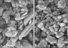
Fig 1
SEM of two pastes × 50 000
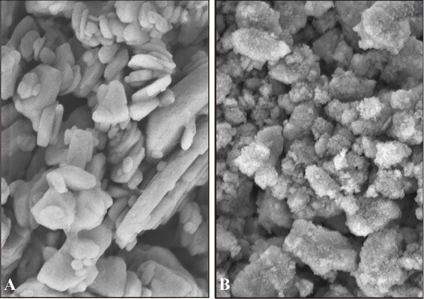
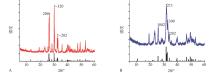
Fig 2
XRD spectra of two pastes
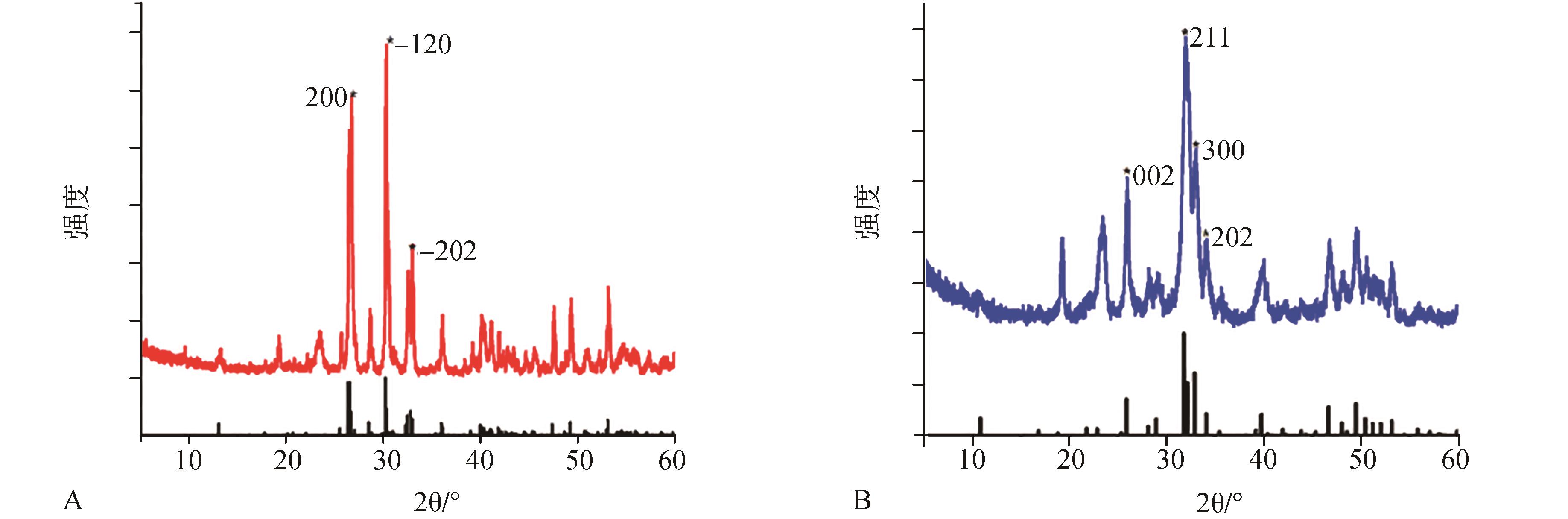
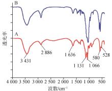
Fig 3
FTIR spectra of two pastes
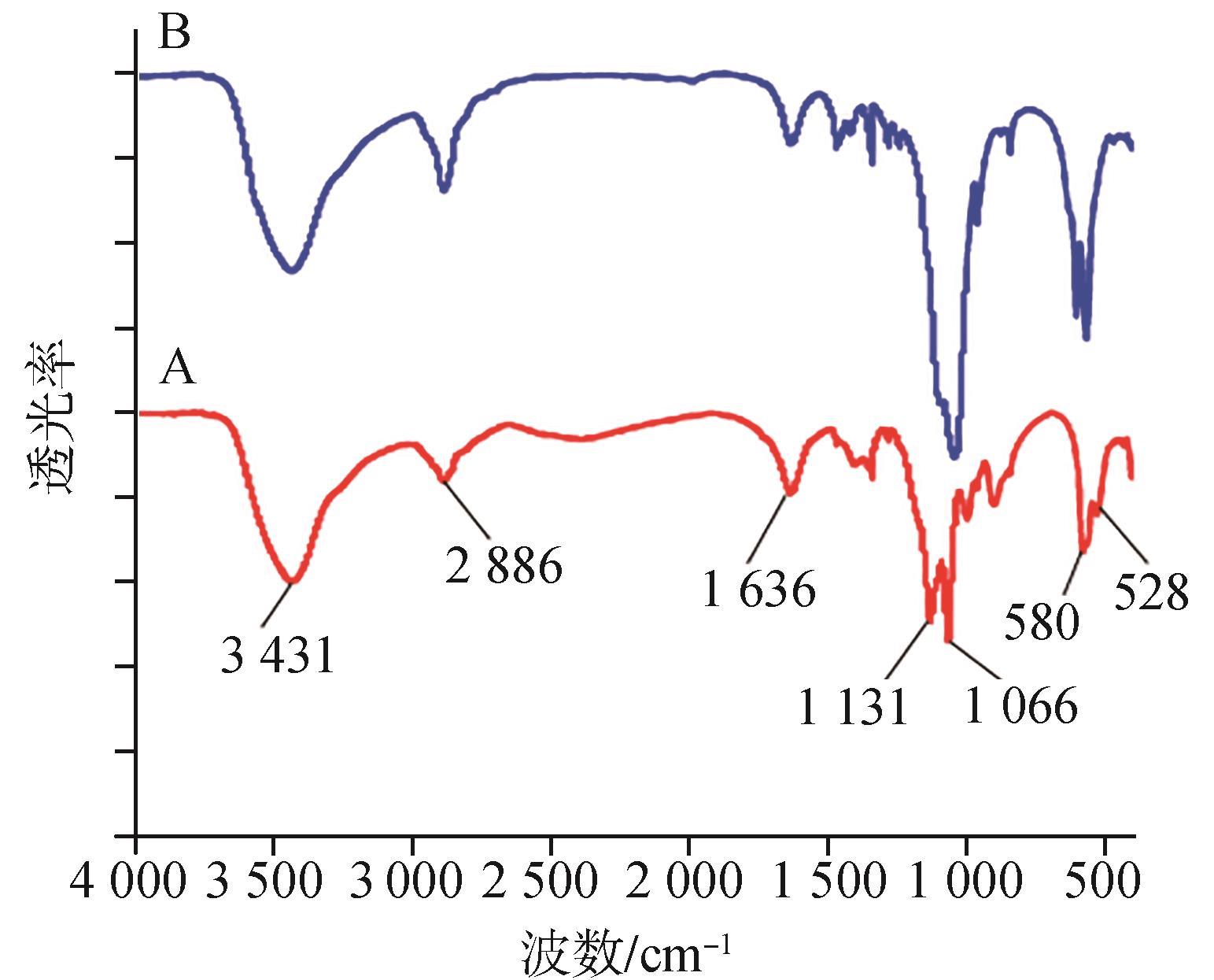
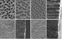
Fig 4
SEM images of dentin slices after demineralization before mineralization and after 7 days of mineralization treated with test reagent

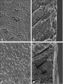
Fig 5
SEM images of remineralized dentin slices after 7 days of mechanical brushing simulating daily brushing

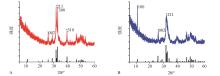
Fig 6
XRD spectra of the surface of the samples after 7 days of mineralization treated with two kinds of mineralized paste

Tab 1
SMH of dentin slices before and after minerali-zation in each group
| 项目 | 空白对照组 | CPP-ACP组 | 三斜磷钙石糊剂组 | 羟磷灰石糊剂组 |
|---|---|---|---|---|
| 矿化前SMH1 | 36.74±3.93 | 37.10±2.19 | 37.24±3.55 | 37.02±3.10 |
| 矿化后SMH2 | 30.40±3.59 | 50.74±1.47 | 51.96±1.30 | 61.18±0.72 |
| ΔSMH | -6.34±7.15 | 13.64±3.18 | 14.72±3.78 | 24.16±3.25 |
| 1 | 中华口腔医学会口腔预防医学专业委员会牙本质敏感专家组 . 牙本质敏感的诊断和防治指南(2019修订版)[J]. 中华口腔医学杂志, 2019, 54(4): 223-227. |
| Expert Committee of Dentin Hypersensitivity, Society of Preventive Dentistry, Chinese Stomatological Association . Guideline for diagnosis, prevention and treatment of dentin hypersensitivity[J]. Chin J Stomatol, 2019, 54(4): 223-227. | |
| 2 | Onwubu SC , Mhlungu S , Mdluli PS . In vitro evaluation of nanohydroxyapatite synthesized from eggshell waste in occluding dentin tubules[J]. J Appl Biomater Funct Mater, 2019, 17(2): 2280800019851764. |
| 3 | Yu J , Yang HY , Li K , et al . A novel application of nanohydroxyapatite/mesoporous silica biocomposite on treating dentin hypersensitivity: an in vitro study[J]. J Dent, 2016, 50: 21-29. |
| 4 | Liu XX , Tenenbaum HC , Wilder RS , et al . Pathogenesis, diagnosis and management of dentin hypersensitivity: an evidence-based overview for dental practitioners[J]. BMC Oral Health, 2020, 20(1): 220. |
| 5 | Liang KN , Xiao SM , Liu HL , et al . 8DSS peptide induced effective dentinal tubule occlusion in vitro [J]. Dent Mater, 2018, 34(4): 629-640. |
| 6 | West NX , Seong J , Davies M . Management of dentine hypersensitivity: efficacy of professionally and self-administered agents[J]. J Clin Periodontol, 2015, 42(): S256-S302. |
| 7 | Rezazadeh F , Dehghanian P , Jafarpour D . Laser effects on the prevention and treatment of dentinal hypersensitivity: a systematic review[J]. J Lasers Med Sci, 2019, 10(1): 1-11. |
| 8 | Wang ZJ , Ma X , Jiang T , et al . The dentin tubule occlusion effects of desensitizing agents and the stability a-gainst acids and brushing challenges[J]. Am J Dent, 2015, 28(3): 128-132. |
| 9 | Wang ZJ , Sa Y , Sauro S , et al . Effect of desensitising toothpastes on dentinal tubule occlusion: a dentine permeability measurement and SEM in vitro study[J]. J De-nt, 2010, 38(5): 400-410. |
| 10 | Jena A , Kala S , Shashirekha G . Comparing the effectiveness of four desensitizing toothpastes on dentinal tubule occlusion: a scanning electron microscope analysis[J]. J Conserv Dent, 2017, 20(4): 269-272. |
| 11 | Ma Q , Wang TD , Meng QF , et al . Comparison of in vitro dentinal tubule occluding efficacy of two different methods using a nano-scaled bioactive glass-containing desensitising agent[J]. J Dent, 2017, 60: 63-69. |
| 12 | Deliormanli AM . Investigation of in vitro mineralization of silicate-based 45S5 and 13-93 bioactive glasses in artificial saliva for dental applications[J]. Ceram Int, 2017, 43(4): 3531-3539. |
| 13 | Ashwini S , Swatika K , Kamala DN . Comparative evaluation of desensitizing efficacy of dentifrice containing 5% fluoro calcium phosphosilicate versus 5% calcium sodium phosphosilicate: a randomized controlled clinical trial[J]. Contemp Clin Dent, 2018, 9(3): 330-336. |
| 14 | Kruppke B , Farack J , Wagner AS , et al . Gelatine modified monetite as a bone substitute material: an in vitro assessment of bone biocompatibility[J]. Acta Biomater, 2016, 32: 275-285. |
| 15 | Nishad KV , Sureshbabu S , Komath M , et al . Synthesis and characterization of low dimensional bioactive monetite by solvent exchange method[J]. Mater Lett, 2017, 209(15): 19-22. |
| 16 | Yu Y , Stevensson B , Pujari-Palmer M , et al . The monetite structure probed by advanced solid-state NMR experimentation at fast magic-angle spinning[J]. Int J Mol Sci, 2019, 20(24): E6356. |
| 17 | Medvecky L , Stulajterova R , Giretova M , et al . Effect of tetracalcium phosphate/monetite toothpaste on dentin re-mineralization and tubule occlusion in vitro [J]. Dent Mater, 2018, 34(3): 442-451. |
| 18 | Shen C , Wu L , Chen Y . Efficient removal of fluoride from drinking water using well-dispersed monetite bundles inlaid in chitosan beads[J]. Chem Eng J, 2016, 303: 391-400. |
| 19 | ten Cate JM , Duijsters PP . Alternating demineralization and remineralization of artificial enamel lesions[J]. Ca-ries Res, 1982, 16(3): 201-210. |
| 20 | Karim BF , Gillam DG . The efficacy of strontium and potassium toothpastes in treating dentine hypersensitivity: a systematic review[J]. Int J Dent, 2013, 2013: 573258. |
| 21 | Fredholm YC , Karpukhina N , Brauer DS , et al . Influence of strontium for calcium substitution in bioactive glasses on degradation, ion release and apatite formation[J]. J R Soc Interface, 2012, 9(70): 880-889. |
| 22 | 陈德敏, 傅远飞, 顾国珍, 等 . 掺锶羟磷灰石固溶体的制备及离解度测定[J]. 中国生物医学工程学报, 2001, 20(3): 278-280. |
| Chen DM , Fu YF , Gu GZ , et al . Preparation and solubility of the solid solution of strontium substituted hydroxyapatite[J]. Chin J Biomed Eng, 2001, 20(3): 278-280. | |
| 23 | Xiong Y , Zhang Y , Liu W , et al . Preparation of Sr-substituted hydroxyapatite nanorods for liquid crystal phase transition[J]. J Wuhan Univ Technol Mat Sci Edit, 2020, 35(2): 441-448. |
| 24 | 傅远飞, 陈德敏 . 掺锶对羟磷灰石细胞毒性的影响[J]. 功能材料, 2008, 39(4): 645-646, 650. |
| Fu YF , Chen DM . Effects of strontium on the cytotoxicity of hydroxyapatite[J]. J Funct Mater, 2008, 39(4): 645-646, 650. | |
| 25 | Xia W , Qin T , Suska F , et al . Bioactive spheres: the way of treating dentin hypersensitivity[J]. ACS Biomater Sci Eng, 2016, 2(5): 734-740. |
| 26 | Wang PP , Li CH , Gong HY , et al . Effects of synthesis conditions on the morphology of hydroxyapatite nano-particles produced by wet chemical process[J]. Powder Technol, 2010, 203(2): 315-321. |
| 27 | 朱庆霞, 徐琼琼, 罗民华 . 碳酸羟磷灰石的生物矿化研究[J]. 陶瓷学报, 2010, 31(2): 234-239. |
| Zhu QX , Xu QQ , Luo MH . Study on biological minera-lization of hydroxyapatite[J]. J Cerma, 2010, 31(2): 234-239. | |
| 28 | 朱庆霞, 刘可春, 李双, 等 . 碳酸羟磷灰石对水中氟离子的吸附研究[J]. 陶瓷学报, 2017, 38(6): 898-903. |
| Zhu QX , Liu KC , Li S , et al . Adsorption of fluoride ions in water by hydroxyapatite[J]. J Cerma, 2017, 38(6): 898-903. | |
| 29 | 罗菁菁, 唐旭炎, 李全利 . 酪蛋白磷酸肽-无定形磷酸钙促进牙再矿化的机制[J]. 国际口腔医学杂志, 2011, 38(6): 662-664, 669. |
| Luo JJ , Tang XY , Li QL . Mechanism of casein phosphopeptide-amorphous calcium phosphate on tooth remineralization[J]. Int J Stomatol, 2011, 38(6): 662-664, 669. |
| [1] | Meng Yuchen, Huang Fan, Wang Silin, Huang Xin, Lu Yi, Pei Dandan. Bonding properties of mild universal adhesives to dentin pretreated with hydroxyapatite-based desensitizing agents [J]. West China Journal of Stomatology, 2022, 40(6): 668-675. |
| [2] | Ting Wei,Xinwei Zhang,Huiqiang Sun,Mengyun Mao. Selective laser sintering and performances of porous titanium implants [J]. West China Journal of Stomatology, 2018, 36(5): 532-538. |
| [3] | Kun Tian, Xiaoyun Feng, Qin Du, Chuhang Liao, Xiaohua. Ren. Study of human leucine-rich amelogenin peptide and its regulation of mineralization by cryogenic transmission electron microscopy [J]. West China Journal of Stomatology, 2017, 35(1): 63-67. |
| [4] | Xu Ke, Zhao Yanhong, Li Hongfa. Fabrication and evaluation of hydroxyapatite-chitosan scaffold via simulated body fluid biomimetic mineralization [J]. West China Journal of Stomatology, 2016, 34(1): 6-11. |
| [5] | Qin Zishun, Yin Lihua, Wang Kaijuan, Liu Qi, Cheng Wenxiao, Gao Peng, Sun Kemo, Zhong Mei, Yu Zhanhai. Effects of Icariin promotion on proliferation and osteogenic differentiation of human periodontal ligament stem cells [J]. West China Journal of Stomatology, 2015, 33(4): 370-376. |
| [6] | Liu Yi, Zhou Rongjing, Wu Hongkun.. Study on the antibacterial activity of four kinds of nano-hydroxyapatite composites against Enterococcus faecalis [J]. West China Journal of Stomatology, 2015, 33(3): 301-305. |
| [7] | Ren Xun, Yao Jing, Du Qin, Liao Chuhang, Tian Kun.. Chitosan-collagen polymer induced remineralization of tooth hard tissue through self-growing methods [J]. West China Journal of Stomatology, 2014, 32(5): 519-524. |
| [8] | Wang Yanmei, He Jiacai, Li Quanli, Shen Jijia. Preparation of sodium alginate-nanohydroxyapatite composite material for bone repair and its biocompatibility [J]. West China Journal of Stomatology, 2014, 32(1): 27-31. |
| [9] | Huang Rui, Liu Peng, Xiao Maode, Zhou Zhi.. A comparative study on apexification using different kinds of materials in dogs [J]. West China Journal of Stomatology, 2013, 31(4): 377-380,384. |
| [10] | Zhao Mengmeng, Wang Qingshan, Wang Shuang, Li Rui.. Experimental study on microleakage between a new nano-hydroxyapatite composite and tooth [J]. West China Journal of Stomatology, 2013, 31(3): 300-302. |
| [11] | Liu Man, Zhang Qiang, Zhou Liwei, Mo Anchun, Li Xiaoyu, Li Jidong. Research on the micro structure of antibacterial nanocomposite membrane and it’s biocompatibility as a guided bone regeneration membrane [J]. West China Journal of Stomatology, 2013, 31(2): 127-130. |
| [12] | Fang Dianji, Guo Yanwei, Li Song, Ning Zhaorong. Research on the adipose-derived stem cells combined with the extract of Eucommiol scaffold material to repair the rabbit mandible defect [J]. West China Journal of Stomatology, 2013, 31(1): 65-69. |
| [13] | Chen Bin1, Wu Wenlei1, Zhang Qiqing2, Huang Xiaofeng3, Chen Xianghua3.. Experimental study on the potential role of BME-10X collagen/hydroxyapatite bone graft in periodontal tissue regeneration of beagle [J]. West China Journal of Stomatology, 2011, 29(05): 542-545. |
| [14] | JIN Qiong1, WANG Xiao -min2, WANG Xiao -fei1, LI Xu -dong2, MA Jian -feng1. The efficacy of collagen-hydroxyapatite composite membrane on bone regeneration [J]. West China Journal of Stomatology, 2011, 29(01): 21-26. |
| [15] | GAO Ying1,2, LI Ji-hua1, LI Yu-bao3, ZUO Yi3, HU Jing1, MA Yong-qing1, WANG Xue-mei1. Reconstruction of critical sized calvarial defects by porous nano-hydroxyapatite/polyamide 6 composite with bone marrow mesenchymal stem cells in rat [J]. West China Journal of Stomatology, 2010, 28(01): 17-20. |
| Viewed | ||||||
|
Full text |
|
|||||
|
Abstract |
|
|||||
 This work is licensed under a Creative Commons Attribution 3.0 License.
