华西口腔医学杂志 ›› 2021, Vol. 39 ›› Issue (1): 38-47.doi: 10.7518/hxkq.2021.01.006
夏恺( ), 孙闻天, 余丽媛, 刘钧()
收稿日期:2020-01-12
修回日期:2020-11-10
出版日期:2021-02-01
发布日期:2021-03-02
通讯作者:
刘钧
E-mail:hsia028@163.com;junliu@scu.edu.cn
作者简介:夏恺,硕士,E-mail:基金资助:
Xia Kai(), Sun Wentian, Yu Liyuan, Liu Jun()
Received:2020-01-12
Revised:2020-11-10
Online:2021-02-01
Published:2021-03-02
Contact:
Liu Jun
E-mail:hsia028@163.com;junliu@scu.edu.cn
Supported by:摘要: 评估不同类型的快速扩弓装置对牙根吸收的影响。 电子检索文献数据库,包括5个英文库及2个中文库,合格文献类型为随机对照试验、临床对照试验、队列研究以及病例-对照研究。由3名研究者进行数据提取,随机对照试验及非随机试验由不同量表进行偏倚风险评估。 初筛的400篇研究中,共有7篇符合标准纳入本系统评价。其中3篇被评为高证据质量,2篇为中等证据质量,2篇低证据质量。现有证据表明,牙支持扩弓装置相较于骨支持扩弓装置会产生更明显的支抗牙根吸收。另外,Haas式腭部基托并不能有效减小牙根吸收的程度,牙支持式扩弓装置间的固位体设计差异(采用带环或铸造框架与支抗牙连接)不会造成牙根吸收发生率及程度的差异。 临床证据表明,相比于其他快扩装置,骨支持快扩装置可产生较小程度的牙根吸收。牙-组织混合支持式(Haas式)与牙支持式(Hyrax式)之间、不同固位体设计的Hyrax式扩弓装置之间所致牙根吸收程度的差异均无统计学意义。
中图分类号:
夏恺, 孙闻天, 余丽媛, 刘钧. 不同快速扩弓装置对牙根吸收影响的系统评价[J]. 华西口腔医学杂志, 2021, 39(1): 38-47.
Xia Kai, Sun Wentian, Yu Liyuan, Liu Jun. Influence of different types of rapid maxillary expansion on root resorption: a systematic review[J]. West China Journal of Stomatology, 2021, 39(1): 38-47.
表 1
检索策略
| 步骤 | Pubmed | Embase | CENTRAL | Web of science | CNKI | CBM | SIGLE |
|---|---|---|---|---|---|---|---|
| 1 | “Palatal Expansion Technique”[Mesh] (2 612) | maxillary expan*.mp. (1 397) | maxillary expan*.mp. OR Palatal Expansion Technique(360) | maxillary expan*(4 646) | 扩弓 (1 038) | 扩弓(697) | Maxillary expan*(5) |
| 2 | maxillary expan* (1 375) | palatal expansion OR palatal expan*.mp.(953) | Palatal Expansion Technique OR palatal expan*.mp.(231) | palatal expan*(3 509) | 牙根吸收(1 666) | 牙根吸收(830) | palatal expan*(2) |
| 3 | palatal expan* (2 766) | “Root Resorption” OR root resorp*.mp.(20 647) | “Root Resorption” OR root resorp*.mp.(409) | root resorp* (8 234) | 1 AND 2 (9) | 1 AND 2 (9) | root resorp*(10) |
| 4 | “Root Resorption” [Mesh](3 343) | 1 OR 2(1 967) | 1 OR 2(397) | 1 OR 2(5 986) | 1 OR 2(6) | ||
| 5 | root resorp*(4 571) | 3 AND 4(58) | 3 AND 4(17) | 3 AND 4(185) | 3 AND 4(0) | ||
| 6 | 1 OR 2 OR 3(3 038) | ||||||
| 7 | 4 OR 5(4 571) | ||||||
| 8 | 6 AND 7(122) |
表 2
Newcastle-Ottawa量表条目及标准
| 项目 | 条目 | 低偏倚风险标准 |
|---|---|---|
| 对象选择 | 暴露队列的代表性 | 完全或基本代表社区群体平均指标 |
| 非暴露队列的选择 | 与暴露队列来自同一社区群体 | |
| 暴露队列的确认方法 | 有可靠记录或调查 | |
| 研究开始前队列中无结局事件 | 是 | |
| 可比性 | 基于设计或分析的可比性 | 研究控制了重要因素或其他混杂因素 |
| 结局 | 结局事件的评估 | 独立、盲法评估或记录 |
| 随访时间是否足够以观察结局事件 | 是 | |
| 随访的充分性 | 无失访或失访数较少不致偏倚或对失访进行描述 |
表 3
纳入文献的基本信息
| 作者 | 研究类型 | 样本 量 | 性别 (男/女) | 年龄/岁 | 扩弓装置类型 | 加力策略 | 上颌扩开宽度/mm | 检测 手段 |
|---|---|---|---|---|---|---|---|---|
| Kayalar 等[ | RCT | 20 | 9/11 | 19.37 | 牙支持式(Hyrax-4,6带环+框架)&骨-牙混合支持式(6带环+腭部微种植钉) | 2次 | 7 | 锥形束CT |
| Dindaro?lu等[ | RCT | 33 | 16/17 | 12.8 | 牙支持式(Hyrax-4,6带环)&牙-组织混合支持式(Haas-4,6带环+亚克力基托) | 0.5 mm | Hyrax,6.55±1.26; Haas,6.32±1.28 | 锥形束CT |
| Yildirim 等[ | 分口试验 | 20 | 9/11 | 14.31 | 牙支持式(亚克力夹板)&骨支持式(腭部微种植钉) | 未知 | 未知 | 微型CT |
| Martins 等[ | 分口试验 | 9 | 4/5 | 15.2 | 牙支持式(Hyrax-4,6带环+框架)&牙支持式(6带环+框架)&对照组(下颌未治疗的前磨牙) | 第1天打开0.8 mm,之后2次/天(0.25 mm/次),共14 d | 6.4 | 组织学 |
| Lemos Rinaldi等[ | 回顾队列 | 45 | 15/30 | 11.3 | 牙支持式(Hyrax)&牙-组织混合支持式(Haas) | 0.4 mm·d-1或0.8 mm·d-1 | 8.0 | 锥形束CT |
| Erverdi 等[ | 非随机同期对照 | 19 | 9/10 | 15.1 | 牙支持式(铸造冠夹板设计)&牙-组织混合支持式(Haas-4,6带环+亚克力基托) | 2次 | 未知 | 组织学 |
| Odenrick 等[ | 非随机同期对照 | 9 | 4/5 | 11.5 | 牙支持式(Hyrax-4,6带环+框架)&牙-组织混合支持式(Haas-4,6带环+亚克力基托+框架) | 1次或2次 | 7 | 组织学 |
表 4
纳入文献的主要结果
| 作者 | 牙根吸收测量指标 | 随访时间 | 扩弓装置类型 | 样本量 | 位点 | 牙根吸收测量结果 | 吸收后修复 | |
|---|---|---|---|---|---|---|---|---|
| Kayalar 等[ | 前磨牙根长/mm:颊/舌侧牙尖至根尖的距离; 第一磨牙根长/mm:根分叉至近颊/远颊/腭根根尖的距离 | 治疗前(T0); 保持14 d后(T1);保持6个月后(T2) | Hyrax式 | 10 | P1B(T0-T1) | 0.10±0.28 | 未知 | |
| P1L(T0-T1) | 0.28±0.28 | |||||||
| M1MB(T0-T1) | 0.12±0.26 | |||||||
| M1DB(T0-T1) | 0.20±0.32 | |||||||
| M1P(T0-T1) | 0.19±0.25 | |||||||
| P1B(T0-T2) | 0.30±0.36 | |||||||
| P1L(T0-T2) | 1.06±0.65 | |||||||
| M1MB(T0-T2) | 0.30±0.25 | |||||||
| M1DB(T0-T2) | 0.65±0.65 | |||||||
| M1P(T0-T2) | 0.42±0.35 | |||||||
| 骨-牙混合支持式 | 10 | P1B(T0-T1) | 0.28±0.57 | |||||
| P1L(T0-T1) | 0.32±0.32 | |||||||
| M1MB(T0-T1) | 0.18±0.21 | |||||||
| M1DB(T0-T1) | 0.25±0.21 | |||||||
| M1P(T0-T1) | 0.22±0.27 | |||||||
| P1B(T0-T2) | 0.36±0.56 | |||||||
| P1L(T0-T2) | 0.50±0.45 | |||||||
| M1MB(T0-T2) | 0.54±0.41 | |||||||
| M1DB(T0-T2) | 0.57±0.38 | |||||||
| M1P(T0-T2) | 0.39±0.22 | |||||||
| 作者 | 牙根吸收测量指标 | 随访时间 | 扩弓装置类型 | 样本量 | 位点 | 牙根吸收测量结果 | 吸收后修复 | |
| Yildirim 等[ | 牙根体积/mm3:治疗后拔除第一前磨牙使用微型CT计算吸收陷窝面积 | 保持3个月后 | Haas式 | 20 | aL | 0.359±0.395 | 未知 | |
| aB | 0.511±0.430 | |||||||
| mL | 0.347±0.522 | |||||||
| mB | 0.676±0.637 | |||||||
| cL | 0.055±0.070 | |||||||
| cB | 0.300±0.513 | |||||||
| 骨支持式 | 20 | aL | 0.025±0.019 | |||||
| aB | 0.050±0.080 | |||||||
| mL | 0.019±0.037 | |||||||
| mB | 0.009±0.018 | |||||||
| cL | 0.010±0.021 | |||||||
| cB | 0.015±0.026 | |||||||
| Lemos Rinaldi等[ | 牙根根长/mm:上颌第一磨牙近中颊尖牙尖至根尖的长度 | 治疗前(T0); 保持6个月后(T1) | Hyrax式(0.4 mm·d-1) | 18 | M1MB(T0-T1) | 0.28±0.07 | 未知 | |
| Haas式(0.4 mm·d-1) | 11 | M1MB(T0-T1) | 0.40±0.07 | |||||
| Haas式(0.8 mm·d-1) | 16 | M1MB(T0-T1) | 0.51±0.05 | |||||
| Dindaro?lu 等[ | 牙根体积/mm3:治疗前后建模计算差值 | 治疗前6个月(T0);加力结束(T1);保持 6月后(T2) | Hyrax式 | 32 | P1(T0-T1) | 40.86±23.28 | 治疗结束6个月后所有位点均观察到修复,但在保持期中仍有活跃的吸收进程 | |
| P2(T0-T1) | 37.64±35.35 | |||||||
| M1(T0-T1) | 83.12±44.04 | |||||||
| P1(T1-T2) | 4.43±24.79 | |||||||
| P2(T1-T2) | 11.65±27.28 | |||||||
| M1(T1-T2) | 20.34±61.07 | |||||||
| P1(T0-T2) | 36.43±24.36 | |||||||
| P2(T0-T2) | 25.99±32.55 | |||||||
| M1(T0-T2) | 32.78±47.80 | |||||||
| Haas式 | 34 | P1(T0-T1) | 31.07±24.39 | |||||
| P2(T0-T1) | 33.60±32.13 | |||||||
| M1(T0-T1) | 72.52±47.63 | |||||||
| P1(T1-T2) | 10.34±24.32 | |||||||
| P2(T1-T2) | 8.66±30.36 | |||||||
| M1(T1-T2) | 25.14±49.69 | |||||||
| P1(T0-T2) | 20.73±30.16 | |||||||
| P2(T0-T2) | 24.94±25.13 | |||||||
| M1(T0-T2) | 47.38±37.51 | |||||||
| Martins 等[ | 治疗结束后拔牙进行组织学检测,测定牙根颈部中部1/3颊舌侧及邻面吸收面积/μm2 | 保持3个月后 | Hyrax式 Hyrax框架式 | 54 54 | P1 P1 | 34,824 27,843 | 69,660 65,531 | 均观察到修复,但仅有1例达到完全修复 |
| Erverdi 等[ | 治疗结束后拔牙进行组织学检测,测定最大吸收陷窝的深度/μm | 保持3个月后 | 牙支持式(夹板式) Haas式 | 16 22 | P1 P1 | 327.50±226.53 337.04±298.55 | 未知 | |
| Odenrick 等[ | 治疗结束后拔牙进行组织学检测,测定累及牙骨质及牙本质的吸收陷窝深度/μm | 保持后(时间不同) | Hyrax式 | 4 | P1或P2 | 平均90~470(Haas较Hyrax大) | 部分位点观察到修复,但无纤维连接 | |
| Haas式 | 5 | |||||||
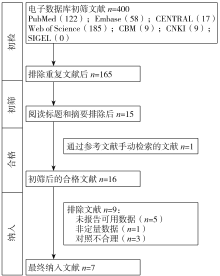
图 1
文献筛选流程图(PRISMA流程图)

表 5
纳入文献质量评价
| 作者 | 研究设计 | 研究类型 | 评级 |
|---|---|---|---|
| Kayalar等[ | RCT | 前瞻性 | A |
| Dindaro?lu等[ | RCT | 前瞻性 | A |
| Yildirim等[ | 分口试验 | 前瞻性 | B |
| Martins等[ | 分口试验 | 前瞻性 | A |
| Lemos Rinaldi等[ | 回顾队列 | 回顾性 | B |
| Erverdi等[ | 非随机同期对照 | 前瞻性 | C |
| Odenrick等[ | 非随机同期对照 | 前瞻性 | C |

图 2
偏倚风险评估A:RCT研究偏倚风险评估;B:非RCT研究偏倚风险评估。

| 1 | Bell RA. A review of maxillary expansion in relation to rate of expansion and patient,s age[J]. Am J Orthod, 1982, 81(1): 32-37. |
| 2 | Conroy-Piskai C, Galang-Boquiren MT, Obrez A, et al. Assessment of vertical changes during maxillary expansion using quad helix or bonded rapid maxillary expander[J]. Angle Orthod, 2016, 86(6): 925-933. |
| 3 | Angell E. Treatment of irregularity of the permanent or adult teeth[J]. Dent Cosmos, 1860, 1(10): 541-544, 599-600. |
| 4 | Haas AJ. Rapid expansion of the maxillary dental arch and nasal cavity by opening the midpalatal suture[J]. Angle Orthod, 1961, 31(2): 73-90. |
| 5 | Araújo MC, Bocato JR, Oltramari PV, et al. Tomographic evaluation of dentoskeletal effects of rapid maxillary expansion using Haas and Hyrax palatal expanders in children: a randomized clinical trial[J]. J Clin Exp Dent, 2020, 12(10): e922-e930. |
| 6 | Weissheimer A, de Menezes LM, Mezomo M, et al. Immediate effects of rapid maxillary expansion with Haas-type and hyrax-type expanders: a randomized clinical trial[J]. Am J Orthod Dentofacial Orthop, 2011, 140(3): 366-376. |
| 7 | Grünheid T, Larson CE, Larson BE. Midpalatal suture density ratio: a novel predictor of skeletal response to rapid maxillary expansion[J]. Am J Orthod Dentofacial Orthop, 2017, 151(2): 267-276. |
| 8 | Kartalian A, Gohl E, Adamian M, et al. Cone-beam computerized tomography evaluation of the maxillary dentoskeletal complex after rapid palatal expansion[J]. Am J Orthod Dentofacial Orthop, 2010, 138(4): 486-492. |
| 9 | Rungcharassaeng K, Caruso JM, Kan JY, et al. Factors affecting buccal bone changes of maxillary posterior teeth after rapid maxillary expansion[J]. Am J Orthod Dentofacial Orthop, 2007, 132(4): 428.e1-428.e8. |
| 10 | Garib DG, Henriques JF, Janson G, et al. Periodontal effects of rapid maxillary expansion with tooth-tissue-borne and tooth-borne expanders: a computed tomography evaluation[J]. Am J Orthod Dentofacial Orthop, 2006, 129(6): 749-758. |
| 11 | Timms DJ, Moss JP. An histological investigation into the effects of rapid maxillary expansion on the teeth and their supporting tissues[J]. Trans Eur Orthod Soc, 1971: 263-271. |
| 12 | Barbagallo LJ, Jones AS, Petocz P, et al. Physical properties of root cementum: part 10. Comparison of the effects of invisible removable thermoplastic appliances with light and heavy orthodontic forces on premolar cementum. A microcomputed-tomography study[J]. Am J Orthod Dentofacial Orthop, 2008, 133(2): 218-227. |
| 13 | Chan E, Darendeliler MA. Physical properties of root cementum: part 7. Extent of root resorption under areas of compression and tension[J]. Am J Orthod Dentofacial Orthop, 2006, 129(4): 504-510. |
| 14 | Bishara SE, Staley RN. Maxillary expansion: clinical implications[J]. Am J Orthod Dentofacial Orthop, 1987, 91(1): 3-14. |
| 15 | Odenrick L, Karlander EL, Pierce A, et al. Surface resorption following two forms of rapid maxillary expansion[J]. Eur J Orthod, 1991, 13(4): 264-270. |
| 16 | Kayalar E, Schauseil M, Kuvat SV, et al. Comparison of tooth-borne and hybrid devices in surgically assisted rapid maxillary expansion: a randomized clinical cone-beam computed tomography study[J]. J Craniomaxillofac Surg, 2016, 44(3): 285-293. |
| 17 | Dindaroğlu F, Doğan S. Evaluation and comparison of root resorption between tooth-borne and tooth-tissue bor-ne rapid maxillary expansion appliances: a CBCT study[J]. Angle Orthod, 2016, 86(1): 46-52. |
| 18 | Baysal A, Karadede I, Hekimoglu S, et al. Evaluation of root resorption following rapid maxillary expansion using cone-beam computed tomography[J]. Angle Orthod, 2012, 82(3): 488-494. |
| 19 | Higgins JPT, Green S. Cochrane handbook for systematic reviews of interventions[M]. Chichester: John Wiley & Sons, Ltd, 2011. |
| 20 | Wells G, Shea B, O'Connell D, et al. The Newcastle-Ottawa Scale (NOS) for assessing the quality of nonrandomised studies in meta-analyses[EB/OL]. [2019-10-15]. http://www.ohri.ca/programs/clinical_epidemiology/oxford.asp. |
| 21 | Guyatt GH, Oxman AD, Vist GE, et al. GRADE: an emerging consensus on rating quality of evidence and strength of recommendations[J]. BMJ, 2008, 336(7650): 924-926. |
| 22 | Erverdi N, Okar I, Kücükkeles N, et al. A comparison of two different rapid palatal expansion techniques from the point of root resorption[J]. Am J Orthod Dentofacial Orthop, 1994, 106(1): 47-51. |
| 23 | Lemos Rinaldi MR, Azeredo F, Martinelli de Lima E, et al. Cone-beam computed tomography evaluation of bone plate and root length after maxillary expansion using too-th-borne and tooth-tissue-borne banded expanders[J]. Am J Orthod Dentofacial Orthop, 2018, 154(4): 504-516. |
| 24 | Martins DC, Souki BQ, Cheib PL, et al. Rapid maxillary expansion: Do banded teeth develop more external root resorption than non-banded anchorage teeth[J]. Angle Orthod, 2016, 86(1): 39-45. |
| 25 | Yildirim M, Akin M. Comparison of root resorption after bone-borne and tooth-borne rapid maxillary expansion evaluated with the use of microtomography[J]. Am J Orthod Dentofacial Orthop, 2019, 155(2): 182-190. |
| 26 | Brezniak N, Wasserstein A. Orthodontically induced inflammatory root resorption. Part Ⅰ: The basic science aspects[J]. Angle Orthod, 2002, 72(2): 175-179. |
| 27 | Braun S, Bottrel JA, Lee KG, et al. The biomechanics of rapid maxillary sutural expansion[J]. Am J Orthod Dentofacial Orthop, 2000, 118(3): 257-261. |
| 28 | Wertz R, Dreskin M. Midpalatal suture opening: a normative study[J]. Am J Orthod, 1977, 71(4): 367-381. |
| 29 | Park JJ, Park YC, Lee KJ, et al. Skeletal and dentoalveolar changes after miniscrew-assisted rapid palatal expansion in young adults: a cone-beam computed tomography study[J]. Korean J Orthod, 2017, 47(2): 77-86. |
| 30 | Persson M, Thilander B. Palatal suture closure in man from 15 to 35 years of age[J]. Am J Orthod, 1977, 72(1): 42-52. |
| 31 | Korbmacher H, Schilling A, Püschel K, et al. Age-dependent three-dimensional microcomputed tomography ana-lysis of the human midpalatal suture[J]. J Orofac Orthop, 2007, 68(5): 364-376. |
| 32 | Knaup B, Yildizhan F, Wehrbein H. Age-related changes in the midpalatal suture. A histomorphometric study[J]. J Orofac Orthop, 2004, 65(6): 467-474. |
| 33 | Choi SH, Shi KK, Cha JY, et al. Nonsurgical miniscrew-assisted rapid maxillary expansion results in acceptable stability in young adults[J]. Angle Orthod, 2016, 86(5): 713-720. |
| 34 | Lin L, Ahn HW, Kim SJ, et al. Tooth-borne vs bone-borne rapid maxillary expanders in late adolescence[J]. Angle Orthod, 2015, 85(2): 253-262. |
| 35 | Kang S, Lee SJ, Ahn SJ, et al. Bone thickness of the palate for orthodontic mini-implant anchorage in adults[J]. Am J Orthod Dentofacial Orthop, 2007, 131(4 Suppl): S74-S81. |
| 36 | Karagkiolidou A, Ludwig B, Pazera P, et al. Survival of palatal miniscrews used for orthodontic appliance anchorage: a retrospective cohort study[J]. Am J Orthod Dentofacial Orthop, 2013, 143(6): 767-772. |
| 37 | Langford SR. Root resorption extremes resulting from clinical RME[J]. Am J Orthod, 1982, 81(5): 371-377. |
| 38 | Wilmes B, Nienkemper M, Drescher D. Application and effectiveness of a mini-implant- and tooth-borne rapid palatal expansion device: the hybrid hyrax[J]. World J Orthod, 2010, 11(4): 323-330. |
| 39 | Gunyuz Toklu M, Germec-Cakan D, Tozlu M. Periodontal, dentoalveolar, and skeletal effects of tooth-borne and tooth-bone-borne expansion appliances[J]. Am J Orthod Dentofacial Orthop, 2015, 148(1): 97-109. |
| 40 | Garrett BJ, Caruso JM, Rungcharassaeng K, et al. Skeletal effects to the maxilla after rapid maxillary expansion assessed with cone-beam computed tomography[J]. Am J Orthod Dentofacial Orthop, 2008, 134(1): 8-9. |
| [1] | 李雨霖, 徐静晨, 蒋晓鸽, 陈嵩. Twin-block矫治器对安氏Ⅱ类错 患者髁突影响的Meta分析[J]. 华西口腔医学杂志, 2023, 41(4): 463-470. 患者髁突影响的Meta分析[J]. 华西口腔医学杂志, 2023, 41(4): 463-470. |
| [2] | 卢倩, 郭柳媚, 毕小琴. 口腔癌患者术后吞咽障碍危险因素的系统评价[J]. 华西口腔医学杂志, 2022, 40(3): 328-334. |
| [3] | 包明哲, 刘伟, 余树容, 门乙, 韩波, 李春洁. 富血小板纤维蛋白在下颌第三磨牙拔除术的应用系统评价与Meta分析[J]. 华西口腔医学杂志, 2021, 39(5): 605-611. |
| [4] | 陈玲, 陈成, 李志勇, 张绮. 口内扫描数字化印模对固定修复临床应用效果的Meta分析[J]. 华西口腔医学杂志, 2021, 39(3): 306-312. |
| [5] | 程锋, 简志杉, 朱莹, 张纯燕, 胡丽, 陈莉莉. 传动直丝弓技术矫正骨性Ⅲ类错牙合的疗效分析[J]. 华西口腔医学杂志, 2020, 38(3): 301-307. |
| [6] | 赵丹,李月恒,杨正艳,蔡婷,吴晓艳,夏雨,周智. 干细胞局部应用对缺损面神经再生效果的系统评价[J]. 华西口腔医学杂志, 2020, 38(1): 59-68. |
| [7] | 刘俊玲,李洪发,闫卉. 对比研究快慢速扩弓治疗对鼻腔及上颌骨结构的影响[J]. 华西口腔医学杂志, 2019, 37(5): 533-536. |
| [8] | 王玉兰,王铁军,柳忠豪. 上颌切牙内收前后牙根及牙槽骨的变化[J]. 华西口腔医学杂志, 2018, 36(6): 638-645. |
| [9] | 刘畅, 王艳, 潘韦霖, 余昌浩, 黄静远, 华成舸. 初始弓丝材料对正畸治疗初始疼痛影响的系统评价与网状Meta分析[J]. 华西口腔医学杂志, 2018, 36(3): 296-300. |
| [10] | 刘媛, 吴龙, 孟凡琦, 侯鑫山, 赵今. 钙钠磷硅酸和硝酸钾牙膏抗牙本质敏感效果的系统评价和Meta分析[J]. 华西口腔医学杂志, 2018, 36(3): 301-307. |
| [11] | 金玥, 陈斌, 泥艳红, 闫福华. 牙周来源的牙周牙髓联合病变和牙周牙髓共存病变患牙牙周治疗时机的系统评价[J]. 华西口腔医学杂志, 2018, 36(2): 167-173. |
| [12] | 汪晓彤, 饶南荃, 谢静, 赵玉鸣, 葛立宏. 酪蛋白磷酸肽-无定型磷酸钙凝胶治疗釉质脱矿效果的系统评价[J]. 华西口腔医学杂志, 2017, 35(6): 629-635. |
| [13] | 黄鑫琪,岑啸,刘钧. 功能矫治对安氏Ⅱ类错 畸形患者髁突位置影响的Meta分析[J]. 华西口腔医学杂志, 2016, 34(6): 589-593. |
| [14] | 孔超 王旭霞 汪倩倩 韩媛媛 赵姝亚 张君. 雷尼酸锶对大鼠上颌快速扩弓影响的实验研究[J]. 华西口腔医学杂志, 2016, 34(4): 336-340. |
| [15] | 喻凤,邓锋,张翼,朱亚玲,张向凤,张赫,王华桥. 骨皮质切开术对比格犬前磨牙压入移动影响的三维形态学研究[J]. 华西口腔医学杂志, 2016, 34(3): 267-271. |
| 阅读次数 | ||||||
|
全文 |
|
|||||
|
摘要 |
|
|||||

