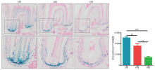
| [1] |
van der Velden U, Amaliya A, Loos BG , et al. Java project on periodontal diseases: causes of tooth loss in a cohort of untreated individuals[J]. J Clin Periodontol, 2015,42(9):824-831.
|
| [2] |
Ramseier CA . Potential impact of subject-based risk factor control on periodontitis[J]. J Clin Periodontol, 2005,32(Suppl 6):283-290.
|
| [3] |
Kretzschmar K, Watt FM . Lineage tracing[J]. Cell, 2012,148(1/2):33-45.
|
| [4] |
Tumbar T . Defining the epithelial stem cell niche in skin[J]. Science, 2004,303(5656):359-363.
|
| [5] |
Braun KM, Niemann C, Jensen UB , et al. Manipulation of stem cell proliferation and lineage commitment: visualisation of label-retaining cells in wholemounts of mouse epidermis[J]. Development, 2003,130(21):5241-5255.
|
| [6] |
Notta F, Doulatov S, Laurenti E , et al. Isolation of single human hematopoietic stem cells capable of long-term multilineage engraftment[J]. Science, 2011,333(6039):218-221.
|
| [7] |
Karhadkar SS, Bova GS, Abdallah N , et al. Hedgehog signalling in prostate regeneration, neoplasia and metastasis[J]. Nature, 2004,431(7009):707-712.
|
| [8] |
Kan C, Chen LJ, Hu YY , et al. Gli1-labeled adult mesenchymal stem/progenitor cells and hedgehog signaling contribute to endochondral heterotopic ossification[J]. Bone, 2018,109:71-79.
|
| [9] |
Shi Y, He GX, Lee WC , et al. Gli1 identifies osteogenic progenitors for bone formation and fracture repair[J]. Nat Commun, 2017,8:2043.
|
| [10] |
Zhao H, Feng JF, Ho TV , et al. The suture provides a niche for mesenchymal stem cells of craniofacial bones[J]. Nat Cell Biol, 2015,17(4):386-396.
|
| [11] |
Miura M, Gronthos S, Zhao M , et al. SHED: stem cells from human exfoliated deciduous teeth[J]. Proc Natl Acad Sci U S A, 2003,100(10):5807-5812.
|
| [12] |
Seo BM, Miura M, Gronthos S , et al. Investigation of multipotent postnatal stem cells from human periodontal ligament[J]. Lancet, 2004,364(9429):149-155.
|
| [13] |
Morsczeck C, Götz W, Schierholz J , et al. Isolation of precursor cells (PCs) from human dental follicle of wisdom teeth[J]. Matrix Biol, 2005,24(2):155-165.
|
| [14] |
Sonoyama W, Liu Y, Fang DJ , et al. Mesenchymal stem cell-mediated functional tooth regeneration in swine[J]. PLoS One, 2006,1(1):e79.
|
| [15] |
Alraies A, Alaidaroos NY, Waddington RJ , et al. Variation in human dental pulp stem cell ageing profiles reflect contrasting proliferative and regenerative capabilities[J]. BMC Cell Biol, 2017,18(1):12.
|

 )
)