华西口腔医学杂志 ›› 2019, Vol. 37 ›› Issue (2): 168-173.doi: 10.7518/hxkq.2019.02.008

• 基础研究 • 上一篇    下一篇

正畸牙压力区牙周膜细胞自噬相关蛋白Beclin-1与微管相关蛋白2轻链3的表达

吕佳岭1,徐洁1,曾锦1,党海霞2,余京泓1,赵娴1,徐晓梅1()   

  1. 1.西南医科大学附属口腔医院正畸科
    2.西南医科大学附属口腔医院口腔医学研究室,泸州 646000
  • 收稿日期:2018-06-15 修回日期:2018-12-13 出版日期:2019-04-01 发布日期:2019-04-28
  • 通讯作者: 徐晓梅 E-mail:648012557@qq.com
  • 作者简介:吕佳岭,硕士,E-mail: 1241940221@qq.com
  • 基金资助:
    泸州市科学技术和人才工作局应用基础研究(2018-JYJ-37);泸州市-西南医科大学联合项目(2016LZXNYD-T04);西南医科大学青年基金(2016136)

Expression of autophagy-related protein Beclin-1 and microtubule-associated protein 2 light chain 3 in periodontal ligament cells in orthodontic tooth pressure areas

Jialing Lü1,Jie Xu1,Jin Zeng1,Haixia Dang2,Jinghong Yu1,Xian Zhao1,Xiaomei Xu1()   

  1. 1. Dept. of Orthodontics, Hospital of Stomatology, Southwest Medical University, Luzhou 646000, China
    2.Stomatology Laboratory, Hospital of Stomatology, Southwest Medical University, Luzhou 646000, China
  • Received:2018-06-15 Revised:2018-12-13 Online:2019-04-01 Published:2019-04-28
  • Contact: Xiaomei Xu E-mail:648012557@qq.com
  • Supported by:
    Applied Basic Research of Office of Science and Technology and Talent Work of Luzhou(2018-JYJ-37);Joint Project of Luzhou-Southwest Medical University(2016LZXNYD-T04);Youth Fund of Southwest Medical University(2016136)

摘要:

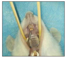
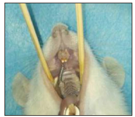
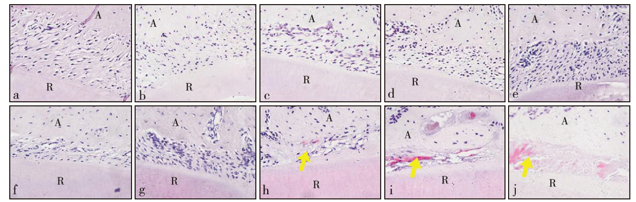
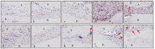
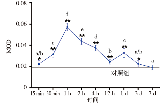
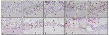
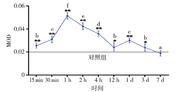
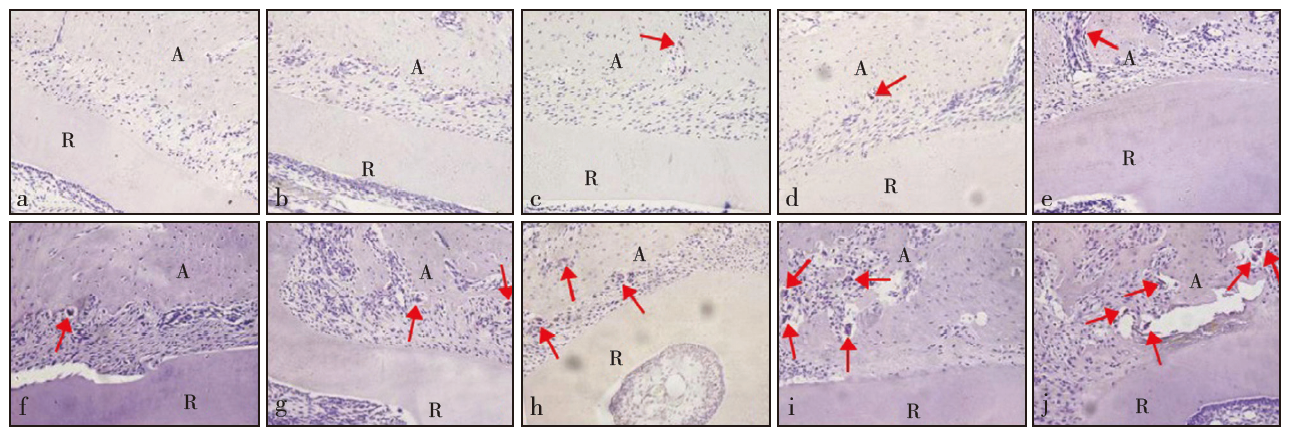
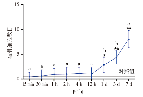
目的 探究正畸牙压力区牙周膜细胞自噬相关蛋白Beclin-1与微管相关蛋白2轻链3(LC3Ⅱ)的表达。方法 将60只雄性SD大鼠随机分为空白对照组和9个实验组,实验组正畸加力0.392 N近中移动右上第一磨牙,加力时间分别为15 min、30 min、1 h、2 h、4 h、12 h、1 d、3 d、7 d,空白对照组不做任何处理。处死大鼠后,行苏木精-伊红(HE)染色观察压力区牙周膜形态学变化、免疫组织化学染色检测Beclin-1与LC3Ⅱ的表达、抗酒石酸酸性磷酸酶(TRAP)染色计数破骨细胞。结果 HE染色显示,加力1 d后压力区牙周膜透明样变出现,并随加力时间延长逐渐加重。免疫组织化学染色显示,实验组Beclin-1和LC3Ⅱ表达均上调,1 h达峰值,随后逐渐降低,1 d时再次增强达一小峰值,后又回降,7 d时降低至基线水平。破骨细胞中也可见Beclin-1和LC3Ⅱ的表达。TRAP染色提示,加力1 d后破骨细胞数量开始增加。结论 自噬或许通过介导牙周膜透明样变发生和影响破骨细胞生物学作用参与正畸牙压力区牙周膜改建的过程。

关键词: 正畸牙移动, 牙周膜, 压力, 自噬

Abstract:

Objective To investigate the expression of autophagy-related protein Beclin-1 and microtubule-associated protein 2 light chain 3 (LC3Ⅱ) in periodontal ligament cells in orthodontic tooth pressure areas. Methods Sixty male SD rats were randomly divided into a blank control group and nine experimental groups. In the experimental groups, 0.392 N orthodontic force was used to move the first right upper molars for 15 min, 30 min, 1 h, 2 h, 4 h, 12 h, 1 d, 3 d, or 7 d. The blank control group did not receive any treatment. The rats were euthanized. Changes in the morphology of the periodontal membrane in the pressure areas were observed through hematoxylin and eosin (HE) staining. The expression levels of Beclin-1 and LC3Ⅱ were detected by immunohistochemical staining, and tartrate-resistant acid phosphatase (TRAP) staining was performed for the counting of osteoclasts. Results The HE stains showed that the hyalinization of the periodontal ligament appeared in the pressure areas after 1 day of exertion and was gradually aggravated. The immunohistochemical stains showed that the expression levels of Beclin-1 and LC3Ⅱ in the experimental groups gradually increased, peaked after 1 h, and then gradually decreased. The expression levels peaked again after 1 d, then decreased to baseline levels at 7 d of exertion. Beclin-1 and LC3Ⅱ were expressed in the osteoclasts. The TRAP stains indicated that the number of osteoclasts started to increase after 1 day. Conclusion Autophagy may participate in the process of periodontal ligament reconstruction in orthodontic tooth pressure areas by mediating the hyalini-zation of periodontal ligament and affecting the biological effects of osteoclasts.

Key words: orthodontic tooth movement, periodontal ligament, pressure, autophagy

中图分类号: