华西口腔医学杂志 ›› 2018, Vol. 36 ›› Issue (6): 619-622.doi: 10.7518/hxkq.2018.06.007

• 基础研究 • 上一篇    下一篇

牙本质玷污层特性对自粘接树脂水门汀粘接强度的影响

刘清,杨玉琼,聂蓉蓉(),孟翔峰   

  1. 南京大学医学院附属口腔医院,南京市口腔医院修复科,南京 210008
  • 收稿日期:2018-01-05 修回日期:2018-04-02 出版日期:2018-12-01 发布日期:2018-12-12
  • 通讯作者: 聂蓉蓉 E-mail:nrr63@sina.com
  • 作者简介:刘清,主任医师,学士,E-mail: 784859119@qq.com
  • 基金资助:
    南京市医学科技发展项目(YKK17142)

Influence of the dentin smear layer trait on the bond strength of the self-adhesive resin cements

Qing Liu,Yuqiong Yang,Rongrong Nie(),Xiangfeng Meng   

  1. Dept. of Prothodontics, Nanjing Stomatological Hospital, Medical School of Nanjing Uni-versity, Nanjing 210008, China
  • Received:2018-01-05 Revised:2018-04-02 Online:2018-12-01 Published:2018-12-12
  • Contact: Rongrong Nie E-mail:nrr63@sina.com
  • Supported by:
    Nanjing Medical Science and Technique Development Foundation(YKK17142)

摘要:

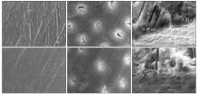
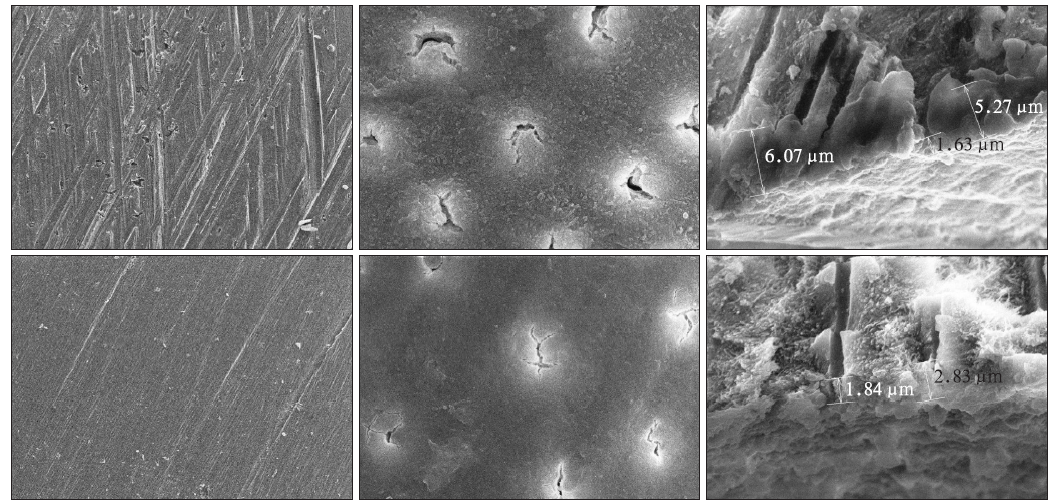
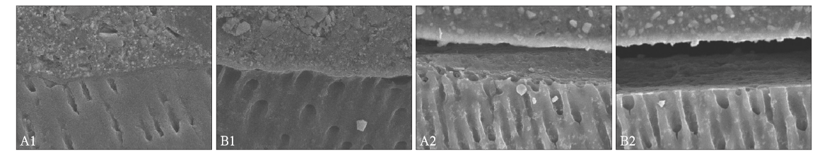
目的 评价牙本质表面玷污层的特性对自粘接型树脂水门汀粘接强度和耐久性的影响。方法 将48颗新鲜无龋的人第三恒磨牙暴露出牙本质表面,分别用标准粒径(105~125 μm)金刚砂车针(A组)和标准粒径+细粒径(25 μm)金刚砂车针进行研磨(B组)。研磨后的牙齿分别与Clearfil SA Cement(CSA)和Multilink Speed(MS)两种自粘接型复合树脂水门汀粘接,制成微拉伸试件,试件分别在水中存储24 h和2年,进行微拉伸强度测试,断裂后的试件使用体视显微镜观察粘接界面并记录断裂模式。采用扫描电子显微镜(SEM)观察研磨后的牙本质表面及牙本质-树脂粘接界面。结果 A组SEM观察到研磨后牙本质表面粗糙,玷污层较厚,牙本质小管口未完全栓塞;B组牙本质表面粗糙程度降低,牙本质小管口完全栓塞,玷污层变薄。CSA和MS的B组初期粘接强度显著低于A组(P<0.05);CSA和MS的粘接强度在水储存2年后出现显著降低(P<0.05),CSA的B组粘接强度显著低于A组(P<0.05),而MS的A组与B组间的粘接强度无明显差异(P>0.05)。结论 牙本质表面玷污层特点及自粘接型树脂水门汀类型都会对粘接强度及耐久性产生影响。

关键词: 自粘接型复合树脂水门汀, 牙本质, 玷污层, 微拉伸强度

Abstract:

Objective This work aimed to evaluate the influence of smear layer on the bonding effectiveness and durability of the self-adhesive resin cements to dentin.Methods A total of 48 fresh caries-free third molars with exposed dentin surface were divided into two groups. The dentin surfaces were treated using a standard grit diamond bur (group A) or further polished using a fine grit diamond bur (group B) and then bonded with either of the two self-adhesive resin cements, namely, Clearfil SA Cement (CSA, Kuraray) and Multilink Speed (MS, Ivoclar Vivadent). After 24 h or 2-year water storage, a microtensile bond strength test was performed.Results In group A, the dentin surface was rough, the smear layer was thick, and the dentin tubule orifice detritus showed low embolism value. In group B, the dentin surface roughness decreased, the embolism pro-portion increased, and the smear layer became thin. The initial bonding strengths of CSA and MS in group A were significantly lower than those in group B (P<0.05). The bonding strengths of CSA and MS significantly decreased after two years of water storage (P<0.05). For CSA, the bond strength of group B was significantly lower than that of group A (P<0.05). For MS, the differences in the bonding strength between groups A and B were insignificant (P>0.05).Conclusion The properties of the smear layer and the types of self-adhesive resin cement used affected the bond strength and durability.

Key words: self-adhesive resin cement, dentin, smear layer, micro-tensile strength

中图分类号: